. 2015 Jan;39(1):29-37.
doi: 10.1016/j.jgr.2014.07.003. Epub 2014 Aug 7.

Ginsenoside fractions regulate the action of monocytes and their differentiation into dendritic cells

Affiliations

Ginsenoside fractions regulate the action of monocytes and their differentiation into dendritic cells

Yeo Jin Lee et al. J Ginseng Res. 2015 Jan.

Abstract

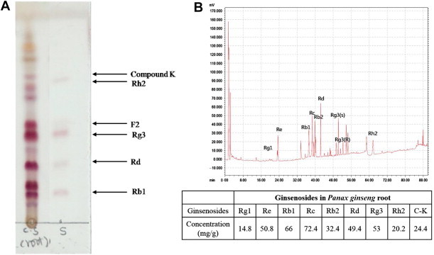
Background: Panax ginseng (i.e., ginseng) root is extensively used in traditional oriental medicine. It is a modern pharmaceutical reagent for preventing various human diseases such as cancer. Ginsenosides-the major active components of ginseng-exhibit immunomodulatory effects. However, the mechanism and function underlying such effects are not fully elucidated, especially in human monocytes and dendritic cells (DCs).

Methods: We investigated the immunomodulatory effect of ginsenosides from Panax ginseng root on CD14(+) monocytes purified from human adult peripheral blood mononuclear cells (PBMCs) and on their differentiation into DCs that affect CD4(+) T cell activity.

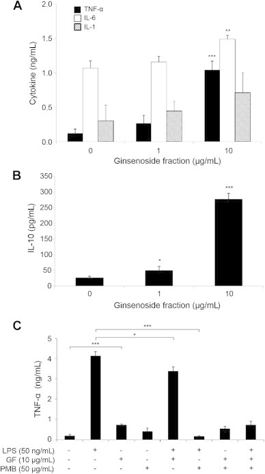
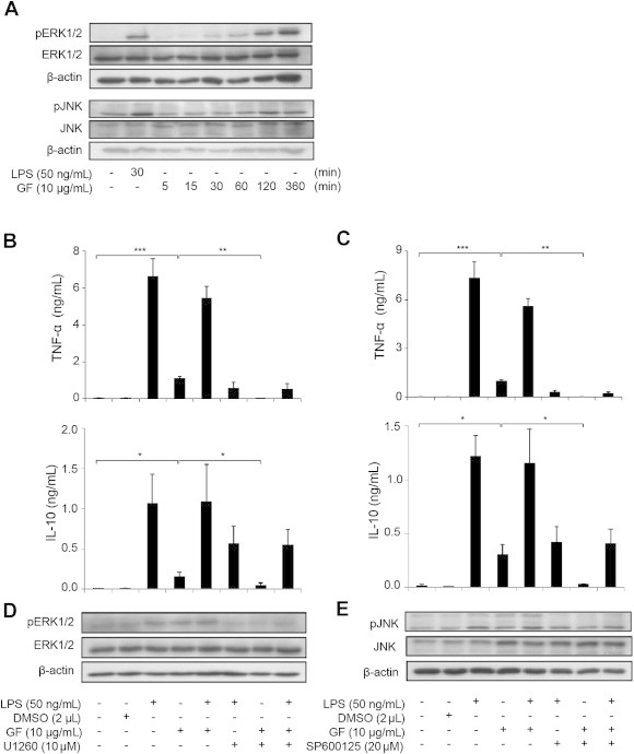
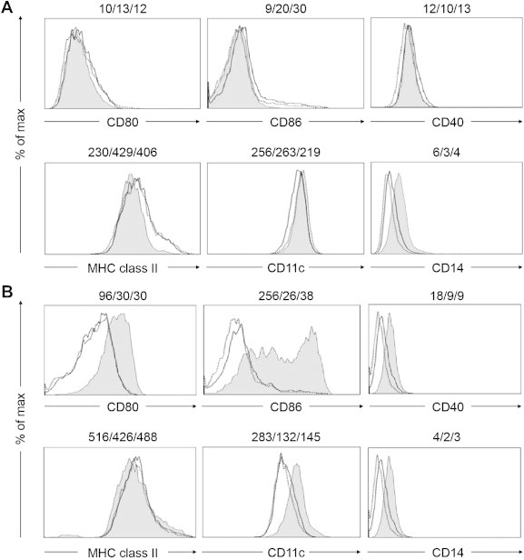
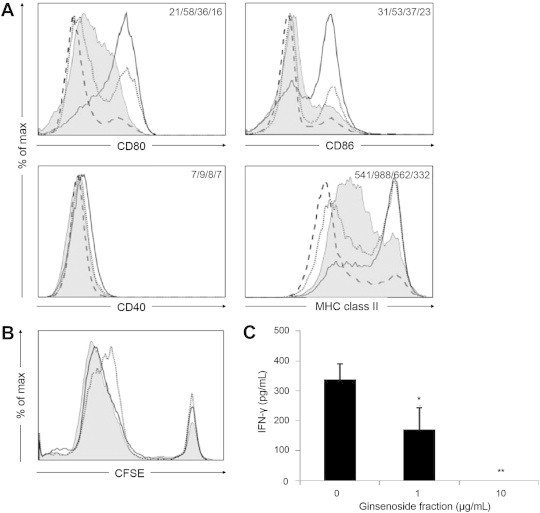
Results: After treatment with ginsenoside fractions, monocyte levels of tumor necrosis factor (TNF)-α, interleukin (IL)-6, and IL-10 increased through phosphorylation of extracellular signal-regulated kinase (ERK)1/2 and c-Jun N-terminal kinase (JNK), but not p38 mitogen-activated protein kinase (MAPK). After treatment with ginsenoside fractions, TNF-α production and phosphorylation of ERK1/2 and JNK decreased in lipopolysaccharide (LPS)-sensitized monocytes. We confirmed that DCs derived from CD14(+) monocytes in the presence of ginsenoside fractions (Gin-DCs) contained decreased levels of the costimulatory molecules CD80 and CD86. The expression of these costimulatory molecules decreased in LPS-treated DCs exposed to ginsenoside fractions, compared to their expression in LPS-treated DCs in the absence of ginsenoside fractions. Furthermore, LPS-treated Gin-DCs could not induce proliferation and interferon gamma (IFN-γ) production by CD4(+) T cells with the coculture of Gin-DCs with CD4+ T cells.

Conclusion: These results suggest that ginsenoside fractions from the ginseng root suppress cytokine production and maturation of LPS-treated DCs and downregulate CD4(+) T cells.

Keywords: CD14+ monocytes; CD4+ T cells; Panax ginseng; dendritic cells; ginsenosides.


Figures

Fig. 1
Fig. 1
Components of crude root ginsenoside. The qualitative analysis of the ginsenoside fractions are by (A) thin-layer chromatography and (B) high-performance liquid chromatography.
Fig. 2
Fig. 2
Apoptosis in CD14+ monocytes in the presence or absence of ginsenoside fractions during their differentiation into dendritic cells. (A and B) For 5 d, the CD14+ monocytes (1 × 106 cells) were cultured with interleukin-4 (IL-4; 500 U/mL) and granulocyte macrophage colony-stimulating factor (GM-CSF; 800 U/mL) in the presence or absence of ginsenoside fractions (1 μg/mL or 10 μg/mL). The expression levels of Annexin V and propidium iodide were measured by flow cytometry. *Indicates statistical significance at p < 0.05, compared to the control group. CON, control; PI, Propidium iodide.
Fig. 3
Fig. 3
Cytokine production by lipopolysaccharide (LPS)-sensitized CD14+ monocytes in the presence or absence of ginsenoside fractions. For 24 h, CD14+ monocytes were treated with ginsenoside fractions (0 μg/mL, 1 μg/mL, or 10 μg/mL). The production of (A) TNF-α, IL-6, IL-1β, and (B) IL-10 was measured by an enzyme-linked immunosorbent assay kit. (C) For 1 h, the cells were pretreated with PMB (50 μg/mL). This was followed by treatment with ginsenoside fractions (10 μg/mL) and/or lipopolysaccharide (LPS; 50 ng/mL) for an additional 24 h. The culture supernatants were collected and the TNF-α levels were measured. Statistical significance is denoted by *p < 0.05, **p < 0.01, and ***p < 0.001, compared to the control group. GF, ginsenoside fraction; IL, interleukin; PMB, polymyxin B; TNF-α, tumor necrosis factor alpha.
Fig. 4
Fig. 4
Activation of ERK1/2 and JNK signaling in lipopolysaccharide (LPS)-sensitized CD14+ monocytes with or without ginsenoside fractions. (A) The CD14+ monocytes were stimulated with LPS (50 ng/mL) for 30 min or with ginsenoside fractions (10 μg/mL) for 0 min, 5 min, 15 min, 30 min, 60 min, 120 min, or 360 min. Forty micrograms of protein from each cell lysate were subjected to Western blot analysis for changes in pERK and pJNK activity. The results are representative of three independent experiments. After 1 h pretreatment with (B and D) 10 μM U0126 or (C and E) 20 μM SP600125, the cells were treated with ginsenoside fractions (10 μg/mL) for 24 h in the presence or absence of LPS (50 ng/mL). (B and C) The production of TNF-α was measured by enzyme-linked immunosorbent assay. (D and E) The phosphorylation of ERK1/2 and JNK was evaluated by Western blot. ERK1/2, extracellular signal-regulated kinase 1/2; GF, ginsenoside fraction; IL-10, interleukin 10; JNK, c-Jun N-terminal kinase; pERK, phosphorylated ERK; TNF-α, tumor necrosis factor alpha.
Fig. 5
Fig. 5
Suppression of the expression of cell surface molecules during dendritic cell differentiation in the presence or absence of ginsenoside fractions. The CD14+ monocytes were treated with interleukin 4 (IL-4; 500 U/mL) and granulocyte macrophage colony-stimulating factor (GM-CSF; 800 U/mL) in the presence or absence of ginsenoside fractions (gray area, 0 μg/mL; dotted line, 1 μg/mL; unbroken line, 10 μg/mL) for (A) 3 d or (B) 5 d. The expression levels of the surface markers CD80, CD86, CD40, MHC class II, CD11c, and CD14 were measured by flow cytometry. The numbers in the panels indicate the mean fluorescence intensity of each ginsenoside molecule at the concentration of 0 μg/mL, 1 μg/mL, or 10 μg/mL, respectively. The data are representative of three independent experiments. MHC, major histocompatibility complex.
Fig. 6
Fig. 6
Dendritic cell (DC) maturation in the presence of ginsenoside fractions and the regulation of CD4+ T cells. (A) The DCs treated with ginsenoside fractions (the unbroken, dotted, and broken lines indicate 0 μg/mL, 1 μg/mL, and 10 μg/mL, respectively) during differentiation were stimulated with lipopolysaccharide (LPS) for 24 h. Their expression of CD80, CD86, CD40, and MHC class II was assessed by flow cytometry. The gray area indicates the untreated DCs (i.e., the control). The numbers in the panels indicate the mean fluorescence intensity (MFI) of the gray area, unbroken line, dotted line, and broken line. (B) During differentiation for 48 h, ethanol-killed Staphylococcus aureus (107 CFU/mL) was primed to DCs treated with ginsenoside fractions (the gray area, unbroken line, and dotted line indicate 0 μg/mL, 1 μg/mL, and 10 μg/mL, respectively). The cells were cocultured with CFSE-labeled CD4+ T cells for 3 d or 5 d. Proliferation was then evaluated by flow cytometry. (C) The interferon gamma (IFN-γ) levels in the supernatants at the end of the 3-d culture were measured by enzyme-linked immunosorbent assay. Statistical significance is denoted at *p < 0.05 and **p < 0.01. CFSE, carboxyfluorescein succinimidyl ester; MHC, major histocompatibility complex.

Similar articles

Cited by

References

    1. Kiefer D., Pantuso T. Panax ginseng. Am Fam Physician. 2003;68:1539–1542. - PubMed
    1. Lee T.K., Johnke R.M., Allison R.R., O'Brien K.F., Dobbs L.J., Jr. Radioprotective potential of ginseng. Mutagenesis. 2005;20:237–243. - PubMed
    1. Kang K.A., Kang J.H., Yang M.P. Ginseng total saponin enhances the phagocytic capacity of canine peripheral blood phagocytes in vitro. Am J Chin Med. 2008;36:329–341. - PubMed
    1. Fan Z.H., Isobe K., Kiuchi K., Nakashima I. Enhancement of nitric oxide production from activated macrophages by a purified form of ginsenoside (Rg1) Am J Chin Med. 1995;23:279–287. - PubMed
    1. Park E.K., Choo M.K., Han M.J., Kim D.H. Ginsenoside Rh1 possesses antiallergic and anti-inflammatory activities. Int Arch Allergy Immunol. 2004;133:113–120. - PubMed