. 2014 Dec 14;5(1):18.
doi: 10.1186/s13293-014-0018-2. eCollection 2014.

Human umbilical endothelial cells (HUVECs) have a sex: characterisation of the phenotype of male and female cells

Affiliations

Human umbilical endothelial cells (HUVECs) have a sex: characterisation of the phenotype of male and female cells

Roberta Addis et al. Biol Sex Differ. .

Abstract

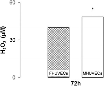
Background: Human umbilical endothelial cells (HUVECs) are widely used to study the endothelial physiology and pathology that might be involved in sex and gender differences detected at the cardiovascular level. This study evaluated whether HUVECs are sexually dimorphic in their morphological, proliferative and migratory properties and in the gene and protein expression of oestrogen and androgen receptors and nitric oxide synthase 3 (NOS3). Moreover, because autophagy is influenced by sex, its degree was analysed in male and female HUVECs (MHUVECs and FHUVECs).

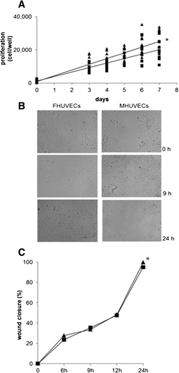
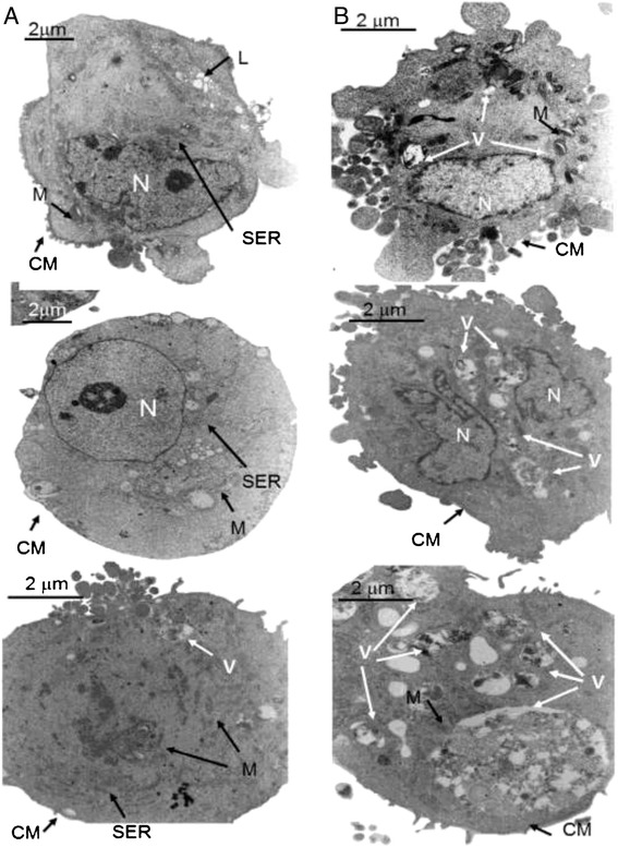
Methods: Umbilical cords from healthy, normal weight male and female neonates born to healthy non-obese and non-smoking women were studied. HUVEC morphology was analysed by electron microscopy, and their function was investigated by proliferation, viability, wound healing and chemotaxis assays. Gene and protein expression for oestrogen and androgen receptors and for NOS3 were evaluated by real-time PCR and Western blotting, respectively, and the expression of the primary molecules involved in autophagy regulation [protein kinase B (Akt), mammalian target of rapamycin (mTOR), beclin-1 and microtubule-associated protein 1 light chain 3 (LC3)] were detected by Western blotting.

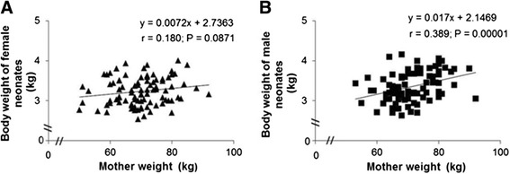
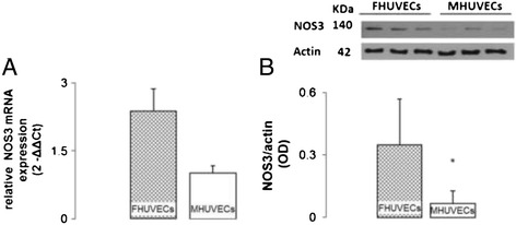
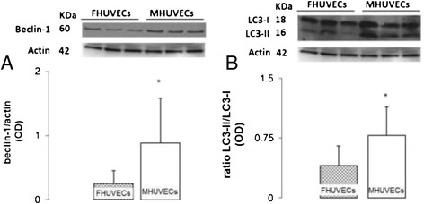
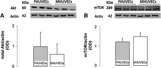
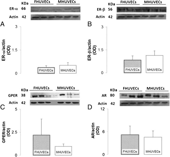
Results: Cell proliferation, migration NOS3 mRNA and protein expression were significantly higher in FHUVECs than in MHUVECs. Conversely, beclin-1 and the LC3-II/LC3-I ratio were higher in MHUVECs than in FHUVECs, indicating that male cells are more autophagic than female cells. The expression of oestrogen and androgen receptor genes and proteins, the protein expression of Akt and mTOR and cellular size and shape were not influenced by sex. Body weights of male and female neonates were not significantly different, but the weight of male babies positively correlated with the weight of the mother, suggesting that the mother's weight may exert a different influence on male and female babies.

Conclusions: The results indicate that sex differences exist in prenatal life and are parameter-specific, suggesting that HUVECs of both sexes should be used as an in vitro model to increase the quality and the translational value of research. The sex differences observed in HUVECs could be relevant in explaining the diseases of adulthood because endothelial dysfunction has a crucial role in the pathogenesis of cardiovascular diseases, diabetes mellitus, neurodegeneration and immune disease.

Keywords: Autophagy; Birth weight; HUVECs; Sex differences.


Figures

Figure 1
Figure 1
Correlation between neonates and maternal weight (kg) stratified by sex. (A, B) Each chart contains the equation of the line, the Spearman product moment correlation coefficient and the P value.
Figure 2
Figure 2
Proliferation (A) and migration of FHUVECs and MHUVECs (B, C). Linear regression analysis (A) of the proliferation of FHUVECs (▲, n = 10) and MHUVECs (▄, n = 7). *The comparison of the two slopes indicates that the slopes are different (P < 0.001). (B) Representative photographs of wound closure at 0, 9 and 24 h taken at × 4 magnification. (C) Mean of basal migratory capacity of MHUVECs (▄, n = 8) and FHUVECs (▲, n = 9). The data are expressed as median percentage of wound closure + MAD compared with the initial area (0 h). *P = 0.024.
Figure 3
Figure 3
Electron microscopic analysis of FHUVECs and MHUVECs. ( A) FHUVECs have little smooth endoplasmic reticulum (SER), abundant lysosomes (L) and a normal cytoplasmic membrane (CM). M and N indicate the mitochondria and nucleus, respectively. (B) MHUVECs have abundant autophagic vacuoles (V) in different stages of digestion and a CM with many blebs. M and N indicate the mitochondria and nucleus, respectively. (A) Magnification × 6,900, scale bars: 2 mm. (B) Magnification × 9,000, scale bars: 2 mm.
Figure 4
Figure 4
H 2 O 2 levels in the supernatants obtained from FHUVECs and MHUVECs. Values are expressed as medians + MAD of 15 independent samples at 72 h (*P = 0.026).
Figure 5
Figure 5
NOS3 mRNA expression and NOS3 protein expression in FHUVECs and MHUVECs. (A) Relative NOS3 mRNA expression. The values are expressed as the means ± SEM (*P = 0.036) of four independent experiments for each sex. (B) Representative Western blot and densitometric analysis of NOS3 expression. The values are expressed as medians + MAD (*P < 0.05) of 17 independent experiments for each sex normalised to actin levels.
Figure 6
Figure 6
Beclin-1 and LC3II to LC3I ratio in FHUVECs and MHUVECs. (A) Representative Western blot and densitometric analysis of beclin-1 expression. (B) Ratio of LC3-II to LC3-I and representative Western blot. Values for each protein are expressed as medians + MAD (*P < 0.05) from at least 18 experiments and normalised to actin levels.
Figure 7
Figure 7
Akt and mTOR expression levels in FHUVECs and MHUVECs. (A) Representative Western blot and densitometric analysis of Akt expression in FHUVECs and MHUVECs. The values are expressed as medians + MAD of at least six independent experiments for each sex. Akt expression was normalised to actin levels. (B) Representative Western blot and densitometric analysis of mTOR expression obtained from FHUVECs and MHUVECs. The data are the means ± SEM of at least six independent experiments for each group normalised to actin.
Figure 8
Figure 8
Protein expression of ERs, GPER and AR in FHUVECs and MHUVECs. (A, B) Representative Western blot and densitometric analysis of ER-α and ER-β expression, respectively, in FHUVECs and MHUVECs. Data are reported as the means ± SEM of at least six independent experiments for each group normalised on actin. (C, D) Representative Western blot and densitometric analysis of GPER and AR obtained from FHUVECs and MHUVECs. Data are reported as medians ± MAD for GPER30 and as means ± SEM for AR of at least four independent experiments for each group normalised on actin.

References

    1. Sena CM, Pereira AM, Seica R. Endothelial dysfunction - a major mediator of diabetic vascular disease. Biochim Biophys Acta (BBA) - Mol Basis Dis. 2013;1832:2216–2231. doi: 10.1016/j.bbadis.2013.08.006. - DOI - PubMed
    1. Xiao L, Liu Y, Wang N. New paradigms in inflammatory signaling in vascular endothelial cells. Am J Physiol Heart Circ Physiol. 2014;306:H317–H325. doi: 10.1152/ajpheart.00182.2013. - DOI - PubMed
    1. Dai R, Ahmed S. Sexual dimorphism of miRNA expression: a new perspective in understanding the sex bias of autoimmune diseases. Ther Clin Risk Manag. 2014;10:151–163. - PMC - PubMed
    1. Lyros E, Bakogiannis C, Liu Y, Fassbender K. Molecular links between endothelial dysfunction and neurodegeneration in Alzheimer’s disease. Curr Alzheimer Res. 2014;11:18–26. doi: 10.2174/1567205010666131119235254. - DOI - PubMed
    1. Legato MJ. Principles of Gender-Specific Medicine: 2. 2. San Diego: Academic Press; 2009.