

Recurrent FGFR3-TACC3 fusion gene in nasopharyngeal carcinoma
- PMID: 25535896
- PMCID: PMC4622012
- DOI: 10.4161/15384047.2014.961874
Recurrent FGFR3-TACC3 fusion gene in nasopharyngeal carcinoma
Abstract
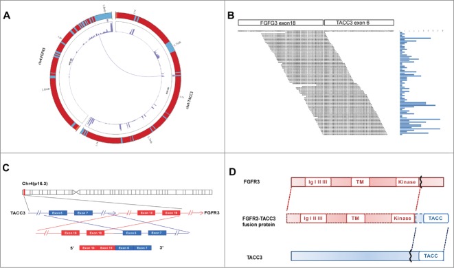
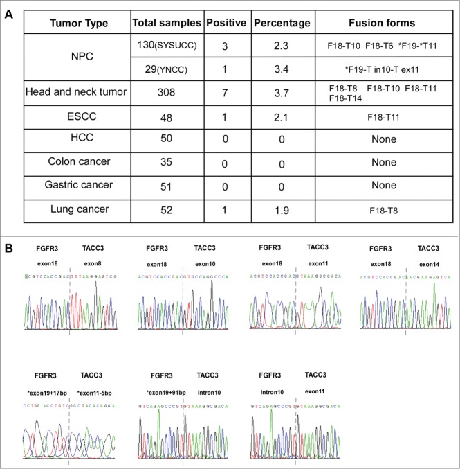
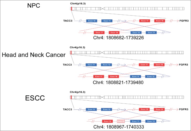
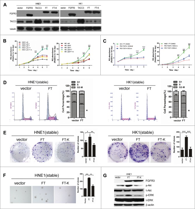
Nasopharyngeal carcinoma (NPC) is one of the most common head and neck malignancies and exhibits regional differences in incidence. Because many fusion genes have been discovered in different types of tumors over the past few years, we aimed to investigate the existence of a fusion gene in primary NPC patients using RNA-seq. In this study, for the first time, we found that fibroblast growth factor receptor 3-transforming acidic coiled-coil-containing protein 3 (FGFR3-TACC3) fusion transcripts are recurrently detected in NPC. The presence of this fusion gene was also detected in head and neck cancer, esophageal squamous cell carcinoma (ESCC), and lung cancer. Furthermore, we found certain new isoforms of the FGFR3-TACC3 fusion transcripts, such as a gene fusion between exon 18 of FGFR3 and exon 6 or exon 14 of TACC3 and agene fusion between exon 19 of FGFR3 and exon 11 of TACC3. In addition, we showed that the FGFR3-TACC3 fusion gene promotes cell proliferation, colony formation, and transforming ability in vitro, whereas the FGFR3-TACC3 K508M mutant or treatment with the FGFR inhibitor PD173074 abrogates these effects, suggesting that FGFR3-TACC3 most likely exerts its effects through activation of FGFR kinase activity. This activation likely leads to the development of NPC. Additionally, FGFR3-TACC3 could trigger activation of the ERK and Akt signaling pathways, whereas FGFR3-TACC3 K508M mutant could not, suggesting that these 2 signaling pathways might be involved in the function of FGFR3-TACC3. Taken together, our data demonstrated the oncogenic role of FGFR3-TACC3 in vitro, indicating that FGFR3-TACC3 may be useful as a diagnostic marker and therapeutic target in cancers.
Keywords: CCND1, cyclin D1; DMSO, dimethyl sulfoxide; DTT, DL-dithiothreitol; FBS, fetal bovine serum; FGFR3, fibroblast growth factor receptor 3; FGFR3-TACC3; LTBR, lymphotoxin β receptor; MTT, 3-(4,5-dimethylthiazol-2-yl)-2,5-diphenyltetrazoliumbromide; NPC; NPC, nasopharyngeal carcinoma; PBS, phosphate-buffered saline; PI, propidium iodide; RT-PCR, reverse transcription-PCR; SDS, sodium dodecyl sulfate; TACC3, transforming acidic coiled-coil-containing protein 3; fusion gene; proliferation; tumorigenesis.
Figures
References
-
- Chan ATC, Teo PML, Johnson PJ. Nasopharyngeal carcinoma. Ann Oncol 2002; 13:1007-15; PMID:12176778; http://dx.doi.org/ 10.1093/annonc/mdf179 - DOI - PubMed
-
- Dolly P H, Johnson PJ. Introduction: nasopharyngeal cancer. Semin Cancerbiol 2002; 12:419.
-
- Kwok Wai L, Ka Fai T, Huang ADP. Focus on nasopharyngeal carcinoma. Cancer C 2004; 5:423-28; http://dx.doi.org/ 10.1016/S1535-6108(04)00119-9 - DOI - PubMed
-
- Razak AR, Siu LL, Liu FF, Ito E, O'Sullivan B, Chan K. Nasopharyngeal carcinoma: the next challenges. Eur J Cancer 2010; 46:1967-78; PMID:20451372; http://dx.doi.org/ 10.1016/j.ejca.2010.04.004 - DOI - PubMed
-
- Tang LQ, Chen QY, Guo SS, Chen WH, Li CF, Zhang L, Lai XP, He Y, Xu YX, Hu DP, et al. The impact of plasma epstein-barr virus DNA and fibrinogen on nasopharyngeal carcinoma prognosis: an observational study. British J Cancer 2014; 111:1102-11; PMID:25051405; http://dx.doi.org/ 10.1038/bjc.2014.393 - DOI - PMC - PubMed
Publication types
MeSH terms
Substances
LinkOut - more resources
Full Text Sources
Other Literature Sources
Molecular Biology Databases
Research Materials
Miscellaneous