. 2014;15(12):1622-34.
doi: 10.4161/15384047.2014.962305.

Enhancement of cellular radiation sensitivity through degradation of Chk1 by the XIAP-XAF1 complex

Affiliations

Enhancement of cellular radiation sensitivity through degradation of Chk1 by the XIAP-XAF1 complex

Kwang Seok Kim et al. Cancer Biol Ther. 2014.

Abstract

X-linked inhibitor of apoptosis (XIAP) and Chk1 are potential molecular targets in radiotherapy. However, their molecular association in the regulation of radiation sensitivity has been rarely studied. Here, we show that XIAP modulates radiation sensitivity by regulating stability of Chk1 in lung cancer cells. Both Chk1 and XIAP are highly expressed in various lung cancer cells. Overexpression of XIAP increased cell survival following genotoxic treatments by preventing downregulation of Chk1. However, XIAP reversed Chk1-protective activity in the presence of XIAP-associated factor 1 (XAF1) by degrading Chk1 via ubiquitination-dependent proteasomal proteolysis. The XIAP-XAF1 complex-mediated Chk1 degradation also required CUL4A and DDB1. Chk1 or XIAP was associated with DDB1 and CUL4A. Depletion of CUL4A or DDB1 prevented the XIAP-XAF1-mediated Chk1 degradation suggesting involvement of a CUL4A/DDB1-based E3 ubiquitin ligase in the process or its collaboration with XIAP E3 ligase activity. Taken together, our findings show that XIAP plays a dual role in modulation of Chk1 stability and cell viability following IR. In the absence of XAF1, XIAP stabilizes Chk1 under IR with corresponding increase of cell viability. By contrast, when XAF1 is overexpressed, XIAP facilitates Chk1 degradation, which leads to enhancement of radiation sensitivity. This selective regulation of Chk1 stability by XIAP and XAF1 could be harnessed to devise a strategy to modulate radiation sensitivity in lung cancer cells.

Keywords: Chk1; Chk1, Checkpoint kinase 1; DDR, DNA damage responses; DNA damage response; IFN-γ, interferon-gamma; IR, ionizing radiation; RING domain; XAF1; XAF1, XIAP-associated factor 1; XIAP; XIAP, X-linked inhibitor of apoptosis; radiation sensitivity; synthetic lethality.


Figures

Figure 1.
Figure 1.
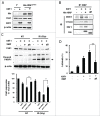
Prevention of Chk1 downregulation by XIAP under genotoxic treatments. (A) Whole cell extracts from WI38, A549, NCI-H1299, and NCI-H460 cells were immunoblotted with XIAP, Chk1, and β-actin antibodies. WI38 is a human diploid lung fibroblast cell line, whereas the others are human lung cancer cell lines. (B) A549 cells were transfected either with vector control or with plasmid construct encoding XIAP and incubated for 36 hr. The cells were harvested after 5 hr following treatments with 10 Gy IR, 500 nM camptothecin (CPT), or 5 mM hydroxyurea (HU). Whole cell extracts were prepared and immunoblotted with the specified antibodies. β-actin antibody was used as a protein loading control. γ-H2AX detection was used as a marker of DNA damage. (C) Graphical representation of the results in B. The relative Chk1 protein levels were normalized to β-actin expression as determined by densitometry. Values are means ± standard deviation (SD) of 3 experiments. **P < 0.01 versus control. (D) Cell survival was measured by MTT assay following genotoxic treatments. A549 cells were transfected either with vector control (control) or with plasmid construct encoding XIAP (XIAP) and treated as in B. The cell survival was normalized against untreated control (NT). Values are means ± SD. Statistical analysis of the results was carried out as in C.
Figure 2.
Figure 2.
Enhancement of cellular radiation sensitivity by depletion of XIAP. (A) A549 cells were transfected with Chk1 construct and/or with SiRNAs against XIAP. Cells were treated with no IR or with 5 Gy of IR. Whole cell extracts were used to detect Chk1 and XIAP by protein gel blot analysis. β-actin was used as a loading control. The band intensity of Chk1 in each lane is normalized against that of the untreated control (lane 1). (B) Clonogenic cell survival was measured for the cells: untreated control (Con), Chk1-overexpressed cells by transient transfection (Chk1), XIAP-depleted cells by the corresponding SiRNA transfection (siXIAP), and Chk1-overexpressed cells with XIAP depletion (Chk1/siXIAP). Cell survival values were normalized to those of the unirradiated cells. (C) Colony formation of the cells, which were treated as in B, is shown.
Figure 3.
Figure 3.
XAF1-mediated degradation of Chk1. (A) A549 cells were transfected with control vector or with plasmid construct encoding XAF1, and incubated for 30 hr. Whole cell extracts were immunoblotted with the indicated antibodies. β-actin was used as a loading control. (B) XAF1- or empty vector-expressed cells were treated for 4 hr with DMSO or with 100μg/ml cycloheximide (CHX). Levels of Chk1 protein were determined by protein gel blot analysis. β-actin was used as a loading control. (C) XAF1- or empty vector-expressed cells were treated with 100μg/ml CHX for the indicated times. Levels of Chk1 protein were determined by protein gel blot analysis (upper panel). Band intensity of Chk1 protein was determined by densitometry, and normalized against the zero hour control, which was set as 100%. Graphical representation of the densitometric analysis of Chk1 allows measurement of Chk1 half-life (Lower panel). (D) XAF1- or empty vector-expressed cells were treated with 10 μM MG132 for 4 hr. Levels of Chk1 protein were determined by western blotting analysis. β-actin was used as a loading control.
Figure 4.
Figure 4.
Ubiquitin-dependent Chk1 proteolysis is mediated by the XIAP-XAF1 complex. (A) A549 cells were transfected with XAF1-encoding construct or with empty vector. Whole cell extracts were prepared, immunoprecipitated with Chk1 antibody, and immunoblotted with XIAP and with XAF1 antibodies. (B) A549 cells were transfected with the constructs encoding the proteins as specified (V, XIAP, XAF1, and XIAP/XAF1). XIAP/XAF1 indicates co-transfection with both constructs. V is an empty vector transfection. Cell extracts were prepared and then immunoblotted (IB) with the indicated antibodies (upper panel). Also Chk1 or XIAP was immunoprecipitated (IP) and co-IP of XAF1 was assessed by protein gel blot analysis (IB) (lower panel). (C) Cells with or without XAF1 overexpression were transfected with XIAP siRNA or scrambled siRNA as a control. Cell extracts were subjected to protein gel blot analysis (IB) (upper panel) and immunoprecipitation (IP) with the indicated antibodies (lower panel). Co-IP of XAF1 was assessed by protein gel blot analysis (IB). (D) A549 cells with or without co-overexpression of XAF1 and XIAP were transfected with a plasmid construct encoding ubiquitin. Ubiquitination of total proteins was determined by protein gel blot analysis of whole cell extracts (left panel). Ubiquitination of endogenous Chk1 was monitored by immunoprecipitation (IP) with antibody against ubiquitin. Co-IP of Chk1 was assessed by protein gel blot analysis (IB) (right panel). (E) A549 cells were transfected with XAF1 alone or co-transfected with XAF1 and XIAP (XAF1/XIAP). Vector (V)-transfection is a control. Cells were fixed, incubated with anti-Chk1 antibody, and stained with Alexa 488-conjugated secondary antibody. Nuclei were stained with DAPI (blue, x1000). Fluorescent images were obtained using a fluorescence microscope.
Figure 5.
Figure 5.
Involvement of DDB1 and CUL4A in the XIAP-XAF1 complex-mediated Chk1 degradation. (A) A549 cells were transfected with constructs that are specified (Vector (V), XIAP, XAF1, XIAP/XAF1). XIAP/XAF1 indicates co-transfection with both constructs. Whole cell extracts were prepared and subjected to protein gel blot analysis (IB) to detect CUL4A, DDB1 and PCNA (upper panel). β-actin was used as a loading control. Immunoprecipitation (IP) was performed by using antibodies against Chk1 or XIAP. Co-IP of DDB1, CUL4A, or PCNA was detected by protein gel blot analysis (lower panel). (B) CUL4A, DDB1 or PCNA was depleted by transfection of the specified SiRNA in A549 cells with or without XAF1 overexpression. Levels of the specified proteins were determined by protein gel blot analysis. Band intensity of Chk1 protein was determined by densitometry, and normalized against that of the non-treated control (NC), which was set as 100%.
Figure 6.
Figure 6.
Role of the RING domain of XIAP in the XIAP-XAF1-mediated Chk1 degradation. (A) Vector (V) or His-XIAPΔRING-expressed cells were transfected with a construct that encodes XAF1. XIAPΔRING is histidine-tagged and detected by western blot analysis with antibody against the tag (His). Chk1, XAF1 and β-actin were also detected by western blot analysis. (B) A549 cells were transfected with constructs that are specified in the figure (XAF1, His-XIAP and ΔR). ΔR indicates XIAPΔRING. Cell extracts were prepared and immunoprecipitated with antibody against XIAP. Co-immunoprecipitated proteins were detected with the specified antibodies by western blot analysis (IB). (C) A549 cells were transfected with constructs that are specified in the figure. Cells were treated with or without 5 Gy IR and incubated for 12 hr. Cells were harvested and whole cell extracts were prepared to detect Chk1 and PARP1 cleavage by western blot analysis (upper panel). An arrow indicates cleaved PARP1. Levels of Chk1 protein in each lane, as determined by densitometry, were normalized to those of β-actin and represented as a graph (lower panel). Values are means ± SD of 3 experiments. **P < 0.01 vs. XIAP/XAF1 coexpression. (D) Measurement of cell death in XAF1-, XIAP/XAF1-, and XIAPΔRING/XAF1-expressed cells following 5 Gy IR. Treated cells were incubated for 24 hr and stained with Annexin V-FITC and propidium iodide (PI). Fluorescence intensity of Annexin V-FITC and propidium iodide was analyzed by flow cytometry. Values are means ± SD of 3 independent experiments. **P < 0.01 vs. XIAP/XAF1 coexpression.
Figure 7.
Figure 7.
Enhancement of radiation sensitivity in interferon gamma-treated lung cancer cells. (A) Indicated lung cancer cells were treated with interferon-gamma (IFN-γ). Whole cell extracts were prepared and subjected to western blot analysis. Antibodies against XAF1, XIAP, and Chk1 were used for the analysis. β-actin was used as a loading control. (B) Graphical representation of levels of Chk1 expression in Figure 7A. Expression of Chk1 protein following IFN-γ treatment was measured by densitometric analysis of Chk1 bands in Figure 7A and normalized against non-treated control (NT) in each cell line. Values are means ± SD with *P < 0.01. (C) Clonogenic cell survival following a combined treatment with IFN-γ and IR in lung cancer cell lines. Cells were treated with 200 Units/ml IFN-γ for 1 hour before IR. IR doses were 0, 1, 2, and 4 Gy. Cell plates were incubated about 2 weeks for colony formation. Cell survival values were normalized to those of the unirradiated cells.

Similar articles

Cited by

References

    1. Sancar A, Lindsey-Boltz LA, Unsal-Kaçmaz K, Linn S. Molecular mechanisms of mammalian DNA repair and the DNA damage checkpoints. Annu Rev Biochem 2004; 73:39-85; PMID; http://dx.doi.org/10.1146/annurev.biochem.73.011303.073723 - DOI - PubMed
    1. Zhao H, Watkins JL, Piwnica-Worms H. Disruption of the checkpoint kinase 1/cell division cycle 25A pathway abrogates ionizing radiation-induced S and G2 checkpoints. Proc Natl Acad Sci USA 2002; 99(23):14795-800; PMID; http://dx.doi.org/10.1073/pnas.182557299 - DOI - PMC - PubMed
    1. Sørensen CS, Syljuåsen RG, Falck J, Schroeder T, Rönnstrand L, Khanna KK, Zhou BB, Bartek J, Lukas J. Chk1 regulates the S phase checkpoint by coupling the physiological turnover and ionizing radiation-induced accelerated proteolysis of Cdc25A. Cancer Cell 2003; 3(3):247-58; PMID; http://dx.doi.org/10.1016/S1535-6108(03)00048-5 - DOI - PubMed
    1. Falck J, Mailand N, Syljuåsen RG, Bartek J, Lukas J. The ATM-Chk2-Cdc25A checkpoint pathway guards against radioresistant DNA synthesis. Nature 2001; 410(6830):842-7; PMID; http://dx.doi.org/10.1038/35071124 - DOI - PubMed
    1. Bao S, Wu Q, McLendon RE, Hao Y, Shi Q, Hjelmeland AB, Dewhirst MW, Bigner DD, Rich JN. Glioma stem cells promote radioresistance by preferential activation of the DNA damage response. Nature 2006;444(7120):756-60; PMID; http://dx.doi.org/10.1038/nature05236 - DOI - PubMed

Publication types

MeSH terms

LinkOut - more resources