. 2014 Dec 23;9(12):e115849.
doi: 10.1371/journal.pone.0115849. eCollection 2014.

Role of hypoxia inducing factor-1β in alcohol-induced autophagy, steatosis and liver injury in mice

Affiliations

Role of hypoxia inducing factor-1β in alcohol-induced autophagy, steatosis and liver injury in mice

Hong-Min Ni et al. PLoS One. .

Abstract

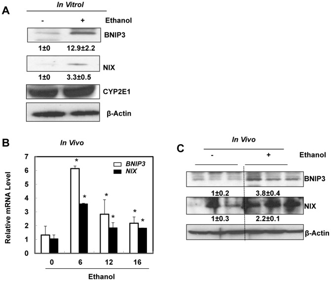
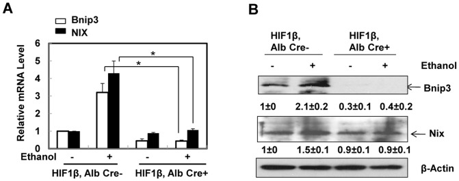
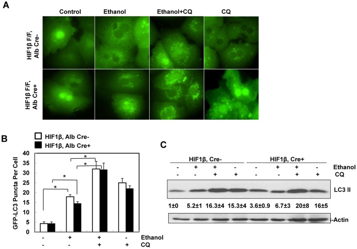
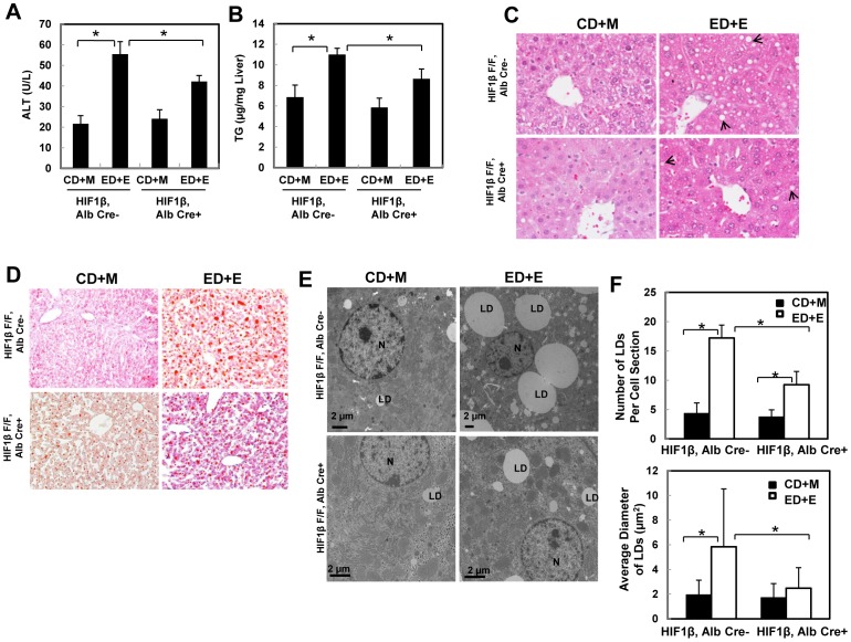
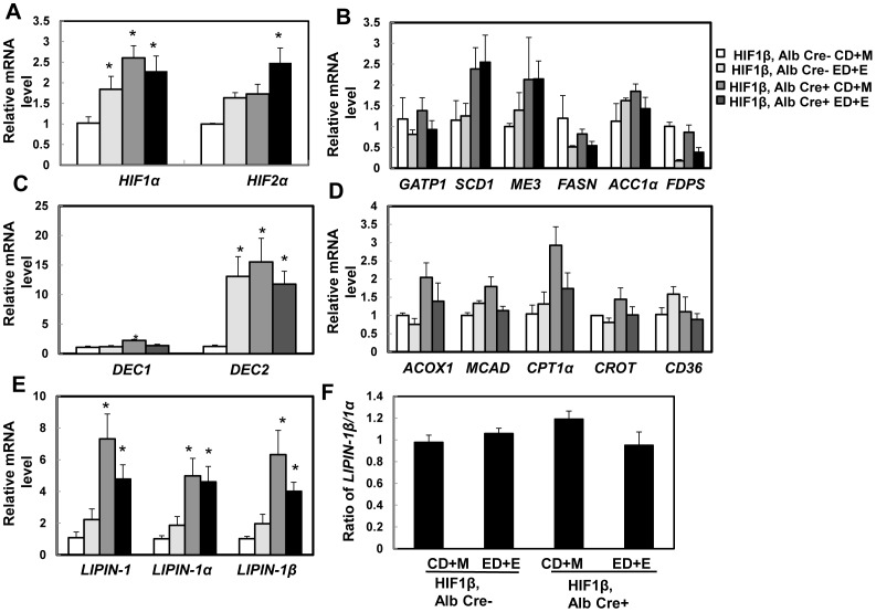
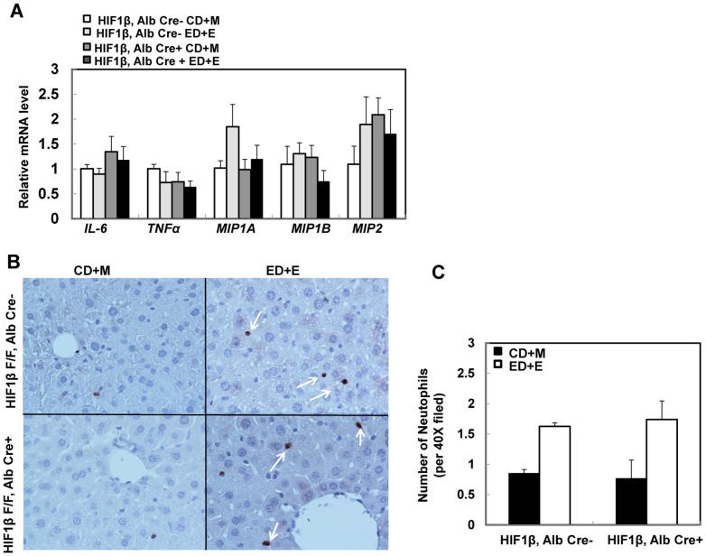
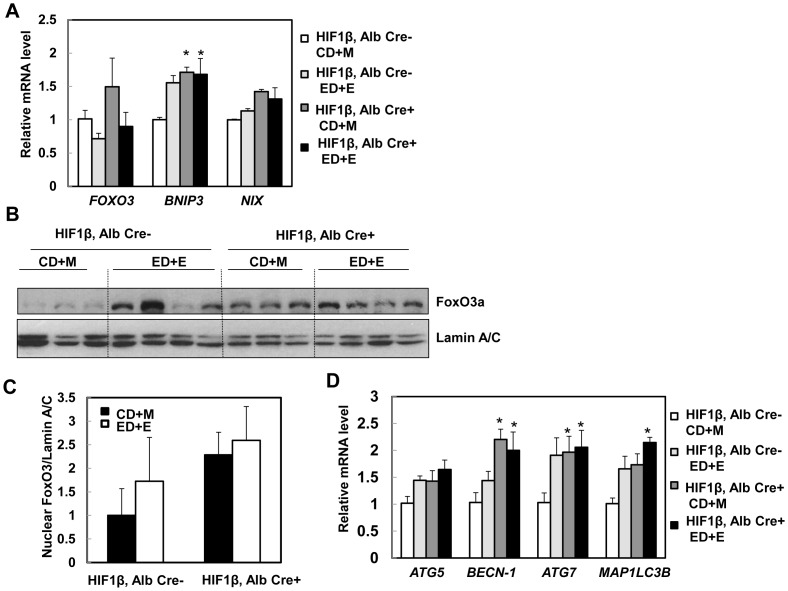
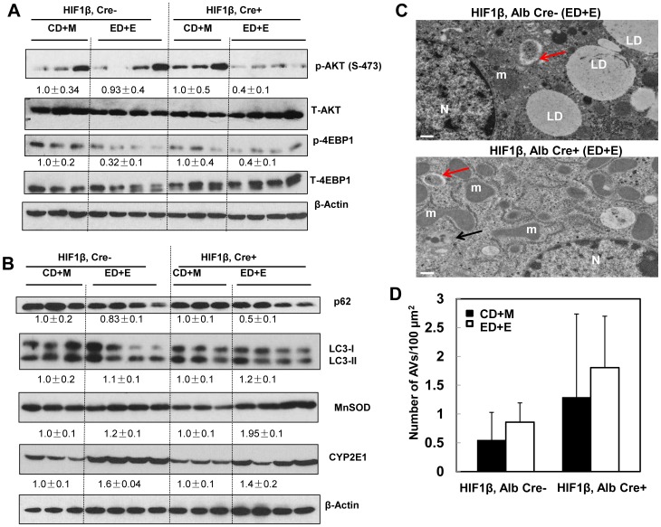
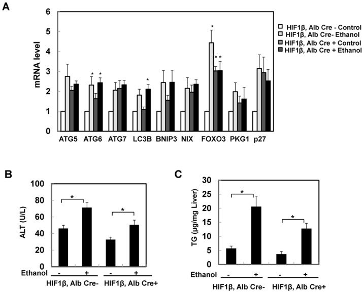
Chronic alcohol causes liver hypoxia and steatosis, which eventually develops into alcoholic liver disease (ALD). While it has been known that alcohol consumption activates hepatic hypoxia inducing factor-1α (HIF-1α), conflicting results regarding the role of HIF-1α in alcohol-induced liver injury and steatosis in mice have been reported. In the present study, we aimed to use hepatocyte-specific HIF-1β knockout mice to eliminate the possible compensatory effects of the single knockout of the 1α subunit of HIF to study the role of HIFs in ALD. C57BL/6 wild type mice were treated with acute ethanol to mimic human binge drinking. Matched wild-type and hepatocyte specific HIF-1β knockout mice were also subjected to a recently established Gao-binge alcohol model to mimic chronic plus binge conditions, which is quite common in human alcoholics. We found that acute alcohol treatment increased BNIP3 and BNIP3L/NIX expression in primary cultured hepatocytes and in mouse livers, suggesting that HIF may be activated in these models. We further found that hepatocyte-specific HIF-1β knockout mice developed less steatosis and liver injury following the Gao-binge model or acute ethanol treatment compared with their matched wild type mice. Mechanistically, protection against Gao-binge treatment-induced steatosis and liver injury was likely associated with increased FoxO3a activation and subsequent induction of autophagy in hepatocyte-specific HIF-1β knockout mice.


Conflict of interest statement

Competing Interests: The authors have declared that no competing interests exist.

Figures

Figure 1
Figure 1. Acute ethanol treatment increased expression of BNIP3 and NIX in primary hepatocytes and mouse liver.
Primary cultured mouse hepatocytes were treated with ethanol (80 mM) for 6 hours. Densitometry was performed and data are presented as a ratio vs control (n = 3). (A) Total cell lysates from cultured hepatocytes were subjected to western blot analysis. (B) Male C57BL/6 mice were treated with either water or ethanol (4.5 g/kg) by gavage for 6, 12 or 16 hours. Hepatic mRNA was isolated and real-time RT-PCR was performed as described in the Materials and Methods. Data are presented as means ± SE (n = 4–6). * p<0.05, vs 0 hour control. One way ANOVA with Scheffé's post hoc test. (C) Mice were treated as in (B) and total liver lysates from the mice (16 hours treatment) were subjected to western blot analysis. Densitometry was performed and data are presented as a ratio of control (n = 3).
Figure 2
Figure 2. Ethanol-induced expression of BNIP3 and NIX was abolished in primary cultured HIF-1β knockout mouse hepatocytes.
(A) Primary cultured wild type and HIF-1β knockout hepatocytes were treated with ethanol (80 mM) for 6 hours. mRNA was isolated from cultured hepatocytes and real-time RT-PCR was performed as described in the Materials and Methods. Data are presented as means ± SE (n = 4). * p<0.05. One way ANOVA with Scheffé's post hoc test. (B) Cells were treated as in (A), total cell lysates were subjected to western blot analysis. Densitometry was performed and data are presented as a ratio vs HIF-1β, Alb Cre- control (n = 3).
Figure 3
Figure 3. Ethanol induced autophagic flux in primary mouse hepatocytes independent of HIF-1β.
Primary cultured wild type and HIF-1β knockout mouse hepatocytes were infected with Ad-GFP-LC3 (100 viral particles per cell) overnight followed by ethanol (80 mM) treatment with or without CQ (20 µM) for 6 hours. (A) Representative fluorescence images of GFP-LC3 from each group are shown. (B) The number of GFP-LC3 puncta per cell was quantified (more than 20 cells were counted from each independent experiment) and data are presented as means ± SE (n = 3 independent experiments). * p<0.05. One way ANOVA analysis with Scheffé's post hoc test. (C) Cells were treated as in (A), total cell lysates were subjected to western blot analysis. Densitometry was performed and data are presented as a ratio of HIF-1β, Alb Cre- control (n = 3).
Figure 4
Figure 4. Hepatocyte-specific HIF-1β knockout mice were resistant to steatosis and liver injury from the Gao binge model.
Age matched male wild type and hepatocyte-specific HIF-1β knockout mice were subjected to Gao-binge treatment. Serum ALT (A) and hepatic TG (B) were measured. Data are presented as means ± SE (n = 3–8). * p<0.05. One way ANOVA with Scheffé's post hoc test. Representative photographs of H &E staining (C) and Oil O Red staining are shown (D). Arrows: hepatic lipid droplets. Representative EM images are shown in (E). M: Mitochondria; N: Nuclei; LD: lipid droplet; Bar: 500 nm. The number and size (average diameter) of LDs per cell section was quantified (F), and data are presented as means ± SE (more than 20 cell sections and 80 LDs). * p<0.05. One way anova analysis with Scheffé's post hoc test.
Figure 5
Figure 5. Expression of lipid metabolism genes after Gao-binge treatment in wild type and hepatocyte-specific HIF-1β knockout mice.
Age matched male wild type and hepatocyte-specific HIF-1β knockout mice were subjected to Gao-binge treatment. Hepatic mRNA was isolated, and real-time RT-PCR was performed as described in the Materials and Methods to quantify expression of HIF(A), lipogenesis genes (B), DEC1 and DEC2 (C), fatty acid oxidation (D), and LIPIN-1 (E). The ratio of LINPIN-1β/LINPIN-1α was calculated based on (E) and is shown in (F). Data are presented as means ± SE (n = 3–5). CD+M: Control diet+maltose; ED+E: Ethanol diet+ethanol binge. * p<0.05, vs HIF-1β CD+M group. One way ANOVA with Scheffé's post hoc test.
Figure 6
Figure 6. Changes in markers of hepatic inflammation in wild type and hepatocyte-specific HIF-1β knockout mice after Gao-binge treatment.
Age matched male wild type and hepatocyte-specific HIF-1β knockout mice were subjected to Gao-binge treatment. Hepatic mRNA was isolated, and real-time RT-PCR was performed as described in the Materials and Methods (A). Data are presented as means ± SE (n = 3–5). Liver tissue was subjected to immunostaining for neutrophils using an anti-Ly6B antibody and representative images are shown in (B, 40x), and the number of neutrophils were quantified from 10 different fields of each mouse. (C). Data are presented as average number of neutrophils in each filed (means ± SE; n = 4).
Figure 7
Figure 7. Changes of FoxO3a in hepatocyte-specific HIF-1β knockout mouse livers.
Matched male wild type and hepatocyte-specific HIF-1β knockout mice were subjected to Gao-binge treatment. Hepatic mRNA was isolated, and real-time RT-PCR was performed as described in the Materials and Methods to quantify the expression of FOXO3, BNIP3 and NIX (A). Data are presented as means ± SE (n = 3–5). * p<0.05, vs HIF-1β CD+M group. One way ANOVA with Scheffé's post hoc test. Mice were treated as in (A), mouse liver nuclear fractions were prepared as described in the Materials and Methods followed by western blot analysis (B) and densitometry analysis (C). Data are presented as means ± SE (n = 3). (D) Mice were treated as in (A), hepatic mRNA was isolated, and real-time RT-PCR was performed as described in the Materials and Methods to quantify autophagy-related genes. Data are presented as means ± SE (n = 3–5). * p<0.05; * p<0.05, vs HIF-1β CD+M group. One way ANOVA with Scheffé's post hoc test.
Figure 8
Figure 8. Inhibition of AKT and mTOR and induction of autophagy in hepatocyte-specific HIF-1β knockout mouse livers after Gao-binge treatment.
Age matched male wild type and hepatocyte-specific HIF-1β knockout mice were subjected to Gao-binge treatment. (A & B) Total liver lysates were subjected to western blot analysis for the indicated proteins. Densitometry was performed and data are presented as a ratio of CD+M control group (n = 3–4). Representative EM images are shown in (C). M: Mitochondria; N: Nuclei; LD: lipid droplet; Bar: 500 nm. Red arrows: autophagosomes, black arrows: autolysosomes. (D) The number of autophagic vacuoles (AVs, including both autophagosomes and autolysosomes) per 100 µm2 cytosol was quantified, and data are presented as means ± SE (more than 15 cell sections).
Figure 9
Figure 9. Hepatocyte-specific HIF-1β knockout mice were resistant to acute ethanol treatment-induced steatosis and liver injury.
Age matched male wild type and hepatocyte-specific HIF-1β knockout mice were subjected to acute ethanol treatment for 16 hours. Hepatic mRNA was isolated, and real-time RT-PCR was performed as described in the Materials and Methods (A). Data are presented as means ± SE (n = 3–5). Serum ALT (A) and hepatic TG (B) were measured. Data are presented as means ± SE (n = 3–5). * p<0.05, vs HIF-1β, Alb Cre- control group or HIF-1β, Alb Cre+ control group, One way ANOVA with Scheffé's post hoc test.

References

    1. Williams JA, Manley S, Ding WX (2014) New advances in molecular mechanisms and emerging therapeutic targets in alcoholic liver diseases. World J Gastroenterol 20:12908–12933. - PMC - PubMed
    1. Gao B, Bataller R (2011) Alcoholic liver disease: pathogenesis and new therapeutic targets. Gastroenterology 141:1572–1585. - PMC - PubMed
    1. Arteel GE, Iimuro Y, Yin M, Raleigh JA, Thurman RG (1997) Chronic enteral ethanol treatment causes hypoxia in rat liver tissue in vivo. Hepatology 25:920–926. - PubMed
    1. Zelickson BR, Benavides GA, Johnson MS, Chacko BK, Venkatraman A, et al. (2011) Nitric oxide and hypoxia exacerbate alcohol-induced mitochondrial dysfunction in hepatocytes. Biochim Biophys Acta 1807:1573–1582. - PMC - PubMed
    1. Rankin EB, Rha J, Selak MA, Unger TL, Keith B, et al. (2009) Hypoxia-inducible factor 2 regulates hepatic lipid metabolism. Mol Cell Biol 29:4527–4538. - PMC - PubMed

Publication types

MeSH terms