. 2014 Dec 23;9(12):e115507.
doi: 10.1371/journal.pone.0115507. eCollection 2014.

Overexpression of CD90 (Thy-1) in pancreatic adenocarcinoma present in the tumor microenvironment

Affiliations

Overexpression of CD90 (Thy-1) in pancreatic adenocarcinoma present in the tumor microenvironment

Jianhui Zhu et al. PLoS One. .

Abstract

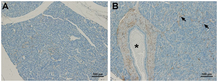
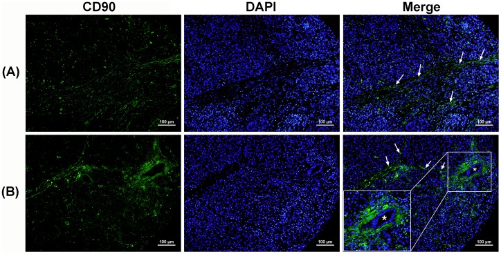
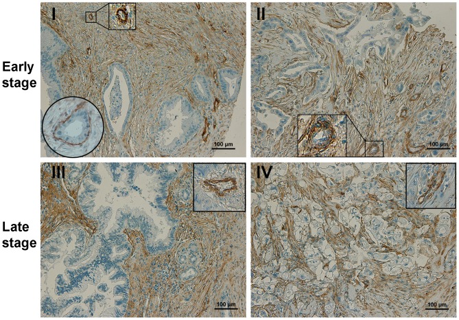
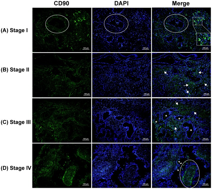
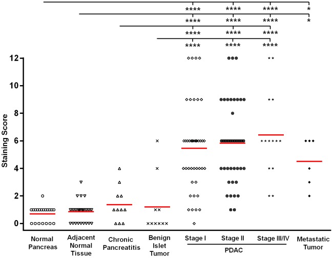
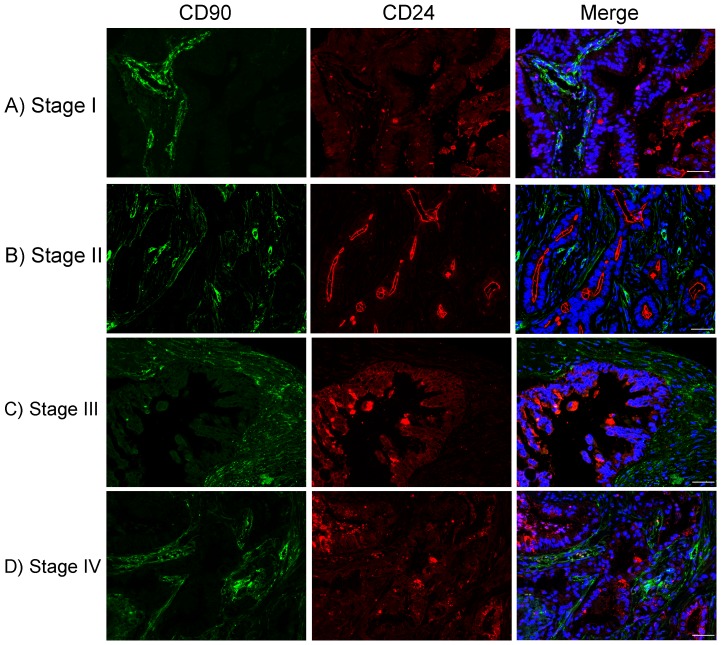
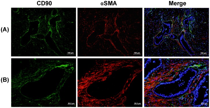
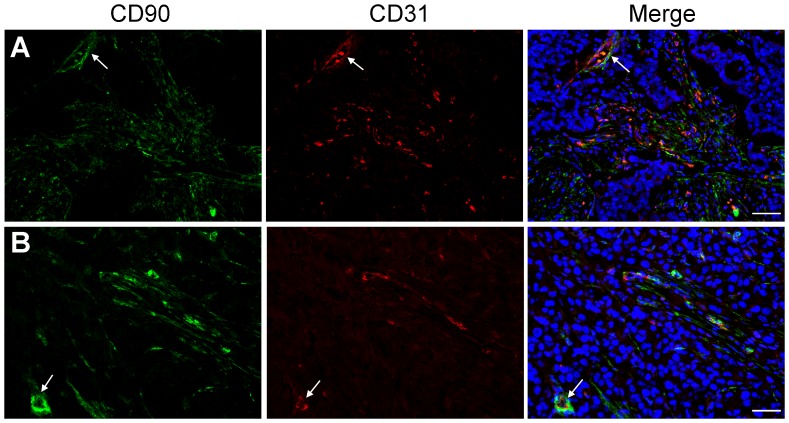
CD90 (Thy-1) plays important roles in oncogenesis and shows potential as a candidate marker for cancer stem cells (CSCs) in various malignancies. Herein, we investigated the expression of CD90 in pancreatic adenocarcinoma (PDAC), with a comparison to normal pancreas and non-malignant pancreatic disease, by immunohistochemical (IHC) analysis of tissue microarrays containing 183 clinical tissue specimens. Statistical analysis was performed to evaluate the correlation between CD90 expression and the major clinicopathological factors after adjustment of age and gender. The IHC data showed that CD90 was significantly overexpressed in PDAC and its metastatic cancers as compared to chronic pancreatitis and benign islet tumors, while it was negative in normal pancreas and 82.7% of adjacent normal pancreas tissues. The abundant CD90 expression was predominantly present in PDAC stroma, such as fibroblasts and vascular endothelial cells, which could serve as a promising marker to distinguish pancreatic adenocarcinoma from normal pancreas and non-malignant pancreatic diseases. Double immunostaining of CD90 with CD24, a CSC marker for PDAC, showed that there was little overlap between these two markers. However, CD90+ fibroblast cells were clustered around CD24+ malignant ducts, suggesting that CD90 may be involved in the tumor-stroma interactions and promote pancreatic cancer development. Furthermore, CD90 mostly overlapped with α-smooth muscle actin (αSMA, a marker of activated pancreatic stellate cells (PSCs)) in PDAC stroma, which demonstrated that CD90+ stromal cells consist largely of activated PSCs. Double immunostaining of CD90 and a vascular endothelial cell marker CD31 demonstrated that CD90 expression on vascular endothelial cells was significantly increased in PDACs as compared to normal pancreas and non-malignant pancreatic diseases. Our findings suggest that CD90 could serve as a promising marker for pancreatic adenocarcinoma where desmoplastic stroma plays an important role in tumor growth and angiogenesis.


Conflict of interest statement

Competing Interests: The authors have declared that no competing interests exist.

Figures

Figure 1
Figure 1. CD90 expression in normal human pancreas.
(A) A representative image of normal pancreas showed negative expression of CD90. (B) Minimal CD90 expression was observed on the connective tissues (arrows). The fibroblasts surrounding the pancreatic main duct (asterisk) showed weak CD90 expression. No expression of CD90 was observed on ducts, acini, islets, and blood vessels. Scale bars  = 100 µm.
Figure 2
Figure 2. Increased CD90 expression (green) was observed on the activated fibroblasts in chronic pancreatitis.
(A) A representative image of pancreatitis tissue showed moderate expression of CD90 in fibroblast cells (indicated by arrows). (B) Fibroblasts around a duct (asterisk) showed distinctly strong expression of CD90 in pancreatitis. The insert is a higher magnification of the ductal area. Nuclei were stained with DAPI (blue). Scale bars  = 100 µm.
Figure 3
Figure 3. Overexpression of CD90 in the stroma of pancreatic adenocarcinoma (PDAC).
In both early (I and II) and late stages (III and IV) of PDACs, CD90 expression was highly increased, which was abundantly present in the activated stroma, including fibroblasts and vascular endothelial cells. The positive CD90+ stromal cells were clustered around the malignant ducts. The circle insert in 3I showed a single layer of CD90+ fibroblasts closely localized around tumor duct. The square inserts in 3I–3IV showed strong CD90 expression on vascular endothelial cells in PDAC stage I, II, III and IV, respectively.
Figure 4
Figure 4. Distribution of CD90 in PDAC of early and late stages evaluated by immunofluorescence staining (green).
DAPI counterstaining was used to visualize nuclei (blue). In early-stage PDACs, CD90 is abundantly expressed on stromal cells, including activated fibroblasts and vascular cells which form the basis for blood vessels (shown in square). Membrane expression of CD90 on the apical cell surface was rarely observed in malignant ducts in late-stage PDACs (marked with a star or arrow). Expression was also observed on endothelial cells (in red circle). Scale bars  = 100 µm.
Figure 5
Figure 5. Comparison of staining score of CD90 expression among normal pancreas, cancer adjacent normal tissue, chronic pancreatitis, benign islet tumor, PDAC of stage I, II, and III/IV, and metastatic tumor.
P-values were determined in ANCOVA by comparing IHC scores between each group, adjusted for age and gender, and the multiple comparisons are accounted for using the Tukey's method. The CD90 staining score was distinctly higher in PDAC of all stages compared to those in non-malignant cases (p<0.0001). Samples with metastatic tumors also showed increased expression of CD90 than in non-malignant cases (p<0.05). (*: p<0.05; ****: p<0.0001).
Figure 6
Figure 6. CD90 (green) and CD24 (red) expression in PDAC of early (A and B) and late (C and D) stages.
DAPI counterstaining was used to visualize nuclei (blue). Scale bars  = 50 µm. There was little overlap between CD90 and CD24. However, CD90+ stromal fibroblasts were localized to tumor glands where CD24 shows strong expression on the cell surface membrane, suggesting that cancer-associated stroma has a function in tumor progression.
Figure 7
Figure 7. Double immunofluoresence staining of CD90 (green) and αSMA (red) on PDAC tissues.
DAPI counterstaining was used to visualize nuclei (blue). Yellow or orange represent the co-localization of CD90 and αSMA. (A) A stage I PDAC specimen exhibited a single layer of CD90+ activated PSCs (yellow) surrounding the duct-like structure. (B) Higher magnification of the stromal area in PDAC displaying an apparent overlap between CD90 and αSMA (yellow or orange in the merged images). The IHC result showed that CD90 mostly overlapped with αSMA in PDAC stroma, demonstrating that CD90+ stromal cells consist largely of activated PSCs.
Figure 8
Figure 8. Coexpression of CD90 (green) and CD31 (red) in PDAC.
DAPI counterstaining was used to visualize nuclei (blue). Scale bars  = 50 µm. The merged images showed that CD90 was co-localized with CD31 in the tumor vasculature (indicated by arrows) of PDACs.

References

    1. Vidal M, Morris R, Grosveld F, Spanopoulou E (1990) Tissue-specific control elements of the Thy-1 gene. EMBO J 9:833–840. - PMC - PubMed
    1. Saalbach A, Wetzig T, Haustein UF, Anderegg U (1999) Detection of human soluble Thy-1 in serum by ELISA. Fibroblasts and activated endothelial cells are a possible source of soluble Thy-1 in serum. Cell Tissue Res 298:307–315. - PubMed
    1. Craig W, Kay R, Cutler RL, Lansdorp PM (1993) Expression of Thy-1 on human hematopoietic progenitor cells. J Exp Med 177:1331–1342. - PMC - PubMed
    1. Dennis JE, Esterly K, Awadallah A, Parrish CR, Poynter GM, et al. (2007) Clinical-scale expansion of a mixed population of bone-marrow-derived stem and progenitor cells for potential use in bone-tissue regeneration. Stem Cells 25:2575–2582. - PubMed
    1. Herrera MB, Bruno S, Buttiglieri S, Tetta C, Gatti S, et al. (2006) Isolation and characterization of a stem cell population from adult human liver. Stem Cells 24:2840–2850. - PubMed

Publication types

MeSH terms