

Factors affecting the ability of the spectral domain optical coherence tomograph to detect photographic retinal nerve fiber layer defects
- PMID: 25536188
- PMCID: PMC4275283
- DOI: 10.1371/journal.pone.0116115
Factors affecting the ability of the spectral domain optical coherence tomograph to detect photographic retinal nerve fiber layer defects
Abstract
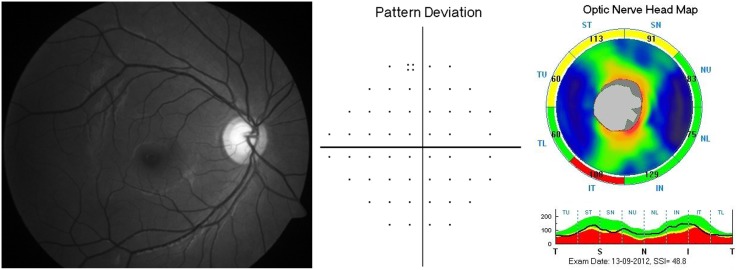
Purpose: To evaluate the ability of normative database classification (color-coded maps) of spectral domain optical coherence tomograph (SDOCT) in detecting wedge shaped retinal nerve fiber layer (RNFL) defects identified on photographs and the factors affecting the ability of SDOCT in detecting these RNFL defects.
Methods: In a cross-sectional study, 238 eyes (476 RNFL quadrants) of 172 normal subjects and 85 eyes (103 RNFL quadrants with wedge shaped RNFL defects) of 66 glaucoma patients underwent RNFL imaging with SDOCT. Logistic regression models were used to evaluate the factors associated with false positive and false negative RNFL classifications of the color-coded maps of SDOCT.
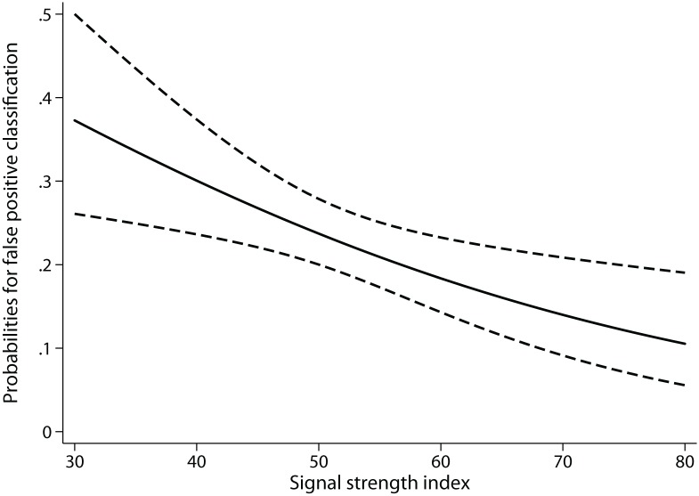
Results: False positive classification at a p value of <5% was seen in 108 of 476 quadrants (22.8%). False negative classification at a p value of <5% was seen in 16 of 103 quadrants (15.5%). Of the 103 quadrants with RNFL defects, 64 showed a corresponding VF defect in the opposite hemisphere and 39 were preperimetric. Higher signal strength index (SSI) of the scan was less likely to have a false positive classification (odds ratio: 0.97, p = 0.01). Presence of an associated visual field defect (odds ratio: 0.17, p = 0.01) and inferior quadrant RNFL defects as compared to superior (odds ratio: 0.24, p = 0.04) were less likely to show false negative classifications.
Conclusions: Scans with lower signal strengths were more likely to show false positive RNFL classifications, and preperimetric and superior quadrant RNFL defects were more likely to show false negative classifications on color-coded maps of SDOCT.
Conflict of interest statement
Figures

Similar articles
-
Ability of cirrus high-definition spectral-domain optical coherence tomography clock-hour, deviation, and thickness maps in detecting photographic retinal nerve fiber layer abnormalities.Ophthalmology. 2013 Jul;120(7):1380-7. doi: 10.1016/j.ophtha.2012.12.048. Epub 2013 Mar 28. Ophthalmology. 2013. PMID: 23541761
-
Ability of different scanning protocols of spectral domain optical coherence tomography to diagnose preperimetric glaucoma.Invest Ophthalmol Vis Sci. 2013 Nov 1;54(12):7252-7. doi: 10.1167/iovs.13-12731. Invest Ophthalmol Vis Sci. 2013. PMID: 24114539
-
Diagnostic classification of macular ganglion cell and retinal nerve fiber layer analysis: differentiation of false-positives from glaucoma.Ophthalmology. 2015 Mar;122(3):502-10. doi: 10.1016/j.ophtha.2014.09.031. Epub 2014 Nov 14. Ophthalmology. 2015. PMID: 25444638
-
Imaging of localized retinal nerve fiber layer defects in preperimetric glaucoma using spectral-domain optical coherence tomography.J Glaucoma. 2014 Mar;23(3):150-9. doi: 10.1097/IJG.0b013e3182707456. J Glaucoma. 2014. PMID: 23059486
-
Peripapillary retinal nerve fiber layer assessment of spectral domain optical coherence tomography and scanning laser polarimetry to diagnose preperimetric glaucoma.PLoS One. 2014 Oct 3;9(10):e108992. doi: 10.1371/journal.pone.0108992. eCollection 2014. PLoS One. 2014. PMID: 25279801 Free PMC article.
Cited by
-
Significance of the disc damage likelihood scale objectively measured by a non-mydriatic fundus camera in preperimetric glaucoma.Clin Ophthalmol. 2015 Nov 20;9:2147-58. doi: 10.2147/OPTH.S93213. eCollection 2015. Clin Ophthalmol. 2015. PMID: 26640365 Free PMC article.
References
-
- Sommer A, Katz J, Quigley HA, Miller NR, Robin AL, et al. (1991) Clinically detectable nerve fiber atrophy precedes the onset of glaucomatous field loss. Arch Ophthalmol 109:77–83. - PubMed
-
- Tuulonen A, Lehtola J, Airaksinen PJ (1993) Nerve fiber layer defects with normal visual fields. Do normal optic disc and normal visual field indicate absence of glaucomatous abnormality? Ophthalmology 100:587–597. - PubMed
-
- Hoyt WF, Frisen L, Newman NM (1973) Fundoscopy of nerve fiber layer defects in glaucoma. Invest Ophthalmol 12:814–829. - PubMed
-
- Jeoung JW, Park KH (2010) Comparison of Cirrus OCT and Stratus OCT on the ability to detect localized retinal nerve fiber layer defects in preperimetric glaucoma. Invest Ophthalmol Vis Sci 51:938–945. - PubMed
-
- Kim NR, Lee ES, Seong GJ, Choi EH, Hong S, et al. (2010) Spectral-domain optical coherence tomography for detection of localized retinal nerve fiber layer defects in patients with open-angle glaucoma. Arch Ophthalmol 128:1121–1128. - PubMed
Publication types
MeSH terms
LinkOut - more resources
Full Text Sources
Other Literature Sources
Medical
