

Effect of environmental enrichment on dopamine and serotonin transporters and glutamate neurotransmission in medial prefrontal and orbitofrontal cortex
- PMID: 25536304
- PMCID: PMC4344853
- DOI: 10.1016/j.brainres.2014.12.034
Effect of environmental enrichment on dopamine and serotonin transporters and glutamate neurotransmission in medial prefrontal and orbitofrontal cortex
Abstract
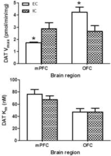
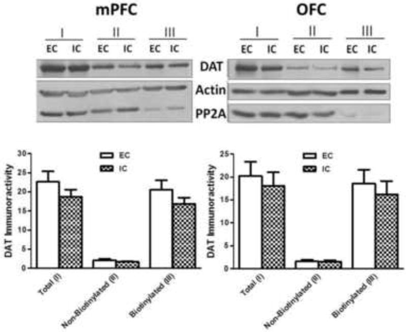
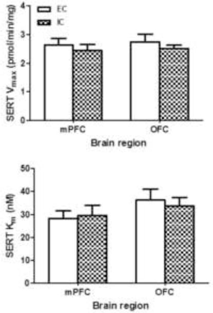
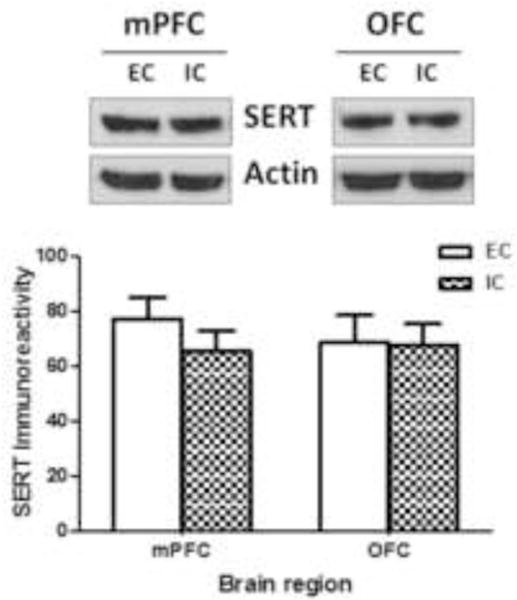
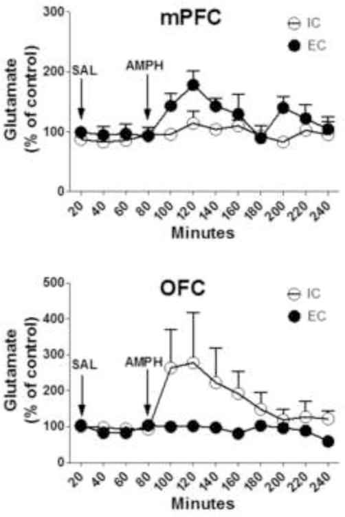
Recent studies have reported that rats raised in an enriched condition (EC) have decreased dopamine transporter (DAT) function and expression in medial prefrontal cortex (mPFC), as well as increased d-amphetamine-induced glutamate release in nucleus accumbens compared to rats raised in an isolated condition (IC). In these previous studies, DAT function and expression were evaluated using mPFC pooled from four rats for each condition to obtain kinetic parameters due to sparse DAT expression in mPFC. In contrast, accumbal glutamate release was determined using individual rats. The current study extends the previous work and reports on the optimization of DAT and serotonin transporter (SERT) functional assays, as well as cell surface expression assays using both mPFC and orbitofrontal cortex (OFC) from individual EC or IC rats. In addition, the effect of d-amphetamine on glutamate release in mPFC and OFC of EC and IC rats was determined using in vivo microdialysis. Results show that environmental enrichment decreased maximal transport velocity (Vmax) for [(3)H]dopamine uptake in mPFC, but increased Vmax for [(3)H]dopamine uptake in OFC. Corresponding changes in DAT cell surface expression were not found. In contrast, Vmax for [(3)H]serotonin uptake and cellular localization of SERT in mPFC and OFC were not different between EC and IC rats. Further, acute d-amphetamine (2mg/kg, s.c.) increased extracellular glutamate concentrations in mPFC of EC rats only and in OFC of IC rats only. Overall, these results suggest that enrichment produces long-lasting alterations in mPFC and OFC DAT function via a trafficking-independent mechanism, as well as differential glutamate release in mPFC and OFC. Rearing-induced modulation of DAT function and glutamate release in prefrontal cortical subregions may contribute to the known protective effects of enrichment on drug abuse vulnerability.
Keywords: Dopamine transporter; Enrichment; Glutamate; Medial prefrontal cortex; Orbitofrontal cortex; Serotonin transporter.
Copyright © 2015 Elsevier B.V. All rights reserved.
Figures
Similar articles
-
Environmental enrichment enhances sensitization to GBR 12935-induced activity and decreases dopamine transporter function in the medial prefrontal cortex.Behav Brain Res. 2004 Jan 5;148(1-2):107-17. doi: 10.1016/s0166-4328(03)00190-6. Behav Brain Res. 2004. PMID: 14684252
-
Environmental enrichment decreases cell surface expression of the dopamine transporter in rat medial prefrontal cortex.J Neurochem. 2005 Jun;93(6):1434-43. doi: 10.1111/j.1471-4159.2005.03130.x. J Neurochem. 2005. PMID: 15935059
-
Effects of environmental enrichment on behavior and dopamine transporter function in medial prefrontal cortex in adult rats prenatally treated with cocaine.Brain Res Dev Brain Res. 2004 Nov 25;153(2):213-23. doi: 10.1016/j.devbrainres.2004.09.001. Brain Res Dev Brain Res. 2004. PMID: 15527889
-
Psychostimulants affect dopamine transmission through both dopamine transporter-dependent and independent mechanisms.Eur J Pharmacol. 2015 Oct 5;764:562-570. doi: 10.1016/j.ejphar.2015.07.044. Epub 2015 Jul 21. Eur J Pharmacol. 2015. PMID: 26209364 Free PMC article. Review.
-
Cocaine and amphetamine-like psychostimulants: neurocircuitry and glutamate neuroplasticity.Dialogues Clin Neurosci. 2007;9(4):389-97. doi: 10.31887/DCNS.2007.9.4/pkalivas. Dialogues Clin Neurosci. 2007. PMID: 18286799 Free PMC article. Review.
Cited by
-
Differential impact of stress and environmental enrichment on corticolimbic circuits.Pharmacol Biochem Behav. 2020 Oct;197:172993. doi: 10.1016/j.pbb.2020.172993. Epub 2020 Jul 11. Pharmacol Biochem Behav. 2020. PMID: 32659243 Free PMC article. Review.
-
The Potential Regulatory Network of Glutamate Metabolic Pathway Disturbance in Chinese Han Withdrawal Methamphetamine Abusers.Front Genet. 2021 Mar 23;12:653443. doi: 10.3389/fgene.2021.653443. eCollection 2021. Front Genet. 2021. PMID: 33833781 Free PMC article.
-
Nature Scenes Counter Mental Fatigue-Induced Performance Decrements in Soccer Decision-Making.Front Psychol. 2022 Apr 29;13:877844. doi: 10.3389/fpsyg.2022.877844. eCollection 2022. Front Psychol. 2022. PMID: 35572319 Free PMC article.
-
Nature exposure might be the intervention to improve the self-regulation and skilled performance in mentally fatigue athletes: A narrative review and conceptual framework.Front Psychol. 2022 Aug 2;13:941299. doi: 10.3389/fpsyg.2022.941299. eCollection 2022. Front Psychol. 2022. PMID: 35983203 Free PMC article. Review.
-
Trust and the poverty trap.Proc Natl Acad Sci U S A. 2017 May 23;114(21):5327-5329. doi: 10.1073/pnas.1704798114. Epub 2017 May 15. Proc Natl Acad Sci U S A. 2017. PMID: 28507150 Free PMC article. No abstract available.
References
-
- Baler RD, Volkow ND. Drug addiction: the neurobiology of disrupted self-control. Trends in Molecular Medicine. 2006;12:559–566. - PubMed
-
- Bardo MT, Klebaur JE, Valone JM, Deaton C. Environmental enrichment decreases intravenous self-administration of amphetamine in female and male rats. Psychopharmacology. 2001;155:278–284. - PubMed
-
- Bardo MT, Valone JM, Robinet PM, Shaw WB, Dwoskin LP. Environmental enrichment enhances the stimulant effect of intravenous amphetamine: search for a cellular mechanism in the nucleus accumbens. Psychobiology. 1999;27:292–299.
-
- Bradford MM. A rapid and sensitive method for the quantitation of microgram quantities of protein utilizing the principle of protein-dye binding. Analytical Biochemistry. 1976;72:248–254. - PubMed
Publication types
MeSH terms
Substances
Grants and funding
LinkOut - more resources
Full Text Sources
Other Literature Sources
