. 2014 Dec 23;9(12):e115402.
doi: 10.1371/journal.pone.0115402. eCollection 2014.

Oxidant exposure induces cysteine-rich protein 61 (CCN1) via c-Jun/AP-1 to reduce collagen expression in human dermal fibroblasts

Affiliations

Oxidant exposure induces cysteine-rich protein 61 (CCN1) via c-Jun/AP-1 to reduce collagen expression in human dermal fibroblasts

Zhaoping Qin et al. PLoS One. .

Abstract

Human skin is a primary target of oxidative stress from reactive oxygen species (ROS) generated from both extrinsic and intrinsic sources. Oxidative stress inhibits the production of collagen, the most abundant protein in skin, and thus contributes to connective tissue aging. Here we report that cysteine-rich protein 61 (CCN1), a negative regulator of collagen production, is markedly induced by ROS and mediates loss of type I collagen in human dermal fibroblasts. Conversely, antioxidant N-acetyl-L-cysteine significantly reduced CCN1 expression and prevented ROS-induced loss of type I collagen in both human dermal fibroblasts and human skin in vivo. ROS increased c-Jun, a critical member of transcription factor AP-1 complex, and increased c-Jun binding to the AP-1 site of the CCN1 promoter. Functional blocking of c-Jun significantly reduced CCN1 promoter and gene expression and thus prevented ROS-induced loss of type I collagen. Targeting the c-Jun/CCN1 axis may provide clinical benefit for connective tissue aging in human skin.


Conflict of interest statement

Competing Interests: The authors have declared that no competing interests exist.

Figures

Figure 1
Figure 1. Oxidative exposure induces CCN1 expression and inhibits type I procollagen in adult human primary dermal fibroblasts.
Fibroblasts were exposed to vehicle (CTRL) or H2O2 on two successive days (ROS), as described in Methods. (a) Oxidative exposure induces endogenous ROS. RedoxSensor Red CC-1, an indicator of ROS level, was visualized and quantified by fluorescence microscopy, as described in Methods. Oxidative exposure induces CCN1 (b) mRNA and (c) protein. Oxidative exposure reduces Type I procollagen (COL-1) (d) mRNA and (e) protein. mRNA levels were quantified by real-time RT-PCR and normalized to an internal control housekeeping gene (36B4). Protein levels were determined by Western blot and normalized to an internal control (β-actin). Inset shows representative blot. Data are means±SEM, N = 3, *p<0.05 vs. CTRL.
Figure 2
Figure 2. CCN1 induction by oxidative exposure mediates type I procollagen reduction in human dermal fibroblasts.
Fibroblasts were exposed to vehicle (CTRL) or H2O2 on two successive days (ROS), as described in Methods. Cells were transfected with control siRNA (CTRL si) or CCN1 siRNA (CCN1 si), and harvested two days later. (a) CCN1 siRNA reduces oxidant-induced CCN1 protein. Knockdown of oxidant-induced CCN1 prevents reduction of type I procollagen (COL-1) (b) mRNA and (c) protein. mRNA levels were quantified by real-time RT-PCR normalized to internal control housekeeping gene (36B4). COL-1 protein levels were determined by Western blot normalized to an internal control (β-actin). Inset shows representative blot. Data are means±SEM, N = 3, *p<0.05 vs. CTRL, **p<0.05 vs. oxidative exposure with CTRL si.
Figure 3
Figure 3. Antioxidant treatment reduces oxidant-induced CCN1 and loss of type I procollagen in human dermal fibroblasts.
Fibroblasts were exposed to vehicle (CTRL) or H2O2 on two successive days (ROS), as described in Methods. Oxidant-exposed human dermal fibroblasts were treated with vehicle (CTRL) or N-acetylcysteine (NAC, 10 µM) for 24 hours. (a) NAC treatment prevents oxidant-induced ROS levels. Oxidation of RedoxSensor Red CC-1 was visualized and quantified by fluorescence microscopy. NAC treatment reduces oxidant-induced CCN1 (b) mRNA and (c) protein. (d) NAC or CCN1 expression attenuates loss of type I procollagen (COL-1) protein by oxidative exposure. mRNA levels were quantified by real-time RT-PCR normalized to an internal control housekeeping gene (36B4). Protein levels were determined by Western blot normalized to an internal control (β-actin). Inset shows representative blot. Data are means±SEM, N = 3, *p<0.05 vs CTRL, **p<0.05 vs CTRL with oxidative exposure, ***p<0.05 vs. NAC with oxidative exposure.
Figure 4
Figure 4. Oxidant exposure activates AP-1 binding site-dependent CCN1 promoter activity in human dermal fibroblasts.
Fibroblasts were exposed to vehicle (CTRL) or H2O2 on two successive days (ROS), as described in Methods. Fibroblasts were transfected with CCN1 promoter/luciferase reporter constructs: 1, wild-type (−883/+1); 2, AP-1 mutation (−883/+1); and 3, AP-1 deletion (605/+1). Luciferase activities were determined two days after transfection. (a) Mutation or deletion of AP-1 binding site abolishes activation of CCN1 promoter by oxidative exposure. Data are means±SEM, N = 3, *p<0.05 vs. CTRL. (b) Nucleotide sequence of the CCN1 proximal promoter. AP-1-binding sequences are marked in box. Heavy underline denotes electrophoretic mobility shift assay (EMSA) probe. Arrow indicates transcription start site. (c) Oxidant-exposure increases protein-binding to AP-1 site in CCN1 promoter. DNA/protein complex was analyzed by EMSA. Arrows indicate specific retarded complexes. Closed triangle indicates free probes. Results are representative of at least three independent experiments.
Figure 5
Figure 5. c-Jun mediates oxidative exposure induction of CCN1 and reduction of type I procollagen in human dermal fibroblasts.
Fibroblasts were exposed to vehicle (CTRL) or H2O2 on two successive days (ROS), as described in Methods. (a) Oxidative exposure induces c-Jun mRNA and (b) total and phospho c-Jun proteins. mRNA levels were quantified by real-time RT-PCR normalized to an internal control housekeeping gene (36B4). Protein levels were determined by Western blot normalized to internal control (β-actin). Inset shows representative blot. (c) Dominant negative c-Jun (DN c-Jun) reduces oxidant activation of CCN1 promoter. Fibroblasts were co-transfected with CCN1 promoter/luciferase reporter construct (−883/+1) and control (CTRL) or DN c-Jun expression vector, and analyzed two days later. (d–f) DN c-Jun reduces oxidant induction of (d) CCN1 mRNA and (e) CCN1 protein. Fibroblasts were transfected with CTRL or DN c-Jun expression vectors, and analyzed two days later. (f) DN c-Jun mitigates loss of type I procollagen (COL-1) by oxidative exposure. Fibroblasts were transfected with CTRL or DN c-Jun alone or together with CCN1 expression vectors, and analyzed two days later. Data are means±SEM, N = 3, *p<0.05 vs. CTRL, **p<0.05 vs. CTRL with oxidative stress, ***p<0.05 vs DN-c-Jun alone.
Figure 6
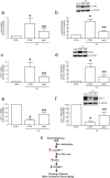
Figure 6. Topical antioxidant treatment blocks induction of c-Jun and CCN1 and loss of type I procollagen in UV-irradiated human skin in vivo.
Human buttocks were treated topically with vehicle (CTRL) or N-acetylcysteine (NAC) 24 hours prior to UV irradiation (2MED). Skin samples were obtained eight hours after UV irradiation, as described in Methods. NAC attenuates UV irradiation induction of c-Jun (a) mRNA and (b) protein, and CCN1 (c) mRNA and (d) protein in human skin. NAC attenuates UV irradiation reduction of type I procollagen (e) mRNA and (f) protein in human skin. mRNA levels were quantified by real-time RT-PCR normalized to an internal control housekeeping gene (36B4). Protein levels were determined by Western blot normalized to an internal control (β-actin). Inset shows representative blot. Data are means±SEM, N = 6, *p<0.05 vs. CTRL no UV, **p<0.05 vs. CTRL + UV. (g) Working model for oxidant driven dermal aging through the c-Jun/CCN1 axis (see “Discussion” for details).

Similar articles

Cited by

References

    1. Harman D (1981) The aging process. Proc Natl Acad Sci U S A 78:7124–7128. - PMC - PubMed
    1. Droge W (2002) Free radicals in the physiological control of cell function. Physiol Rev 82:47–95. - PubMed
    1. Scharffetter-Kochanek K, Brenneisen P, Wenk J, Herrmann G, Ma W, et al. (2000) Photoaging of the skin from phenotype to mechanisms. Exp Gerontol 35:307–316. - PubMed
    1. Yaar M, Gilchrest BA (2007) Photoageing: mechanism, prevention and therapy. Br J Dermatol 157:874–887. - PubMed
    1. Fisher GJ, Quan T, Purohit T, Shao Y, Cho MK, et al. (2009) Collagen fragmentation promotes oxidative stress and elevates matrix metalloproteinase-1 in fibroblasts in aged human skin. Am J Pathol 174:101–114. - PMC - PubMed

Publication types

MeSH terms