

Curcumin ameliorates streptozotocin-induced liver damage through modulation of endoplasmic reticulum stress-mediated apoptosis in diabetic rats
- PMID: 25536420
- PMCID: PMC4389763
- DOI: 10.3109/10715762.2014.999674
Curcumin ameliorates streptozotocin-induced liver damage through modulation of endoplasmic reticulum stress-mediated apoptosis in diabetic rats
Abstract
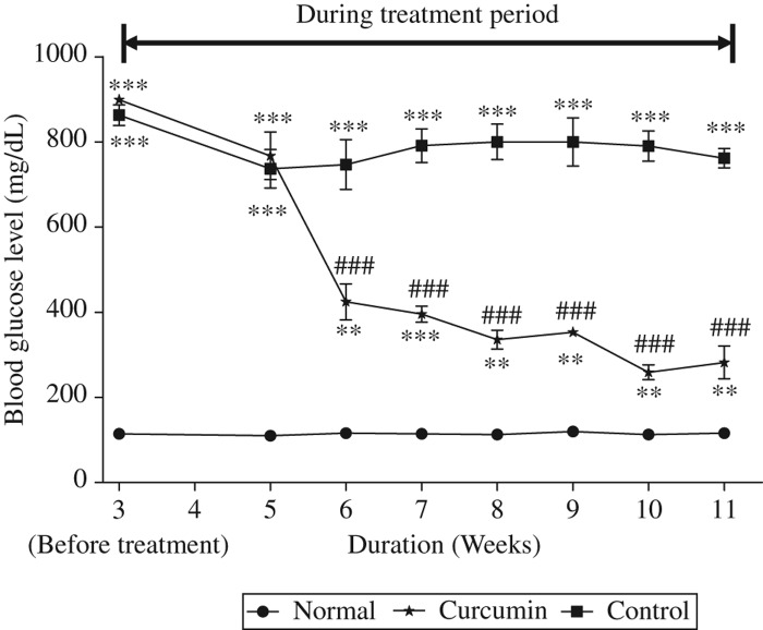
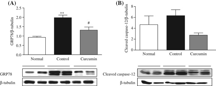
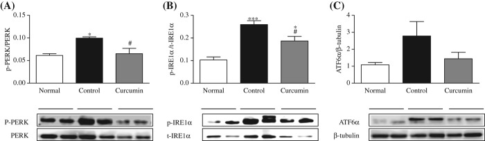
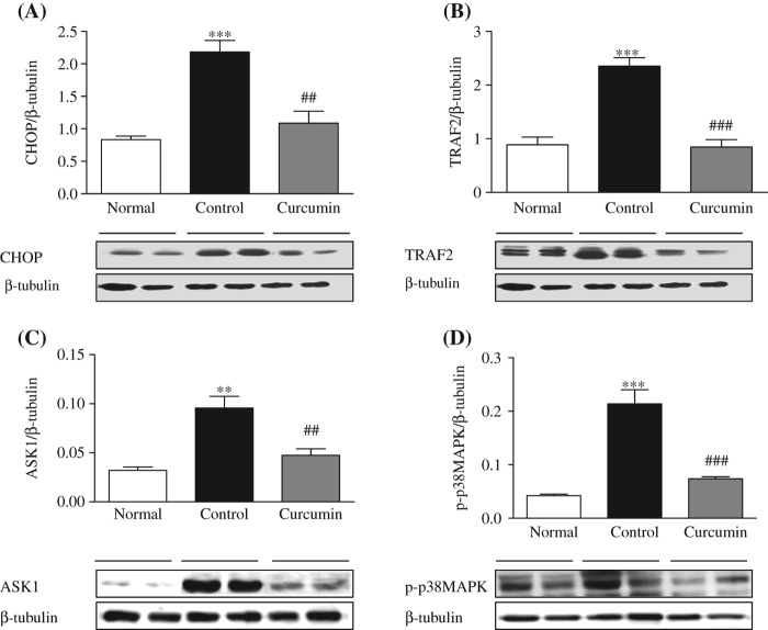
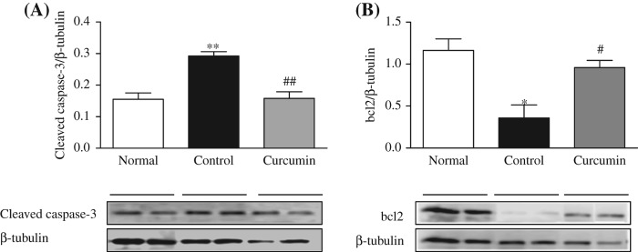
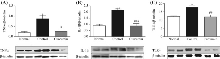
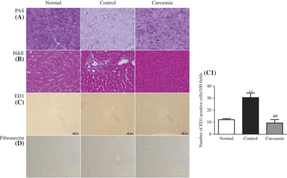
We investigated the effect of curcumin on liver injury in diabetic rats induced by streptozotocin (STZ) through modulation of endoplasmic reticulum stress (ERS) and unfolded protein response (UPR). Experimental diabetes was induced by a single intraperitoneal injection of STZ (55 mg/kg), and curcumin was given at 100 mg/kg by gavage for 56 days. We observed that curcumin improved the morphological and histopathological changes, significantly decreased hepatic ERS marker protein: glucose-regulated protein 78, and improved liver function in diabetic rats. Moreover, treatment with curcumin markedly decreased the sub-arm of the UPR signaling protein such as phospho-double-stranded RNA-dependent protein kinase-like ER kinase, CCAAT/enhancer-binding protein homologous protein, tumor necrosis factor receptor-associated factor 2, and inositol-requiring enzyme1α; and inhibited tumor necrosis factor α, interleukin 1β, phospho-p38 mitogen-activated protein kinase, and apoptosis signal-regulating kinase 1 in liver tissues of diabetic rats. Apoptotic and anti-apoptotic signaling proteins, such as cleaved caspase-3 and B-cell lymphoma 2, were significantly increased and decreased, respectively in diabetic rats; curcumin treatment prevented all of these alterations. In summary, our results indicate that curcumin has the potential to protect the diabetic liver by modulating hepatic ERS-mediated apoptosis, and provides a novel therapeutic strategy for the diabetic liver damage.
Keywords: apoptosis; curcumin; diabetes mellitus; endoplasmic reticulum stress; liver.
Figures
Similar articles
-
Curcumin attenuates oxidative stress induced NFκB mediated inflammation and endoplasmic reticulum dependent apoptosis of splenocytes in diabetes.Biochem Pharmacol. 2017 Nov 1;143:140-155. doi: 10.1016/j.bcp.2017.07.009. Epub 2017 Jul 13. Biochem Pharmacol. 2017. PMID: 28711624
-
Attenuation of Endoplasmic Reticulum Stress-Mediated Liver Damage by Mulberry Leaf Diet in Streptozotocin-Induced Diabetic Rats.Am J Chin Med. 2016;44(1):87-101. doi: 10.1142/S0192415X16500063. Am J Chin Med. 2016. PMID: 26916916
-
Therapeutic potential of aquatic Stevia extract in alleviating endoplasmic reticulum stress and liver damage in streptozotocin-induced diabetic rats.Mol Biol Rep. 2024 Sep 18;51(1):993. doi: 10.1007/s11033-024-09907-6. Mol Biol Rep. 2024. PMID: 39292293
-
Decoding cell death signals in liver inflammation.J Hepatol. 2013 Sep;59(3):583-94. doi: 10.1016/j.jhep.2013.03.033. Epub 2013 Apr 6. J Hepatol. 2013. PMID: 23567086 Review.
-
Curcumin and its analogues protect from endoplasmic reticulum stress: Mechanisms and pathways.Pharmacol Res. 2019 Aug;146:104335. doi: 10.1016/j.phrs.2019.104335. Epub 2019 Jun 29. Pharmacol Res. 2019. PMID: 31265891 Review.
Cited by
-
Curcumin Inhibits the PERK-eIF2α-CHOP Pathway through Promoting SIRT1 Expression in Oxidative Stress-induced Rat Chondrocytes and Ameliorates Osteoarthritis Progression in a Rat Model.Oxid Med Cell Longev. 2019 May 16;2019:8574386. doi: 10.1155/2019/8574386. eCollection 2019. Oxid Med Cell Longev. 2019. PMID: 31223428 Free PMC article.
-
TCM Formula Xiaoyaosan Decoction Improves Depressive-Like Behaviors in Rats with Type 2 Diabetes.Evid Based Complement Alternat Med. 2015;2015:415243. doi: 10.1155/2015/415243. Epub 2015 Oct 5. Evid Based Complement Alternat Med. 2015. PMID: 26508978 Free PMC article.
-
Curcumin protects rats against gentamicin-induced nephrotoxicity by amelioration of oxidative stress, endoplasmic reticulum stress and apoptosis.Pharm Biol. 2022 Dec;60(1):491-500. doi: 10.1080/13880209.2022.2037663. Pharm Biol. 2022. PMID: 35188833 Free PMC article.
-
Phenylpropenoic Acid Glucoside from Rooibos Protects Pancreatic Beta Cells against Cell Death Induced by Acute Injury.PLoS One. 2016 Jun 14;11(6):e0157604. doi: 10.1371/journal.pone.0157604. eCollection 2016. PLoS One. 2016. PMID: 27299564 Free PMC article.
-
Curcumin ameliorates chronic obstructive pulmonary disease by modulating autophagy and endoplasmic reticulum stress through regulation of SIRT1 in a rat model.J Int Med Res. 2019 Oct;47(10):4764-4774. doi: 10.1177/0300060519869459. Epub 2019 Sep 12. J Int Med Res. 2019. PMID: 31510839 Free PMC article.
References
-
- Das AV, Padayatti PS, Paulose CS. Effect of leaf extract of Aegle marmelose (L.) Correa ex Roxb. on histological and ultrastructural changes in tissues of streptozotocin induced diabetic rats. Indian J Exp Biol. 1996;34:341–345. - PubMed
-
- Latry P, Bioulac-Sage P, Echinard E, Gin H, Boussarie L, Grimaud JA, Balabaud C. Perisinusoidal fibrosis and basement membrane-like material in the livers of diabetic patients. Hum Pathol. 1987;18:775–780. - PubMed
-
- Harrison SA. Liver disease in patients with diabetes mellitus. J Clin Gastroenterol. 2006;40:68–76. - PubMed
-
- Houstis N, Rosen ED, Lander ES. Reactive oxygen species have a causal role in multiple forms of insulin resistance. Nature. 2006;440:944–948. - PubMed
-
- Ozcan U, Cao Q, Yilmaz E, Lee AH, Iwakoshi NN, Ozdelen E, et al. Endoplasmic reticulum stress links obesity, insulin action, and type 2 diabetes. Science. 2004;306:457–461. - PubMed
Publication types
MeSH terms
Substances
LinkOut - more resources
Full Text Sources
Other Literature Sources
Medical
Research Materials