. 2015 Feb;92(2):44.
doi: 10.1095/biolreprod.114.124677. Epub 2014 Dec 23.

Placental growth factor influences maternal cardiovascular adaptation to pregnancy in mice

Affiliations

Placental growth factor influences maternal cardiovascular adaptation to pregnancy in mice

Kristiina L Aasa et al. Biol Reprod. 2015 Feb.

Abstract

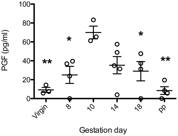
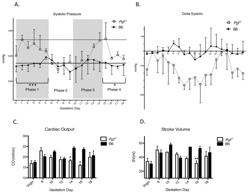
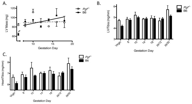
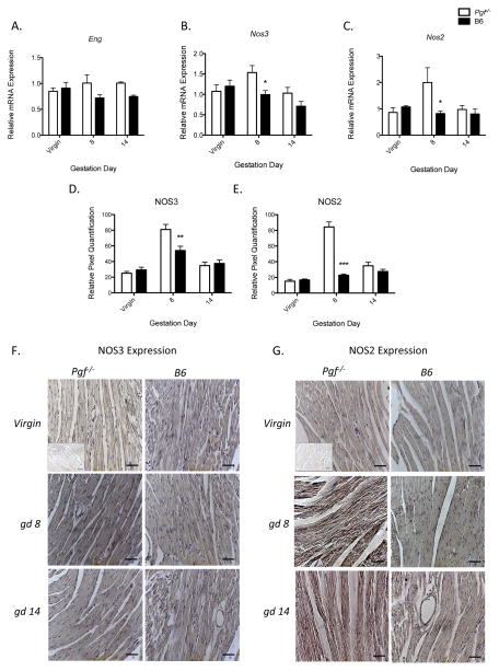
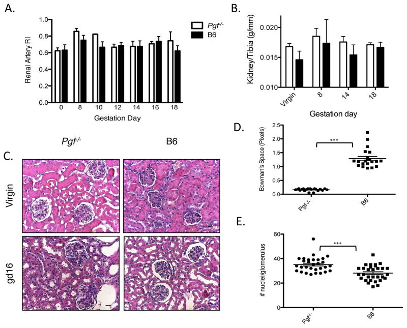
In healthy human pregnancies, placental growth factor (PGF) concentrations rise in maternal plasma during early gestation, peak over Weeks 26-30, then decline. Because PGF in nongravid subjects participates in protection against and recovery from cardiac pathologies, we asked if PGF contributes to pregnancy-induced maternal cardiovascular adaptations. Cardiovascular function and structure were evaluated in virgin, pregnant, and postpartum C56BL/6-Pgf(-) (/) (-) (Pgf(-) (/) (-)) and C57BL/6-Pgf(+/+) (B6) mice using plethysmography, ultrasound, quantitative PCR, and cardiac and renal histology. Pgf(-/-) females had higher systolic blood pressure in early and late pregnancy but an extended, abnormal midpregnancy interval of depressed systolic pressure. Pgf(-/-) cardiac output was lower than gestation day (gd)-matched B6 after midpregnancy. While Pgf(-) (/) (-) left ventricular mass was greater than B6, only B6 showed the expected gestational gain in left ventricular mass. Expression of vasoactive genes in the left ventricle differed at gd8 with elevated Nos expression in Pgf(-) (/) (-) but not at gd14. By gd16, Pgf(-) (/) (-) kidneys were hypertrophic and had glomerular pathology. This study documents for the first time that PGF is associated with the systemic maternal cardiovascular adaptations to pregnancy.

Keywords: cardiac remodeling; cardiovascular risk; fetal growth; placenta; ultrasound.


Conflict of interest statement

Declaration of Interest:

Peter Carmeliet holds patents on the use PGF as a cardioprotectant.

Figures

Figure 1
Figure 1
Circulating PGF concentrations in B6 mice using ELISA (n=3–5 pregnancies/time-point). PGF was below detectable levels in all Pgf−/− dams (not shown). PGF concentrations in B6 females peaked at gd10, the only time-point that showed a significant difference from virgin levels. Gd10 levels were not significantly higher than levels at gd14; but were higher than all other time points measured. Significance determined by one-way ANOVA with Tukey’s post-test. * p≤0.05 from gd10, **p≤0.001 from gd10.
Figure 2
Figure 2
Systolic arterial pressure (SAP) (A–B) was analyzed in trained Pgf−/− and control females across gestation using tail cuff plethysmography (n=4 B6, n=3 Pgf−/−). (A) Raw SAP over pregnancy was segregated into phases according to typical blood pressure fluctuations in normal mouse pregnancy (29). In phases 1 (gd0-5) and phase 4 (gd15-18), SAP was higher in Pgf−/− than B6 dams. Baseline (pre-pregnancy) values are indicated for Pgf−/− (horizontal dotted line) and B6 (horizontal solid line). Pre-pregnancy SAP was higher in Pgf−/− versus control females. (B) SAP was normalized to baseline values and represented as a change from baseline over pregnancy. Fluctuations in SAP in Pgf−/− dams appear to follow the same trends as B6 dams, however, magnitude of fluctuations appear more pronounced. The nadir appearing at gd9 in control mice appears to be prolonged to several days in Pgf−/− (A–B). PGF-related functional adaptations of the maternal heart. Maternal cardiac systolic function (B–C) was assessed using echocardiography (n=3–5/group/time-point). (C) Cardiac output (CO) increases over gestation in B6 dams but not Pgf−/− dams (by linear regression). PGF has an affect on CO in pregnancy, with decreased CO seen in Pgf−/− females, reaching significance at gd14 and gd16. (D) Stroke volume (SV) increases across gestation only in B6 dams (by linear regression). SV is lower overall in Pgf−/− females; differences at specific gds were reached at gd16. Changes over time were determined by linear regression with a statistically non-zero slope (C–D). Differences between groups were determined by Two-way ANOVA with Bonferroni’s post-test (A–D). *P≤0.05, **P≤0.01, ***P≤0.001 versus age-matched B6.
Figure 3
Figure 3
PGF deficiency is accompanied by cardiac structural differences across pregnancy and post-partum. (A) Left ventricular (LV) mass, determined by echocardiography (n=3–5/group/time-point), increased over gestation in B6 but not Pgf−/− dams. Total heart wet weight and chamber weights were determined at euthanasia (n=3–5/group/time-point) (B–C). Weights were normalized to tibia length (unchanged between groups; p=0.17) to account for differences in body weight initially and over the course of pregnancy (B–C). (B) Normalized LV mass appeared increased in Pgf−/− dams compared to controls across pregnancy and post-partum, the difference was not significant (p=0.088). (C) Normalized heart mass was greater in Pgf−/− versus B6 dams throughout pregnancy and post-partum (p=0.049) but the differences did not reach significance at any specific time-point. Changes over time were determined by linear regression with a statistically non-zero slope (A). Differences between groups were determined by Two-way ANOVA with Bonferroni’s post-test (B–C).
Figure 4
Figure 4
Upregulated gene expression in left ventricular (LV) tissue of Pgf−/− mice at midgestation. mRNA expression was determined using qPCR and normalized to expression of Gapdh (n=4/group/time-point) (A–C). Differences in LV NOS2 and NOS3 protein levels were measured by IHC (D–G). (A) Relative endoglin (Eng) expression was increased overall compared to B6 controls (p=0.039) but did not reach significance at any specific time-point. (B) Relative Nos3 (eNOS) expression was increased in Pgf−/− LV compared to B6. Differences at specific time points were significant at gd8. (C) Relative LV Nos2 (iNOS) expression was increased in Pgf−/− compared to B6 only at gd8. (D) NOS3 protein expression showed a non-statistically significant increase in Pgf−/− LV compared to B6 overall (p=0.09); gestation-day specific differences reached significance at gd8 (p<0.01). (E) NOS2 protein expression was also increased in Pgf−/− LV tissue versus B6 control both overall (p<0.0001) and at gd8 (p<0.001). (F–G) Representative micrographs from virgin, gd8 and gd14 Pgf−/− and B6 LV showing IHC staining for NOS3 and NOS2 with hematoxylin counter-stain. Quantitative differences in IHC staining were determined by relative pixel density. Inlets represent negative isotype control (F–G). Differences between groups determined by Two-way ANOVA with Bonferroni’s post-test (A–C). *P≤0.05. **P≤0.01, ***P<0.001. Scale bars represent 20μm.
Figure 5
Figure 5
Renal hypertrophy and structural abnormalities are present in pregnancies lacking PGF. (A) Renal artery RI in Pgf−/− versus B6 did not reach a statistically significant difference (n=3–5/group/time-point, p=0.11). (B) Total kidney wet-weight was measured at euthanasia and normalized to tibia length (n=3–5/group/time-point). Kidney weight increased in Pgf−/− dams over pregnancy and post-partum compared to B6 controls (p=0.0015). Differences at specific gd reached significance by 30 days post-partum (pp30). (C) Representative sections from non-pregnant (NP) and gd16 Pgf−/− and B6 dams. (D) Bowman’s capsular space (urinary space) was measured in randomly selected cortical glomeruli by subtracting glomerular area from total corpuscle surface area (n=3/group/time-point). Urinary space was significantly decreased in Pgf−/− kidneys versus controls. (E) Cellularity was measured in 10 random glomeruli; cellularity was increased in gd16 Pgf−/− versus B6 kidneys. Scale bars=20μm (C). Differences between groups were determined by Two-way ANOVA with Bonferroni’s post-test (A–B). Differences in urinary space and glomerular cellularity at gd16 were determined by Student’s T-Test (D–E). ***P≤0.0001.
Figure 6
Figure 6
Maternal CV consequences of PGF deficiency have negligible fetal impact. High-frequency ultrasound was used for velocimetry of the uterine and umbilical arteries (n=3–5 dams/group/time-point; n=4 fetuses/dam) (A–C). (A) Uterine RI is increased in Pgf−/− dams compared to B6 across gestation (p=0.013) but did not reach significance at any specific gd. (B) Fetal heart rate was measured between peaks on PW Doppler cine loops of the umbilical artery. Fetal heart rate did not differ between genotypes at any gestational time-point measured. BPM= Beats per minute. (C) Umbilical artery RI did not differ between genotypes across gestation. (D) Litter size did not differ between Pgf−/− and B6 pregnancies. Differences between groups were assessed by Two-way ANOVA with Bonferroni’s post-test (A–D).

References

    1. Vrachnis N, Kalampokas E, Sifakis S, Vitoratos N, Kalampokas T, Botsis D, Iliodromiti Z. Placental growth factor (PlGF): a key to optimizing fetal growth. J Matern Fetal Neonatal Med. 2013;26:995–1002. - PubMed
    1. Iwasaki H, Kawamoto A, Tjwa M, Horii M, Hayashi S, Oyamada A, Matsumoto T, Suehiro S, Carmeliet P, Asahara T. PlGF repairs myocardial ischemia through mechanisms of angiogenesis, cardioprotection and recruitment of myo-angiogenic competent marrow progenitors. PLoS One. 2011;6:e24872. - PMC - PubMed
    1. Carnevale D, Lembo G. Placental growth factor and cardiac inflammation. Trends Cardiovasc Med. 2012;22:209–212. - PubMed
    1. Luttun A, Tjwa M, Carmeliet P. Placental growth factor (PlGF) and its receptor Flt-1 (VEGFR-1): novel therapeutic targets for angiogenic disorders. Ann N Y Acad Sci. 2002;979:80–93. - PubMed
    1. Craici I, Wagner S, Garovic VD. Preeclampsia and future cardiovascular risk: formal risk factor or failed stress test? Ther Adv Cardiovasc Dis. 2008;2:249–259. - PMC - PubMed

Publication types

Grants and funding