Review
. 2015 Feb;114(2):94-109.
doi: 10.1016/j.ymgme.2014.12.001. Epub 2014 Dec 9.

Therapies for the bone in mucopolysaccharidoses

Affiliations
Review

Therapies for the bone in mucopolysaccharidoses

Shunji Tomatsu et al. Mol Genet Metab. 2015 Feb.

Abstract

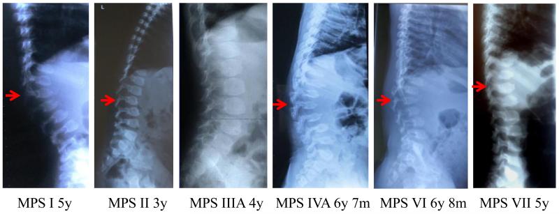
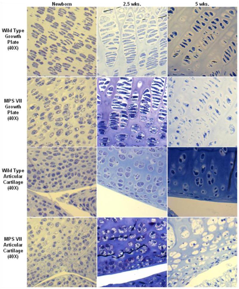
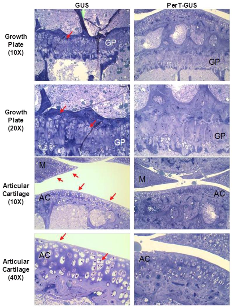
Patients with mucopolysaccharidoses (MPS) have accumulation of glycosaminoglycans in multiple tissues which may cause coarse facial features, mental retardation, recurrent ear and nose infections, inguinal and umbilical hernias, hepatosplenomegaly, and skeletal deformities. Clinical features related to bone lesions may include marked short stature, cervical stenosis, pectus carinatum, small lungs, joint rigidity (but laxity for MPS IV), kyphoscoliosis, lumbar gibbus, and genu valgum. Patients with MPS are often wheelchair-bound and physical handicaps increase with age as a result of progressive skeletal dysplasia, abnormal joint mobility, and osteoarthritis, leading to 1) stenosis of the upper cervical region, 2) restrictive small lung, 3) hip dysplasia, 4) restriction of joint movement, and 5) surgical complications. Patients often need multiple orthopedic procedures including cervical decompression and fusion, carpal tunnel release, hip reconstruction and replacement, and femoral or tibial osteotomy through their lifetime. Current measures to intervene in bone disease progression are not perfect and palliative, and improved therapies are urgently required. Enzyme replacement therapy (ERT), hematopoietic stem cell transplantation (HSCT), and gene therapy are available or in development for some types of MPS. Delivery of sufficient enzyme to bone, especially avascular cartilage, to prevent or ameliorate the devastating skeletal dysplasias remains an unmet challenge. The use of an anti-inflammatory drug is also under clinical study. Therapies should start at a very early stage prior to irreversible bone lesion, and damage since the severity of skeletal dysplasia is associated with level of activity during daily life. This review illustrates a current overview of therapies and their impact for bone lesions in MPS including ERT, HSCT, gene therapy, and anti-inflammatory drugs.

Keywords: Anti-inflammatory drug; Enzyme replacement therapy; Gene therapy; Hematopoietic stem cell transplantation; Mucopolysaccharidoses; Skeletal dysplasia.


Figures

Figure 1
Figure 1
Dorsolumbar spine X-ray pictures of patients with MPS (arrows show the apex of the kyphosis). MPS I (severe): A high lumbar kyphosis is seen at L2. The apical ovoid vertebral body has a prominent anteroinferior beaking with hypoplasia of the anterosuperior aspect. The prominent posterior scalloping of the lumbar vertebrae is observed. Hypoplasia of the superior facets is seen. Similar features are seen to a less extent in the levels above and below this vertebra. As a result of these skeletal anomalies, the patient has retrolisthesis between the vertebrae at the apex of the kyphosis. MPS II (severe): The inferior beaking of ovoid vertebrae at lumbar distinguishes the abnormal condition from MPS IVA. The mild posterior scalloping of the lumbar vertebrae with widening of interpediculate spaces is observed. MPS II is radiologically similar to MPS I (Hurler syndrome); however, the bone deformity is mild with a slower rate of progression. MPS III (severe): MPS III shows the mildest skeletal deformity among all types of MPS. Ovoid vertebrae at lumbar are seen with mild widening of interpediculate spaces. The bone deformity is mildest with a slower rate of progression among all types of MPS. MPS IVA (severe): MPS IVA shows the most severe skeletal deformity among all types of MPS. Prominent lumbar kyphosis is seen at L2. Universal platyspondyly shows a central anterior beaking in contrast to the inferior, anterior beaking seen in MPS I (Hurler syndrome). Marked increase of interpediculate spaces (most severe), multiple vertebral subluxations (most severe), and small sacrum are seen. Bone mineral density is low. MPS VI (severe): Prominent kyphosis is seen at L2. Universal platyspondyly shows a central anterior beak in contrast to the inferior, anterior beaking seen in MPS I (Hurler syndrome). The moderate posterior scalloping of the lumbar vertebrae is observed. Hypoplastic lumbar vertebrae (L1-L5) with characteristic superior notch (L4 and L5), multiple vertebral subluxations, marked increase of interpediculate spaces, and small sacrum are seen. Bone mineral density is low. MPS VII (severe): Kyphosis is seen at L1. Moderate ovoid vertebrae at lumbar distinguish the abnormal condition from MPS IVA. The lumbar vertebrae with widening of interpediculate spaces are observed. Universal platyspondyly of dorsolumbar vertebrae is observed with a mild central anterior beaking. The patient with MPS VII here shows radiologically less severity compared with MPS IVA.
Figure 2
Figure 2
Age-dependent change of storage vacuoles at an early stage in cartilage of MPS VII mouse model. Left panel: 2-3 days old, middle panel: 2.5 weeks old, right panel: 5 weeks old. Clear vacuoles are observed in most chondrocytes at birth. Vacuoles in chondrocytes increase in number with age and storage materials are fully accumulated at the age of 5 weeks old. Vacuolization and disorganization of the column structure in articular cartilage is more progressive than seen in epiphyseal cartilage (toluidine blue-stained 0.5-μm-thick sections: X 40).
Figure 3
Figure 3
Bone pathology of iliac crest in a 17-year-old patient with MPS IV after clinical trial and 6 months extension study of ERT (toluidine blue-stained 0.5-μm-thick sections: X 100).
Figure 4
Figure 4
Histopathology of the knee joint of 17 weeks-old IV GUS and PerT-GUS treated MPS VII mice (ERT started at 5 weeks old). Images are of the growth plate and articular cartilage. PerT-GUS treated mouse shows substantial reduced number of vacuolated chondrocytes compared with native GUS treated mouse. Arrows show vacuolated cells in the growth plate, articular cartilage and meniscus area. AC: articular cartilage, GP: growth plate, M: meniscus. Toluidine blue-stained 0.5-μm-thick sections. Adapted from Rowan DJ, Tomatsu S, Grubb JH, et al. Long circulating enzyme replacement therapy rescues bone pathology in mucopolysaccharidosis VII murine model. Mol Genet Metab 2012 107(1-2):161-72.
Figure 5
Figure 5
Three-dimensional micro-CT reconstructions of knee joints of wild-type, untreated MPS VII, and PerT-GUS treated MPS VII mice intraperitoneally (IP) (IP 2 mg/kg ERT started at day 2-3 and continued weekly until the autopsy). Each picture shows unsectioned bone (left side) or sagittal-sectioned bone (right side). Cross sections are sagittal through the midlines. The long arrows identify areas of thickened cortical bone. The short arrows identify abnormal exophytic bone formations on articular surfaces. Ages of wild-type and untreated MPS VII mice are 5, 23, and 36 weeks old. Ages of 2 mg/kg PerT-GUS treated mice are 27, 41, and 57 weeks-old. Adapted from Rowan DJ, Tomatsu S, Grubb JH, et al. Long circulating enzyme replacement therapy rescues bone pathology in mucopolysaccharidosis VII murine model. Mol Genet Metab 2012 107(1-2):161-72.
Figure 6
Figure 6
Growth plate histology of 8-weeks-old MPS IVA mouse treated with bone-targeting enzyme. Newborn ERT started at day 2, and weekly ERT continued for 8 weeks. Vacuolated storage is substantially reduced in a treated mouse with MPS IVA. Toluidine blue-stained 0.5-μm-thick sections (X 100). Adapted from Tomatsu S, Montaño AM, Oikawa H et al. Enzyme replacement therapy in newborn mucopolysaccharidosis IVA mice: early treatment rescues bone lesions? Mol Genet Metab Jun 4. pii: S1096-7192(14)00185-1. doi: 10.1016/j.ymgme.2014.05.013. [Epub ahead of print].
Figure 7
Figure 7
A 13-years-old Hurler patient: 10 years post-HSCT X-ray of spine and pathology at lumbar spine. Left (X-ray): Severe humpback of L2 is seen. The patient underwent spinal surgery at 13.5 years of age. Mild invagination of the superior and inferior endplates of the lower dorsal vertebrae is observed, and the vertebrae appear flattened. Moderate posterior scalloping of the lumbar vertebrae is observed. There is a mild dextroscoliosis of the lumbar spine. Appearance of the spine is not significantly changed since 6 years of age. Deformity of spine is much milder than that seen in an untreated patient with a severe form. Right (pathology from surgical remnants): no storage vacuoles appear in chondrocytes of lumbar spine at 13.5 years of age and the size and morphology of chondrocytes are normal.
Figure 8
Figure 8
X-ray photographs with age in a patient with MPS IVA after BMT. Lateral view of thoracolumbar vertebrae a: pre-BMT (left) and three years later post-BMT (right). Platyspondylia and anterior beaking of thoracolumbar vertebra increase slightly in size, and the margin of vertebra becomes clear. One year later post-BMT, BMD at L2-4 increases from 0.372 to 0.548 (g/cm2), and it is maintained at the level of 0.48 ± 0.054 for the following 9 years. Adapted from Chinen Y, Higa T, Tomatsu S, et al. Long-term therapeutic efficacy of allogenic bone marrow transplantation in a patient with mucopolysaccharidosis IVA. Mol Genet Metab Rep 1 (2014) 31-41.
Figure 9
Figure 9
Mechanism of multiple-AAA targeting system. Viral capsid in the right panel has multiple copies of D8 integrated into capsid proteins, showing the retargeting of gene vector to bone (hydroxyapatite in the mineral region) schematically.
Figure 10
Figure 10
Appearance of foam cells/macrophages/vacuolated cells in tissues in autopsied specimens from a 20-year-old male MPS IVA patient1. Left; Bone marrow in the vertebrae shows foam cells and vacuolated osteoblasts (40x), right; Trachea shows ballooned vacuolated chondrocytes (100x). Stained with toluidine blue (0.5 μm; light microscopy).
Figure 11
Figure 11
Hypothesis for role of PPS on reduction of GAGs, suppression of inflammation, and promotion of chondrogenesis in chondrocytes RUNX2: runt-related transcription factor 2, MAP: Mitogen-activated protein, NF-κB: nuclear factor-kappa B.

References

    1. Neufeld E, Muenzer J. The mucopolysaccharidoses. In: Scriver C, Beaudet A, Sly W, Valle D, editors. The Metabolic and Molecular Bases of Inherited Disease. McGraw-Hill; New York, NY: 2001. pp. 3421–3452.
    1. Tomatsu S, Montaño A, Oikawa H, Giugliani R, Harmatz P, Smith M, Orii T. Impairment of Body Growth in Mucopolysaccharidoses, In Handbook of Growth and Growth Monitoring in Health and Disease 1. Springer; New York: 2012. pp. 2091–2117.
    1. Patel P, Suzuki Y, Maeda M, Yasuda E, Shimada T, Orii KE, Orii T, Tomatsu S. Growth charts for patients with Hunter syndrome. Mol. Genet. Metab. Rep. 2014;1:5–18. - PMC - PubMed
    1. Tomatsu S, Montaño AM, Oikawa H, Smith M, Barrera L, Chinen Y, Thacker MM, Mackenzie WG, Suzuki Y, Orii T. Mucopolysaccharidosis type IVA (Morquio A disease): clinical review and current treatment. Curr. Pharm. Biotechnol. 2011;12:931–945. - PubMed
    1. Rodriguez M, Miller TL, Mackenzie WG, Ditro C, Chidekel AS, Shaffer TH. Characteristics of Impulse Oscillometry and Thoracoabdominal Motion in Children with Thoracic Cage Disorders. Pediatr. Pulmonol. 2010;45:679–686. - PMC - PubMed

Publication types

Substances