. 2015 Jan;9(1):15-20.
doi: 10.1016/j.jash.2014.10.009. Epub 2014 Oct 23.

Prevalence of angiotensin II type 1 receptor (AT1R)-activating autoantibodies in primary aldosteronism

Affiliations

Prevalence of angiotensin II type 1 receptor (AT1R)-activating autoantibodies in primary aldosteronism

Hongliang Li et al. J Am Soc Hypertens. 2015 Jan.

Abstract

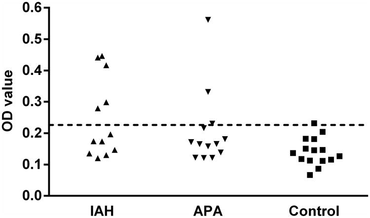
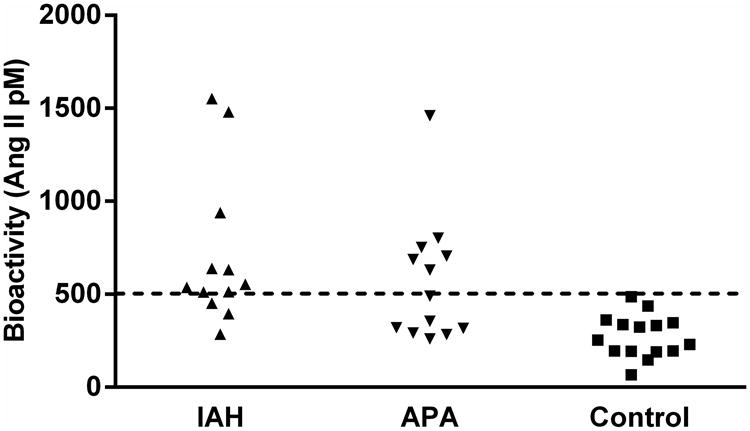
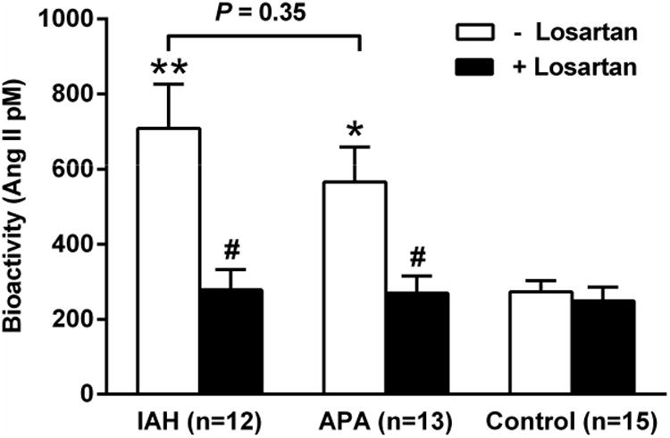
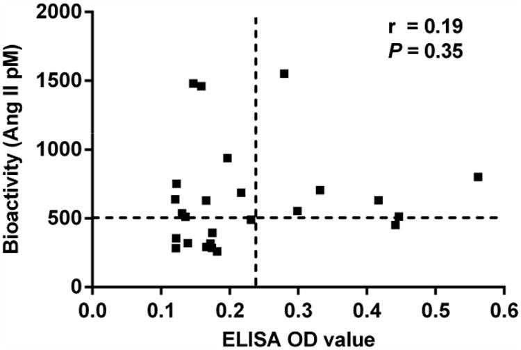
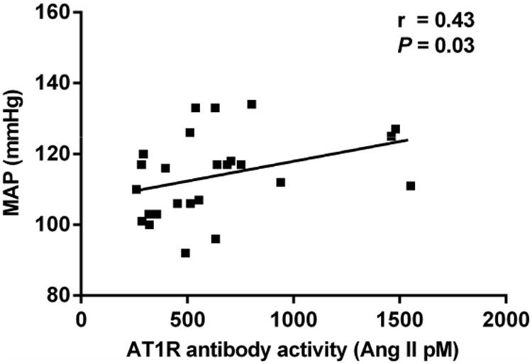
Autoantibodies to the angiotensin II type 1 receptor (AT1R) have been reported in patients with primary aldosteronism, including aldosterone producing adenoma (APA) and idiopathic adrenal hyperplasia (IAH). Sera from 25 primary aldosteronism subjects (12 with IAH and 13 with APA) and 15 normotensive control subjects were assayed for AT1R autoantibodies by enzyme-linked immunosorbent assay and an AT1R-transfected cell-based bioassay. Nine of 12 IAH subjects (75%) and six of 13 APA subjects (46%) were positive for AT1R autoantibodies in the bioactivity assay. The mean AT1R autoantibody activity for the IAH and APA subjects was significantly greater than controls (P < .001 and P < .01, respectively), and this in vitro activity was suppressed by the AT1R blocker losartan. None of the controls had significant AT1R autoantibody activity. Enzyme-linked immunosorbent assay values were less sensitive but were positive in some subjects with IAH and APA. The mean arterial pressure of these primary aldosteronism subjects correlated modestly with AT1R autoantibody activity. These data confirm the presence of active AT1R autoantibodies in a high percentage of subjects with primary aldosteronism irrespective of their underlying etiology. These observations have both pathophysiological and clinical implications.

Keywords: Aldosterone–producing adenoma; cell-based bioassay; hypertension; idiopathic adrenal hyperplasia.


Conflict of interest statement

Conflict of interest: None

Figures

Figure 1
Figure 1. ELISA detection of AT1R autoantibodies in primary aldosteronism patients and normotensive control subjects
IAH, idiopathic adrenal hyperplasia (n=12), closed upward triangles; APA, aldosterone producing adenoma (n=13), closed downward triangles; control, normotensive control (n=15), closed squares. The dashed line is the threshold derived from the mean optical density (OD) values + 2SD of the normotensive controls.
Figure 2
Figure 2. The effect of sera from each primary aldosteronism patient and normotensive control subject on AT1R activation in transfected Chinese hamster ovary (CHO) cells
IAH (n=12), closed upward triangles; APA (n=13), closed downward triangles; control (n=15), closed squares. Data are expressed in Ang II equivalent dosages (mean±SEM). The dashed line is the threshold derived from the mean values + 2SD of the normotensive controls.
Figure 3
Figure 3. The mean effect of sera from primary aldosteronism patients and normotensive control subjects in the absence and presence of AT1R blockade on AT1R activation in transfected CHO cells
Both IAH and APA had a highly significant increase of AT1R activity compared to the normotensive controls (*P<0.01, **P<0.001). There was no significant difference in AT1R activity between the two primary aldosteronism groups. The AT1R blocker losartan (10 μM) effectively blocked the autoantibody activity in each group of the primary aldosteronism patients (#P<0.01). Although the activity in IAH and APA dropped significantly with losartan, there was no significant change in the low level of activity in the control subjects.
Figure 4
Figure 4. Correlation analysis between ELISA and cell-based bioassay for AT1R autoantibodies
The vertical and horizontal dashed lines represent the cut-off values (mean + 2SD of controls) for ELISA and bioassay positivity, respectively. No significant correlation was detected between ELISA OD values and bioactivity values.
Figure 5
Figure 5. Correlation analysis between patient mean arterial pressure (MAP) and AT1R antibody activity measured in AT1R-trasnfected CHO cells
MAP was computed from blood pressures taken at the time of bilateral adrenal vein sampling. There was a significant correlation between the patient MAP and their concurrent serum AT1R antibody activity.

References

    1. Conn JW, Louis LH. Primary aldosteronism, a new clinical entity. Ann Intern Med. 1956;44:1–15. - PubMed
    1. Funder JW. The genetic basis of primary aldosteronism. Curr Hypertens Rep. 2012;14:120–4. - PubMed
    1. Al-Salameh A, Cohen R, Desailloud R. Overview of the genetic determinants of primary aldosteronism. Appl Clin Genet. 2014;7:67–79. - PMC - PubMed
    1. Choi M, Scholl UI, Yue P, Bjorklund P, Zhao B, Nelson-Williams C, et al. K+ channel mutations in adrenal aldosterone-producing adenomas and hereditary hypertension. Science. 2011;331:768–72. - PMC - PubMed
    1. Azizan EA, Murthy M, Stowasser M, Gordon R, Kowalski B, Xu S, et al. Somatic mutations affecting the selectivity filter of KCNJ5 are frequent in 2 large unselected collections of adrenal aldosteronomas. Hypertension. 2012;59:587–91. - PubMed

Publication types

LinkOut - more resources