. 2015 Jan;22(1):180-6.
doi: 10.1107/S1600577514020360. Epub 2015 Jan 1.

Upgrade of MacCHESS facility for X-ray scattering of biological macromolecules in solution

Affiliations

Upgrade of MacCHESS facility for X-ray scattering of biological macromolecules in solution

Alvin Samuel Acerbo et al. J Synchrotron Radiat. 2015 Jan.

Abstract

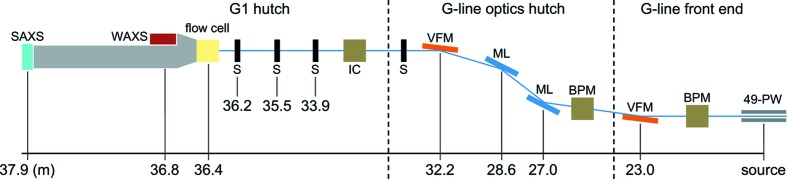
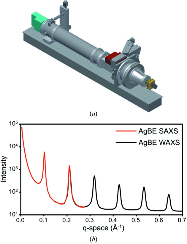
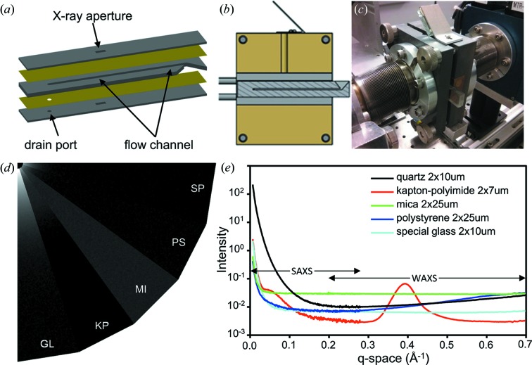
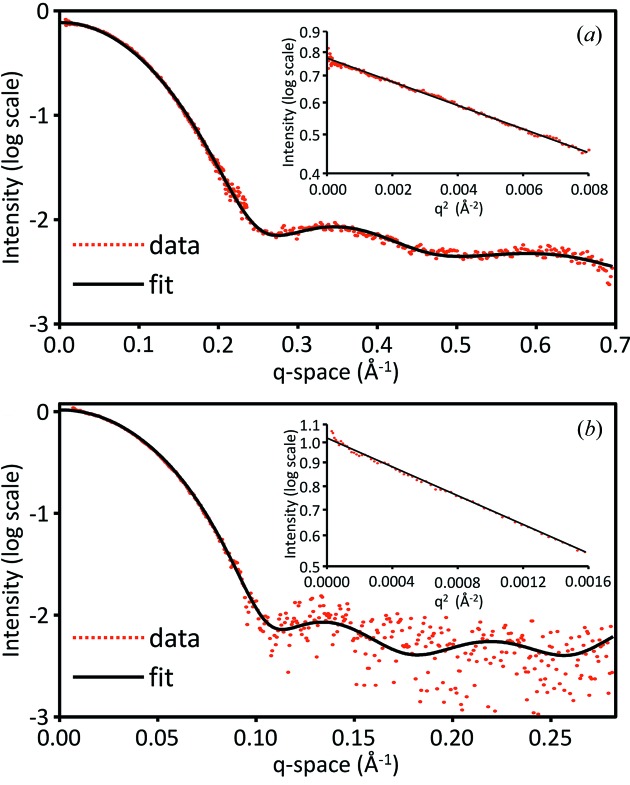
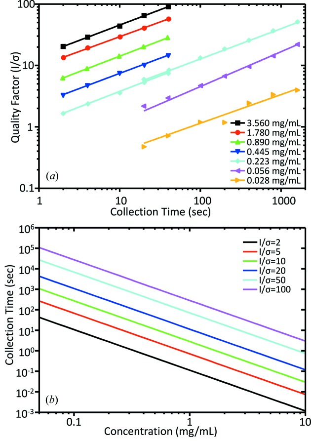
X-ray scattering of biological macromolecules in solution is an increasingly popular tool for structural biology and benefits greatly from modern high-brightness synchrotron sources. The upgraded MacCHESS BioSAXS station is now located at the 49-pole wiggler beamline G1. The 20-fold improved flux over the previous beamline F2 provides higher sample throughput and autonomous X-ray scattering data collection using a unique SAXS/WAXS dual detectors configuration. This setup achieves a combined q-range from 0.007 to 0.7 Å(-1), enabling better characterization of smaller molecules, while opening opportunities for emerging wide-angle scattering methods. In addition, a facility upgrade of the positron storage ring to continuous top-up mode has improved beam stability and eliminated beam drift over the course of typical BioSAXS experiments. Single exposure times have been reduced to 2 s for 3.560 mg ml(-1) lysozyme with an average quality factor I/σ of 20 in the Guinier region. A novel disposable plastic sample cell design that incorporates lower background X-ray window material provides users with a more pristine sample environment than previously available. Systematic comparisons of common X-ray window materials bonded to the cell have also been extended to the wide-angle regime, offering new insight into best choices for various q-space ranges. In addition, a quantitative assessment of signal-to-noise levels has been performed on the station to allow users to estimate necessary exposure times for obtaining usable signals in the Guinier regime. Users also have access to a new BioSAXS sample preparation laboratory which houses essential wet-chemistry equipment and biophysical instrumentation. User experiments at the upgraded BioSAXS station have been on-going since commissioning of the beamline in Summer 2013. A planned upgrade of the G1 insertion device to an undulator for the Winter 2014 cycle is expected to further improve flux by an order of magnitude.

Keywords: MacCHESS; SAXS; high throughput.


Figures

Figure 1
Figure 1
Schematic of the beamline setup at the MacCHESS BioSAXS beamline G1. A pair of vertical focusing mirrors and multilayer monochromators split a 2 mrad fan from the wiggler into the endstation G1 hutch. Abbreviations: 49-pole wiggler (49-PW), beam-position monitor (BPM); vertical-focusing mirror (VFM); multilayer monochromator (ML); slits (S); ion chamber (IC).
Figure 2
Figure 2
(a) Illustration of the aluminium flight tube (grey), SAXS (cyan) and WAXS (red) detectors as well as the BioSAXS sample flow cell (yellow). (b) Full SAXS and WAXS scattering curves of AgBE, showing an overlap between 0.20 and 0.28 Å−1.
Figure 3
Figure 3
(a) Schematic of the BioSAXS flow cell, showing the basic design elements. Two outer layers of PMMA (grey) provide structural support to the flow cell, while the central channel of PMMA contains the flow channel and effectively sets the X-ray path length. Sandwiched between the PMMA layers are X-ray transparent window layers of 25 µm-thick polystyrene (yellow). (b) Side view of the BioSAXS flow cell enclosed in the flow cell holder. A mirror on top of the flow cell holder allows a camera to capture video of the area of the flow cell where X-ray exposure occurs. (c) Configuration of the BioSAXS flow cell and flow cell holder at beamline G1. (d) Composition of detector image wedges of five candidate window materials in real space (equivalent q-range from 0 to 0.25 Å−1). High scattering intensities are indicated in white, low scattering intensities in black. Abbreviations: GL = quartz, KP = kapton, MI = mica, PS = polystyrene, SP = special glass. (e) Background-corrected SAXS and WAXS scattering curves of the five candidate window materials.
Figure 4
Figure 4
(a) Scattering plot of 3.560 mg ml−1 lysozyme taken from an average of 20 successive 2 s exposures (red). A CRYSOL fit (black) using PDB 1lyz overlays well with the scattering data. Guinier analysis (inlay) resolves an R g of 1.43 nm and an average I/σ of 65. (b) Scattering plot of 0.390 mg ml−1 glucose isomerase taken from an average of 20 successive 2 s exposures (red). A CRYSOL fit (black) using PDB 1oad overlays well with the scattering data up to 0.10 Å−1. Guinier analysis (inlay) resolves an R g of 3.32 nm with an average I/σ of 210.
Figure 5
Figure 5
(a) Scattering data quality factor (QF), I/σ, as a function of data collection time for a range of dilutions of lysozyme. Data points for individual dilutions (indicated by markers) were best described using a power function (solid line). (b) Predicted correlation between sample concentration and estimated required data collection time for arbitrary QF for lysozyme.

References

    1. Blanchet, C. E., Zozulya, A. V., Kikhney, A. G., Franke, D., Konarev, P. V., Shang, W., Klaering, R., Robrahn, B., Hermes, C., Cipriani, F., Svergun, D. I. & Roessle, M. (2012). J. Appl. Cryst. 45, 489–495.
    1. Cho, H. S., Dashdorj, N., Schotte, F., Graber, T., Henning, R. & Anfinrud, P. (2010). Proc. Natl Acad. Sci. USA, 107, 7281–7286. - PMC - PubMed
    1. Daugherty, M. D., Booth, D. S., Jayaraman, B., Cheng, Y. & Frankel, A. D. (2010). Proc. Natl Acad. Sci. USA, 107, 12481–12486. - PMC - PubMed
    1. David, G. & Pérez, J. (2009). J. Appl. Cryst. 42, 892–900.
    1. Gillilan, R., Cook, M., Temnykh, G., Møller, M. & Nielsen, S. (2013). Trans. Am. Crystallogr. Assoc. Symp. 44, 40–50.

Publication types