

miR-122 is a unique molecule with great potential in diagnosis, prognosis of liver disease, and therapy both as miRNA mimic and antimir
- PMID: 25537773
- PMCID: PMC4439190
- DOI: 10.2174/1566523214666141224095610
miR-122 is a unique molecule with great potential in diagnosis, prognosis of liver disease, and therapy both as miRNA mimic and antimir
Abstract
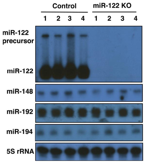
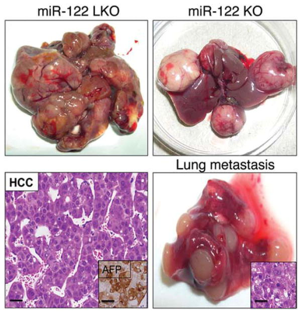
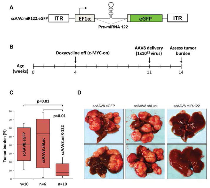
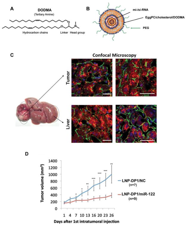
miR-122, a completely conserved liver-specific miRNA in vertebrates, is essential for the maintenance of liver homeostasis. This 22 nucleotide RNA regulates diverse functions such as cholesterol, glucose and iron homeostasis, lipid metabolism and infection of hepatitis C virus (HCV) and of the parasitic protozoa, Leishmania donovani. It is the first miRNA that underwent successful clinical trials in HCV infected patients. In contrast, miR-122 expression is reduced in nonalcoholic steatohepatitis (NASH) patients, and in a subset of hepatocellular carcinoma (HCC) patients including hepatitis B virus (HBV) positive patients with highly invasive and metastatic cancer. Studies in mice genetically depleted of miR-122 have highlighted its critical role in liver biology. These mice progressively develop steatohepatitis, fibrosis and hepatocellular cancer, establishing it as a bona fide tumor suppressor. Additionally, delivery of miR-122 using a viral vector or liposomal nanoparticles resulted in liver tumor suppression in animal models. These results suggest miR-122 supplementation might be beneficial in NASH or HBV positive HCC patients. Furthermore, circulating miR-122 has emerged as a sensitive biomarker for liver injury. The ability of miR-122 to promote differentiation of embryonic and adult stem cells to hepatocyte-like cells in vitro suggests its potential role in driving the hepatic differentiation program. In this review, we will discuss the role of miR-122 in liver physiology and the deleterious consequences of its loss of function, its role as a sensitive biomarker for liver injury and therapeutic target. Development of novel technologies for targeted delivery of miR-122 to tumor cells and for direct monitoring of miR-122 in biological fluids is urgently needed for translating the basic research to the bedside. This review focuses on miR-122, the most abundant hepatic miRNA, in the context of liver health and diseases.
Conflict of interest statement
The authors confirm that this article content has no conflict of interest.
Figures
References
-
- Chang J, Nicolas E, Marks D, et al. miR-122, a mammalian liver-specific microRNA, is processed from hcr mRNA and may downregulate the high affinity cationic amino acid transporter CAT-1. RNA Biol. 2004;1(2):106–13. - PubMed
-
- Lagos-Quintana M, Rauhut R, Yalcin A, Meyer J, Lendeckel W, Tuschl T. Identification of tissue-specific microRNAs from mouse. Curr Biol. 2002;12(9):735–9. - PubMed
-
- Wienholds E, Kloosterman WP, Miska E, et al. MicroRNA expression in zebrafish embryonic development. Science. 2005;309(5732):310–1. - PubMed
-
- Filipowicz W, Grosshans H. The liver-specific microRNA miR-122: biology and therapeutic potential. Prog Drug Res. 2011;67:221–38. - PubMed
Publication types
MeSH terms
Substances
Grants and funding
LinkOut - more resources
Full Text Sources
Other Literature Sources
Medical
