

Case Reports
doi: 10.1007/s12105-014-0598-5.
Epub 2014 Dec 24.
Allergic Fungal Sinusitis
Affiliations
- PMID: 25537829
- PMCID: PMC4651931
- DOI: 10.1007/s12105-014-0598-5
Item in Clipboard
Case Reports
Allergic Fungal Sinusitis
Head Neck Pathol.
2015 Dec.
Abstract
A 42 year old male presents with worsening pain and an increase in thick chronic drainage of the left sinus. Image studies show complete opacification of the left frontal sinus, left sphenoid sinus, and the left maxillary sinus. The patient was taken to the operating room and tissue for microscopic evaluation was obtained. The microscopic findings were classic for allergic fungal sinusitis: areas of alternating mucinous material and inflammatory cell debris and abundant Charcot-Leyden crystals. Cultures were performed and the patient began steroid therapy and desensitization therapy.
Keywords: Congestion; Desensitization therapy; Sinus drainage; Tide lines.
Figures
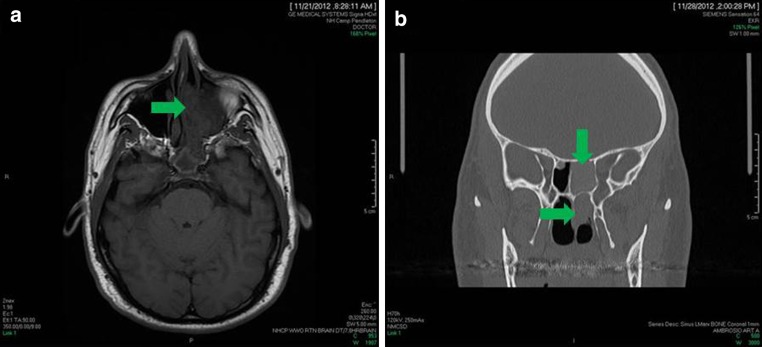
a Magnetic resonance image (MRI) shows boney demineralization and an opacification of the left nasal cavity and sinuses. b Computed tomography (CT) also shows opacification of the sinuses, however, other entities were considered on the differential diagnosis to include sinonasal inflammatory polyps and a neoplasm
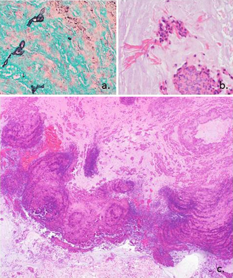
a Grocott’s methenamine silver (GMS) stain highlights the scant fungal elements seen in the specimen. b This high power image shows degenerated inflammatory cells and eosinophils with Charcot–Leyden crystals. c Hematoxylin and eosin stain shows alternating ripples of eosinophils and neutrophils creating a “tide line” or “tree ring” appearance. This low power picture is classic for allergic fungal sinusitis
References
-
- Thompson, et al. Diagnostic pathology: head and neck. Manitoba: Amirsys Publishing Inc; 2011.
-
- Huchton, et al. Allergic fungal sinusitis: an otorhinolaryngologic perspective. Allergy Asthma Proc. 2013;24(5):307–311. - PubMed
-
- Schubert: Allergic fungal sinusitis. Clin Rev Allergy Immunol. 1080-0549/06/205-216. 2006. - PubMed
Publication types
MeSH terms
Substances
LinkOut - more resources
Full Text Sources
Other Literature Sources
Medical
