

Eagle's Syndrome
- PMID: 25537830
- PMCID: PMC4651924
- DOI: 10.1007/s12105-014-0599-4
Eagle's Syndrome
Abstract
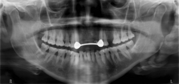
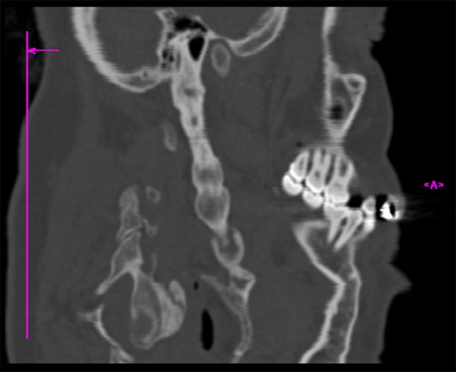
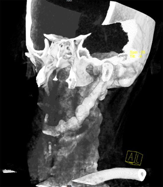
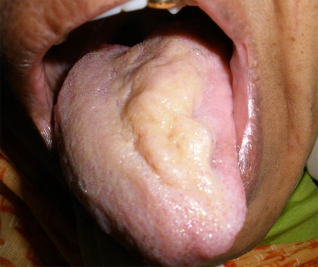
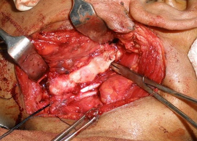
Eagle's Syndrome (ES) refers to a symptomatic anomaly due to elongation of the styloid process or mineralization of the styloid complex. If not diagnosed timely and treated properly, elongation of the styloid process or the hyper-mineralization of the stylohyoid ligament may eventually lead to complete ossification of the stylohyoid complex. Non-specific head and neck symptoms of the ES may pose diagnostic challenges to the clinician. Therefore it is crucial to include ES among differential diagnosis when evaluating patients with similar head and neck symptoms. Once the diagnosis is confirmed, treatment plan should be tailored in accordance with the individual requirements of the case and performed without delay. Both pharmacological and surgical methods have been described for the treatment of the patients with ES. However for those who suffer from persistent symptoms, surgical removal of the elongated styloid process is the treatment of choice and can be done with an intraoral or an extraoral approach. The aim of this work is to present unusual clinical symptoms and radiologic findings of ES due to complete ossification of the stylohyoid complex. The importance of a correct diagnosis and appropriate treatment are highlighted.
Keywords: Eagle’s Syndrome; Hypoglossal nerve palsy; Ossification; Stylohyoid.
Figures
References
-
- Bensoussan Y, Letourneau-Guillon L, Ayad T. A typical presentation of Eagle syndrome with hypoglossal nerve palsy and Horner syndrome. Head Neck. 2014;36(12):E136–8. - PubMed
Publication types
MeSH terms
Supplementary concepts
LinkOut - more resources
Full Text Sources
Other Literature Sources
