. 2015 Feb 13;290(7):4330-42.
doi: 10.1074/jbc.M114.628438. Epub 2014 Dec 23.

IgLON cell adhesion molecules are shed from the cell surface of cortical neurons to promote neuronal growth

Affiliations

IgLON cell adhesion molecules are shed from the cell surface of cortical neurons to promote neuronal growth

Ricardo Sanz et al. J Biol Chem. .

Abstract

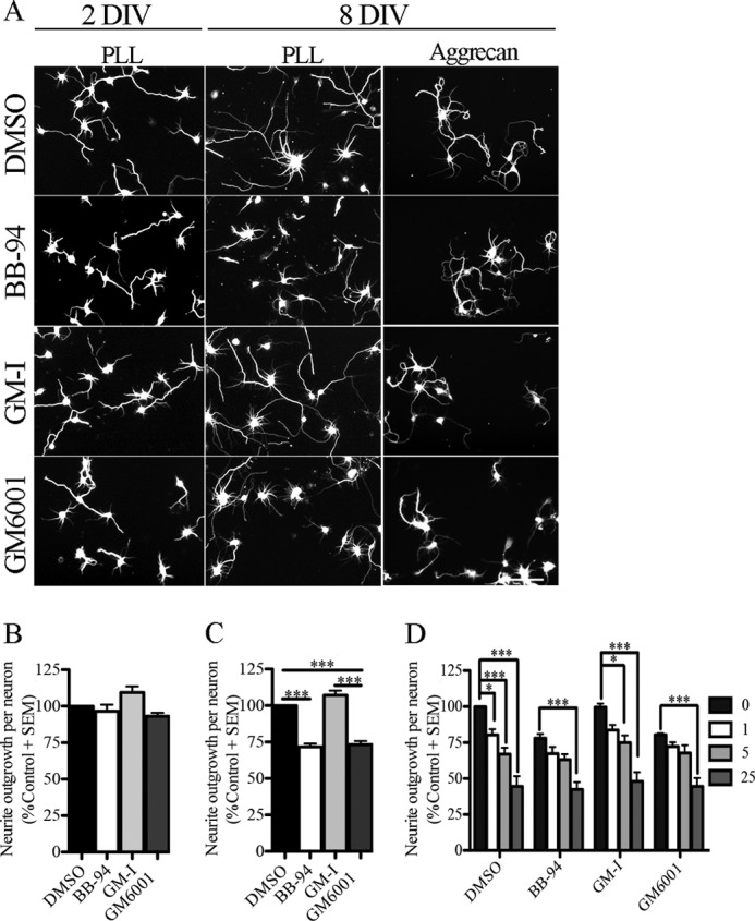
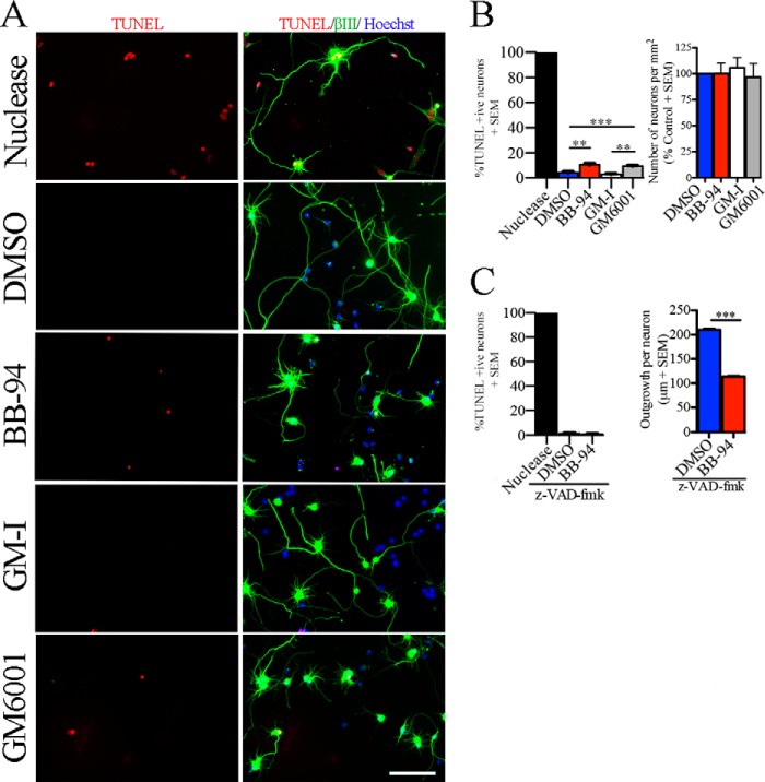
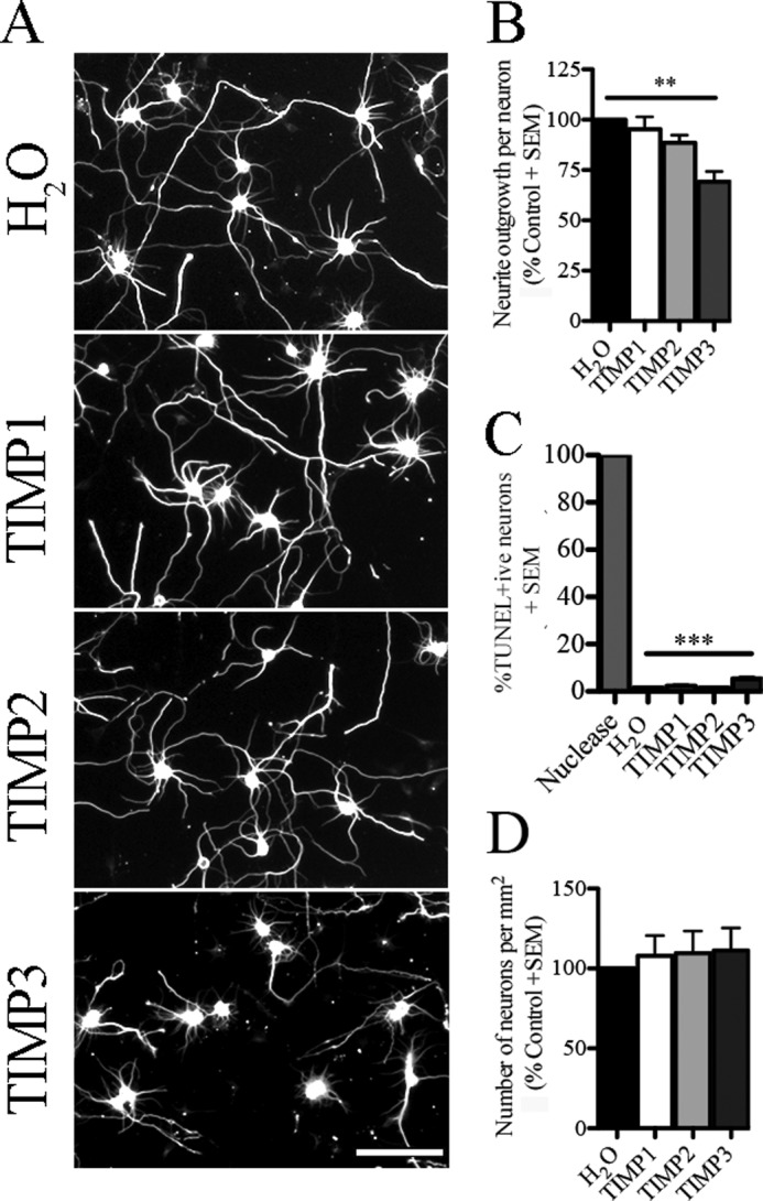
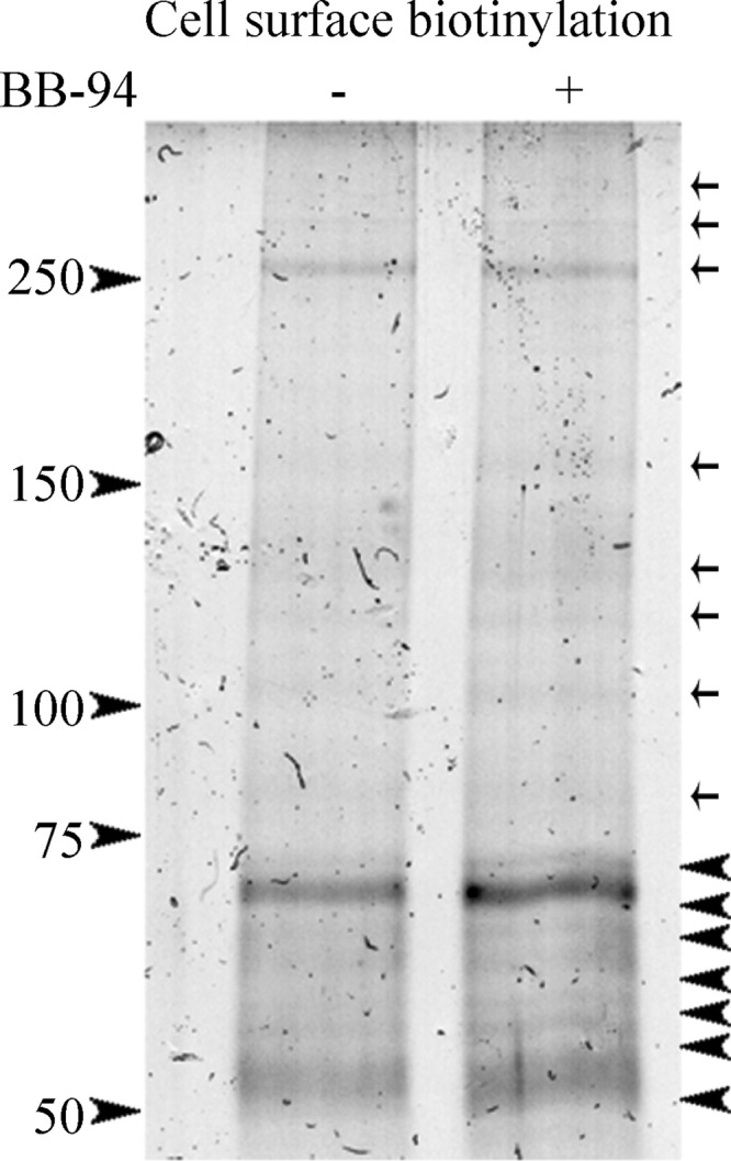
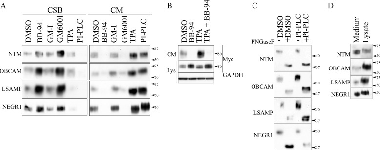
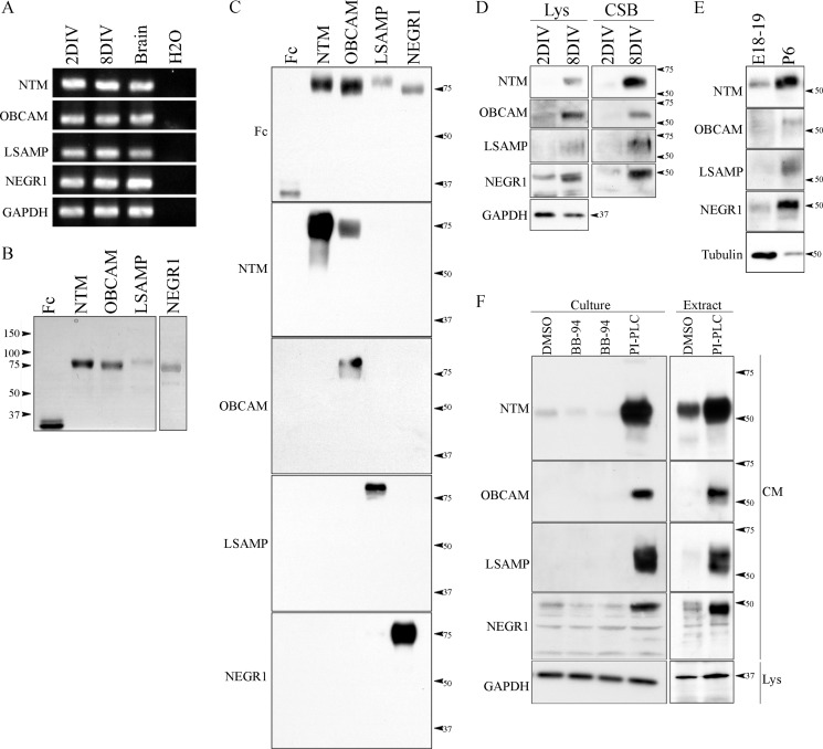
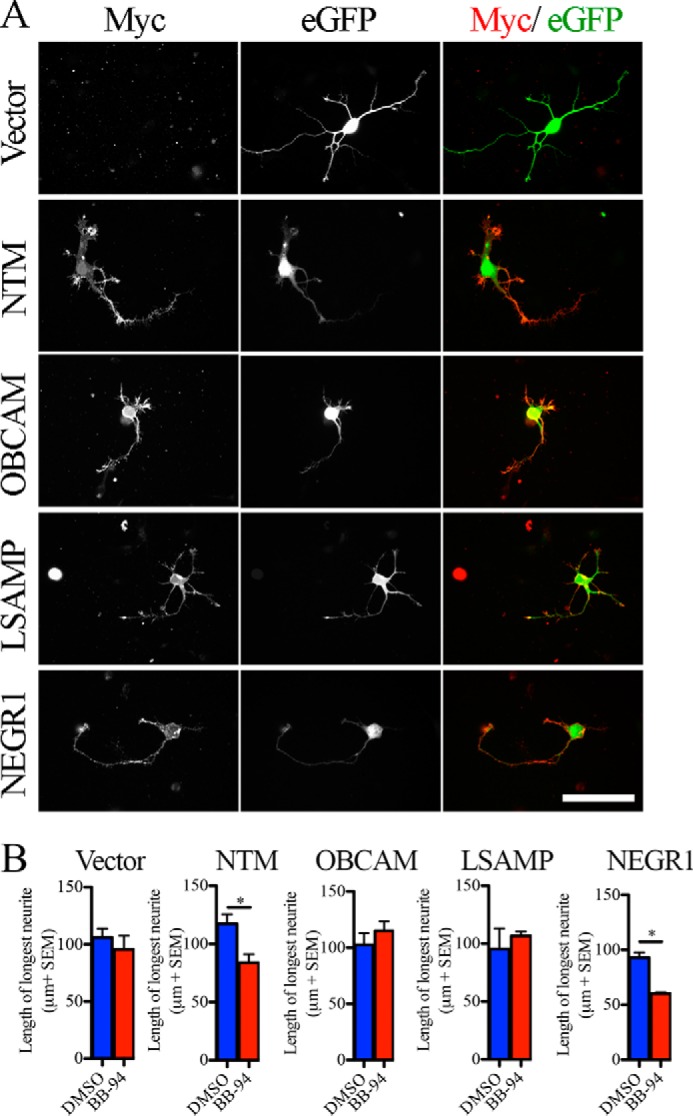
Matrix metalloproteinases and a disintegrin and metalloproteinases are members of the zinc endopeptidases, which cleave components of the extracellular matrix as well as cell surface proteins resulting in degradation or release of biologically active fragments. Surface ectodomain shedding affects numerous biological processes, including survival, axon outgrowth, axon guidance, and synaptogenesis. In this study, we evaluated the role of metalloproteinases in regulating cortical neurite growth. We found that treatment of mature cortical neurons with pan-metalloproteinase inhibitors or with tissue inhibitors of metalloproteinase-3 reduced neurite outgrowth. Through mass spectrometry, we characterized the metalloproteinase-sensitive cell surface proteome of mature cortical neurons. Members of the IgLON family of glycosylphosphatidylinositol-anchored neural cell adhesion molecules were identified and validated as proteins that were shed from the surface of mature cortical neurons in a metalloproteinase-dependent manner. Introduction of two members of the IgLON family, neurotrimin and NEGR1, in early embryonic neurons was sufficient to confer sensitivity to metalloproteinase inhibitors in neurite outgrowth assays. Outgrowth experiments on immobilized IgLON proteins revealed a role for all IgLON family members in promoting neurite extension from cortical neurons. Together, our findings support a role for metalloproteinase-dependent shedding of IgLON family members in regulating neurite outgrowth from mature cortical neurons.

Keywords: ADAM; Axon; Cell Adhesion; Matrix Metalloproteinase (MMP); Neuron; Shedding; Tissue Inhibitor of Metalloproteinase (TIMP).


Figures

FIGURE 1.
FIGURE 1.
Pan-metalloproteinase inhibitors repress neurite outgrowth in mature cortical neurons. A, rat cortical neurons from two developmental stages (2 DIV and 8 DIV) seeded on a PLL and an aggrecan (5 μg/ml) substrate and treated with a vehicle control (DMSO), metalloproteinase inhibitors (BB-94 and GM601), and a GM6001-Inactive control (GM-I). Neuronal projections were visualized with βIII-tubulin staining. B and C, outgrowth quantification from 2-DIV (B) and 8-DIV (C) cortical neurons. Outgrowth was normalized to the DMSO control for each experiment. D, outgrowth quantification from 8-DIV cortical neurons seeded on PLL and aggrecan substrates (1, 5, and 25 μg/ml) and treated with DMSO, BB-94, GM-I, and GM6001. Outgrowth data were normalized to DMSO control on PLL substrate. n = 4–8 from independent cultures. Data shown as mean ± S.E. *, p < 0.05; ***, p < 0.001 by one- and two-way ANOVA, followed by Bonferroni post hoc test. Scale bar, 100 μm.
FIGURE 2.
FIGURE 2.
Metalloproteinase inhibitors inhibit growth independently of adhesion or cell death. A, TUNEL assay was performed in aged rat cortical neurons (8 DIV) seeded on a PLL substrate and treated with a vehicle control (DMSO), metalloproteinase inhibitors (BB-94 and GM6001), and GM-I. Neurons were stained with anti-βIII-tubulin, Hoechst and TUNEL. As a positive control for the TUNEL assay, neurons were pretreated with a DNA nuclease. B, total number of neurons and apoptotic neurons present after indicated treatments. For TUNEL assays, the data are shown as a percentage of TUNEL-positive neurons. For total number of neurons, data have been normalized to control DMSO. C, TUNEL assay and neurite outgrowth quantification of mature cortical neurons treated with DMSO and BB-94 in the presence of the pan-caspase inhibitor (Z-VAD-fmk). n = 3–4 from independent cultures. Data are shown as mean ± S.E. **, p < 0.01; ***, p < 0.001 by one-way ANOVA followed by Bonferroni post hoc test (B) and Student's t test (C). Scale bar, 100 μm.
FIGURE 3.
FIGURE 3.
ADAM family member promotes neurite outgrowth in aged cortical neurons. A, aged cortical neurons (8 DIV) seeded on a PLL substrate and treated with control (H2O) or endogenous metalloproteinase inhibitors (TIMP1–3). Neuronal projections were visualized with βIII-tubulin staining. B, neurite outgrowth quantification of aged cortical neurons exposed to soluble recombinant TIMPs and control conditions. Data have been normalized to the control condition. C, TUNEL assay was performed in aged cortical neurons treated with TIMPs or H2O control. DNA nuclease was used as a positive control. Data are shown as a percentage of TUNEL-positive neurons. D, number of neurons present on the PLL substrate after treatment with soluble TIMPs and control condition. n = 3–4 from independent cultures. Data are mean ± S.E. **, p < 0.01; ***, p < 0.001 by one-way ANOVA followed by Bonferroni post hoc test. Scale bar, 100 μm.
FIGURE 4.
FIGURE 4.
Proteomic analysis in cortical neurons identifies IgLON family members as novel metalloproteinase substrates. A cell surface biotinylation was performed on mature cortical neurons treated with a control condition (DMSO) and metalloproteinase inhibitor (BB-94). Cells were washed, and cell surface biotinylated proteins were precipitated with streptavidin beads, separated by SDS-PAGE, and stained with Coomassie Brilliant Blue stain. Seven up-regulated bands (arrowheads) in the BB-94 treatment compared with control conditions were excised and sent for a proteomic analysis. Arrows define protein bands that are insensitive to metalloproteinase activity.
FIGURE 5.
FIGURE 5.
IgLON family members are shed by metalloproteinases. A, to validate the processing of IgLON family members, myc-IgLON constructs were expressed in COS-7 cells. Biotinylated cell surface proteins (CSB) and conditioned medium (CM) of transduced COS-7 cells were analyzed by Western blotting after treatment with DMSO, GM-I, BB-94, GM6001, TPA, or PI-PLC. B, to validate the processing of IgLON family member by metalloproteinase activity, the medium of COS-7 cells expressing the Myc-tagged IgLON family member NEGR1 was analyzed after treatment with TPA in the presence or absence of BB-94. C, to identify the size of the cleaved IgLON fragment, the medium of COS-7 cells, transduced with myc-IgLON constructs and treated with DMSO and PI-PLC, was concentrated and deglycosylated with peptide:N-glycosidase F (PNGaseF). D, conditioned media and lysates of COS-7 cells transduced with myc-IgLON constructs were analyzed by SDS-PAGE and Western blotting with IgLON-specific antibodies. Western blots are representative of 3–6 independent experiments.
FIGURE 6.
FIGURE 6.
IgLON expression and processing is developmentally regulated in cortical neurons. A, reverse transcription PCR (RT-PCR) was performed in cortical neurons from two developmental stages (2 and 8 DIV). B, recombinant IgLON-Fc proteins were purified from the conditioned media of transfected HEK293T cells and analyzed by SDS-PAGE and Coomassie Brilliant Blue stain. C, specificity of commercially available IgLON antibodies was validated by Western blot against soluble recombinant IgLON-Fc proteins. D and E, developmental expression of IgLON family members was assayed in cortical neurons. Lysates (Lys) and biotinylated cell surface proteins (CSB) were collected from 2- and 8-DIV cortical neurons and lysates from the cortex of embryonic (E18–19) and postnatal (P6) stage rats. The expression of IgLON family members was determined by Western blot using commercially available IgLON antibodies. F, to validate processing of IgLON family members by metalloproteinase activity in cortical neurons, the medium of aged cortical neurons and the medium of membrane extracts from postnatal stages of brain cortex were collected. Commercially available IgLON antibodies were used to detect cleaved IgLON fragments in the medium. Anti-GAPDH and tubulin antibodies were used as loading controls. Western blots are representative of 3–4 independent experiments.
FIGURE 7.
FIGURE 7.
Surface IgLON expression sensitizes 2-DIV cortical neurons to metalloproteinase inhibitors. A, Myc-IgLON family members were co-electroporated with enhanced GFP (eGFP) in dissociated E18–19 rat cortical neurons. Surface myc-IgLON expression was detected with anti-Myc antibody in the absence of permeabilization. B, outgrowth assay was performed in enhanced GFP-positive cortical neurons transduced with myc-IgLON constructs treated with DMSO or BB-94. n = 3–4 from independent cultures. Data are mean ± S.E., *, p < 0.05, by Student's t test. Scale bar, 50 μm.
FIGURE 8.
FIGURE 8.
Immobilized IgLON family members promote neurite outgrowth in cortical neurons. A, outgrowth quantification of mature cortical neurons seeded on Fc-IgLON substrates (10, 50, and 100 μg/ml). Data were normalized to control Fc substrate. B, outgrowth quantification of mature cortical neurons seeded on IgLON substrates (100 μg/ml) and exposed to DMSO or BB-94. Data are shown as mean ± S.E., *, p < 0.05; **, p < 0.01; ***, p < 0.001 by one-way (B) and two-way (C) ANOVA, followed by Bonferroni post hoc test. C, schematic representation of the role of IgLON processing in neurite outgrowth.

References

    1. Kolodkin A. L., Tessier-Lavigne M. (2011) Mechanisms and molecules of neuronal wiring: a primer. Cold Spring Harb. Perspect. Biol. 3, a001727. - PMC - PubMed
    1. Nash M., Pribiag H., Fournier A. E., Jacobson C. (2009) Central nervous system regeneration inhibitors and their intracellular substrates. Mol. Neurobiol. 40, 224–235 - PubMed
    1. Hooper N. M., Karran E. H., Turner A. J. (1997) Membrane protein secretases. Biochem. J. 321, 265–279 - PMC - PubMed
    1. Maretzky T., Schulte M., Ludwig A., Rose-John S., Blobel C., Hartmann D., Altevogt P., Saftig P., Reiss K. (2005) L1 is sequentially processed by two differently activated metalloproteases and presenilin/γ-secretase and regulates neural cell adhesion, cell migration, and neurite outgrowth. Mol. Cell. Biol. 25, 9040–9053 - PMC - PubMed
    1. Romi E., Gokhman I., Wong E., Antonovsky N., Ludwig A., Sagi I., Saftig P., Tessier-Lavigne M., Yaron A. (2014) ADAM metalloproteases promote a developmental switch in responsiveness to the axonal repellant Sema3A. Nat. Commun. 5, 4058. - PubMed

Publication types

MeSH terms