. 2015 Mar 15;21(6):1466-76.
doi: 10.1158/1078-0432.CCR-14-2072. Epub 2014 Dec 23.

Impaired self-renewal and increased colitis and dysplastic lesions in colonic mucosa of AKR1B8-deficient mice

Affiliations

Impaired self-renewal and increased colitis and dysplastic lesions in colonic mucosa of AKR1B8-deficient mice

Yi Shen et al. Clin Cancer Res. .

Abstract

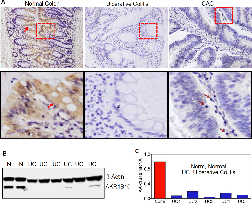
Purpose: Ulcerative colitis and colitis-associated colorectal cancer (CAC) is a serious health issue, but etiopathological factors remain unclear. Aldo-keto reductase 1B10 (AKR1B10) is specifically expressed in the colonic epithelium, but downregulated in colorectal cancer. This study was aimed to investigate the etiopathogenic role of AKR1B10 in ulcerative colitis and CAC.

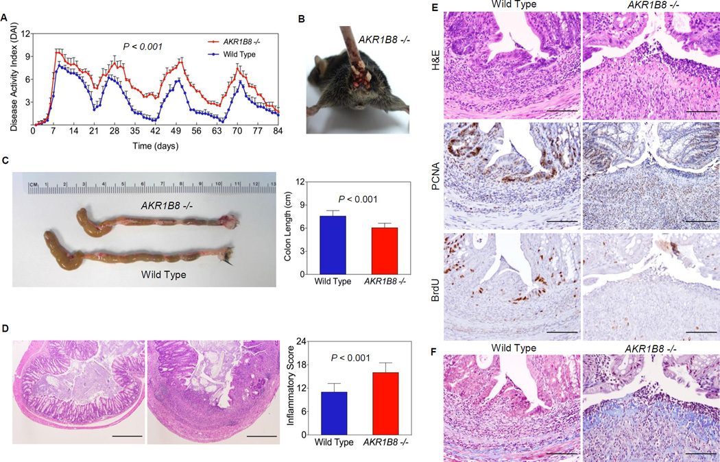
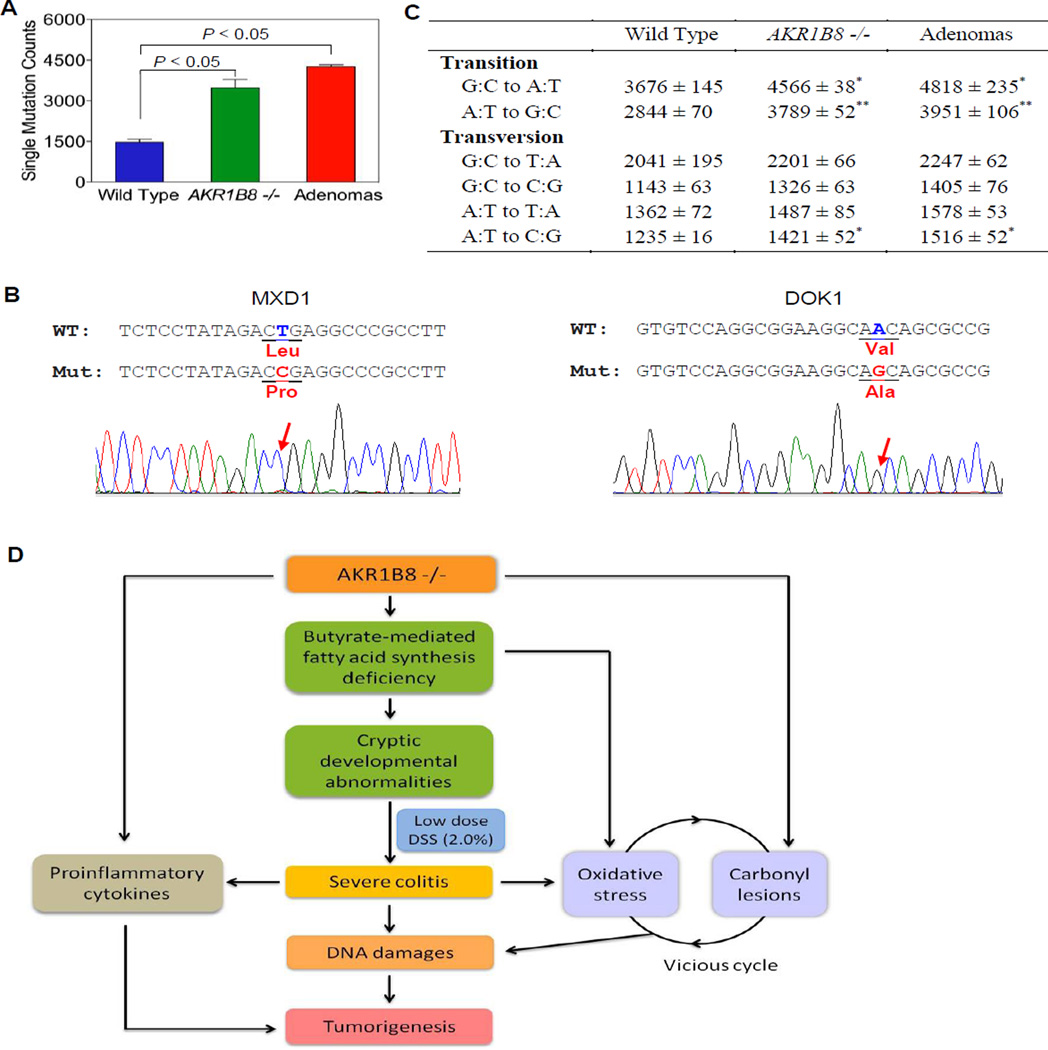
Experimental design: Ulcerative colitis and CAC biopsies (paraffin-embedded sections) and frozen tissues were collected to examine AKR1B10 expression. Aldo-keto reductase 1B8 (the ortholog of human AKR1B10) knockout (AKR1B8(-/-)) mice were produced to estimate its role in the susceptibility and severity of chronic colitis and associated dysplastic lesions, induced by dextran sulfate sodium (DSS) at a low dose (2%). Genome-wide exome sequencing was used to profile DNA damage in DSS-induced colitis and tumors.

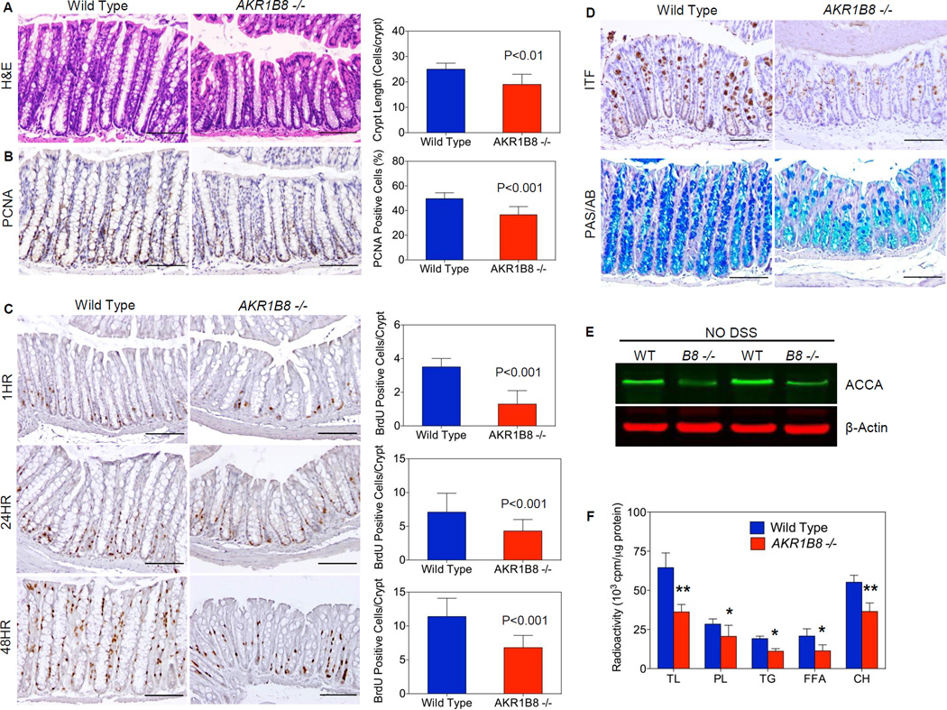
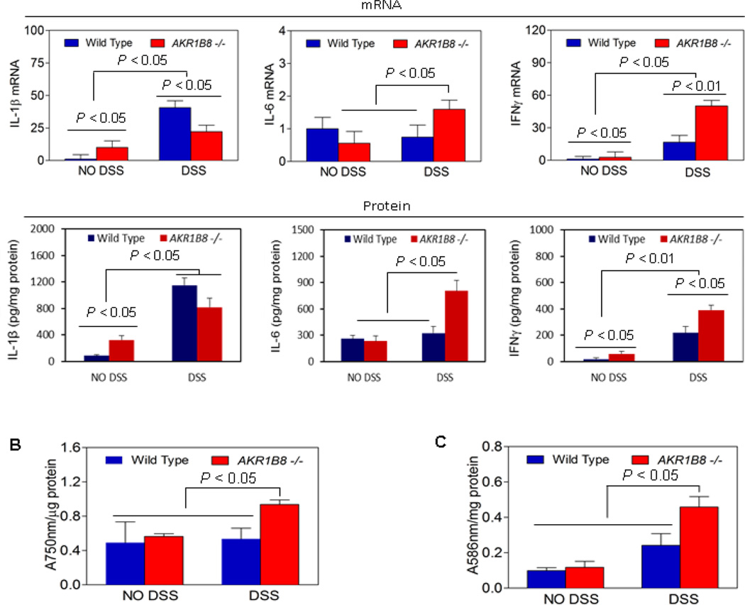
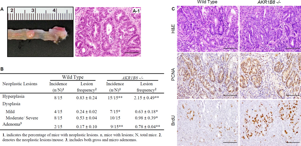
Results: AKR1B10 expression was markedly diminished in over 90% of ulcerative colitis and CAC tissues. AKR1B8 deficiency led to reduced lipid synthesis from butyrate and diminished proliferation of colonic epithelial cells. The DSS-treated AKR1B8(-/-) mice demonstrated impaired injury repair of colonic epithelium and more severe bleeding, inflammation, and ulceration. These AKR1B8(-/-) mice had more severe oxidative stress and DNA damage, and dysplasias were more frequent and at a higher grade in the AKR1B8(-/-) mice than in wild-type mice. Palpable masses were seen in the AKR1B8(-/-) mice only, not in wild-type.

Conclusions: AKR1B8 is a critical protein in the proliferation and injury repair of the colonic epithelium and in the pathogenesis of ulcerative colitis and CAC, being a new etiopathogenic factor of these diseases.


Conflict of interest statement

Conflicts of Interest: Authors declare no conflicts of financial interests.

Figures

Figure 1
Figure 1. AKR1B10 expression in normal, ulcerative colitis and cancerous colon tissues
(A) Immunohistochemistry in paraffin-embedded sections of normal colon, ulcerative colitis and associated tumor tissues. AKR1B10 protein was detected in normal colonic epithelium (arrows), but not in ulcerative colitis and tumor tissues. Lower panel: amplification of the squared areas. Purple arrows indicate infiltrated mononuclear cells in CAC. Scale bar, 100µm. (B) Western blot. N, normal colon tissues; UC, ulcerative colitis. (C) Real-time RT-PCR. The AKR1B10 mRNA level in normal colon is set up at 1.0, and AKR1B10 mRNA levels in ulcerative colitis tissues are presented as fold over normal. GAPDH mRNA was detected as an internal control.
Figure 2
Figure 2. AKR1B8 deficiency interrupts growth and homeostasis of mouse colonic crypts
(A) H&E histology (n=30 each); (B) PCNA expression (n=5 each); (C) BrdU labeling at 1, 24, and 48 hours (n=5 each); (D) Upper panel, ITF expression; and lower panel, Alcian blue and Periodic acid Schiff staining (n=5 each). Scale bar indicates 100 µm. (E) Western blot of acetyl-CoA carboxylase-α (ACCA) protein; (F) Incorporation of 14C-butyric acid into total lipids and various species: phospholipids (PL), triglycerides (TG), free fatty acids, and cholesterol (CH). n=5 each; *, P<0.05 and **, P<0.01 compared to wild type. Statistical significance was tested by One-way Anova test.
Figure 3
Figure 3. DSS-induced colitis and mucosal lesion repair
Wild type and AKR1B8 −/− mice (n=15 each) were administered with dextran sulfate sodium in drink water to induce colitis as described in Materials and Methods. (A) Disease activity index; (B) Rectal bleeding; (C) Colon length; and (D) Histological inflammatory lesions; (E) Upper panel, H&E histology; middle panel, PCNA expression; and lower panel, BrdU labeling (1 hour). (F) Masson Trichrome staining, showing collagen deposit at the ulcer. Scale bar, 100µm.
Figure 4
Figure 4. Proinflammatory cytokines, oxidative stress, and carbomyl levels in AKR1B8 −/− mice
(A) Expression of cytokines IL-1β, IL-6, and IFNγ. Upper panel: mRNA levels; lower panel: protein levels. Data indicate mean ± SD, n= 5. (B) Oxidative stress (n=5 each). (C) Lipid peroxides (n=5 each). Statistical significance was tested by Student’s t test.
Figure 5
Figure 5. Colitis-associated dysplastic lesions in AKR1B8
Wild type and AKR1B8 −/− mice (n=15 each) were administered with dextran sulfate sodium in drink water to induce colitis as described in Materials and Methods. (A) A palpable tumor (arrow) from an AKR1B8 −/− mouse. (A-1), H&E histology. (B) Summary of colitis-associated dysplastic lesions. *, P<0.05 and **, P<0.01 compared to wild type control. Statistical significance was tested by Student’s t test. (C) Grade and aggressiveness of dysplasia: Upper panel, H&E histology; middle panel, PCNA expression; and lower panel, BrdU labeling (1 hour). Scale bar, 100µm.
Figure 6
Figure 6. DNA damage in AKR1B8 −/− mice
Genome-wide Exome sequencing analyses of WT and AKR1B8 −/− colitis mucosa and tumors (n=2 each) were conducted as described in Materials and Methods. (A) Single homozygous nucleotide mutants. (B) Conventional DNA sequencing of DOX1 and MXD1 tumor suppressor genes, confirming the point mutations. (C) Transitional and transversional point mutations. *, P<0.05 and **, P<0.01 compared to wild type control. Statistical significance was tested by Student’s t test. (D) Hypothetic model of AKR1B8 in colitis and associated dysplastic lesions. AKR1B8 deficiency reduces long chain fatty acid/lipid synthesis from butyrate, which diminishes biomembrane assembly and cell proliferation. The AKR1B8 deficiency also leads to accumulation of toxic carbonyl compounds, which, together with oxidative stress, forms a vicious cycle, leading to DNA damage and subsequent dysplastic lesions. AKR1B8 may also affect proinflammatory cytokine expression via a mechanism unknown yet.

References

    1. Danese S, Fiocchi C. Ulcerative colitis. N Engl J Med. 2011;365:1713–1725. - PubMed
    1. Xavier RJ, Podolsky DK. Unravelling the pathogenesis of inflammatory bowel disease. Nature. 2007;448:427–434. - PubMed
    1. Karin M, Greten FR. NF-kappaB: linking inflammation and immunity to cancer development and progression. Nat Rev Immunol. 2005;5:749–759. - PubMed
    1. Danese S, Malesci A, Vetrano S. Colitis-associated cancer: the dark side of inflammatory bowel disease. Gut. 2011;60:1609–1610. - PubMed
    1. Meira LB, Bugni JM, Green SL, Lee CW, Pang B, Borenshtein D, et al. DNA damage induced by chronic inflammation contributes to colon carcinogenesis in mice. The Journal of clinical investigation. 2008;118:2516–2525. - PMC - PubMed

Publication types

MeSH terms

Substances

LinkOut - more resources