Age-related joint space narrowing independent of the development of osteoarthritis of the shoulder
- PMID: 25538427
- PMCID: PMC4262869
- DOI: 10.4103/0973-6042.145213
Age-related joint space narrowing independent of the development of osteoarthritis of the shoulder
Abstract
Purpose: It is commonly accepted that the glenohumeral joint space remains unchanged until the onset of osteoarthritis, at which point progressive degenerative changes, and joint space narrowing occur. The aim of this study was to evaluate the radiographic width of the glenohumeral joint space in patients of different ages: Those with otherwise normal radiographs, those with a history of instability, those with calcific tendonitis, and those with a radiologic diagnosis of osteoarthritis.
Materials and methods: In this retrospective study, two independent investigators measured the glenohumeral joint width on true anteroposterior and axillary views of standardized shoulder radiographs taken from 2002 to 2009. The digital image resolution was 0.01 mm. Group I comprised 60 patients with normal shoulder radiographs, Group II comprised 53 patients with instability but normal radiographs, Group III comprised 109 patients with radiologically proven calcific tendonitis, and Group IV comprised 120 patients with manifest osteoarthritis.
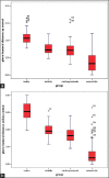
Results: The interobserver reliability (r) was 0.621-0.862. The mean joint space width was significantly different among Groups I-IV (central anteroposterior: 4.28 ± 0.75 mm, 3.12 ± 0.73 mm, 2.87 ± 0.80 mm, and 1.47 ± 1.07 mm, respectively; P = 0.001; central axillary: 6.12 ± 1.09 mm, 3.92 ± 0.77 mm, 3.34 ± 0.84 mm, and 1.08 ± 1.12 mm, respectively; P = 0.001). There was a significant negative correlation between the joint space width and age at all measured levels in both projections (P < 0.001).
Conclusions: The glenohumeral joint space width decreases with increasing age beginning in early adulthood, and this effect is enhanced by osteoarthritis.
Level of evidence: Level II, retrospective study.
Keywords: Aging; cartilage; joint space; osteoarthritis; radiograph; shoulder.
Conflict of interest statement
Figures


References
-
- Habermeyer P, Engel G. Endoprothetik. In: Habermeyer P, editor. Schulterchirurgie. 3rd ed. München, Jena: Elsevier, Urban & Fischer; 2005. pp. 497–553.
-
- Matsen FA, Rockwood CA, Wirth MA, Lippitt SB, Parsons M. Gleno humeral arthritis and its management. In: Rockwood CA, Matsen FA, Wirth MA, Lippitt SB, editors. The Shoulder. Philadelphia: Saunders; 2004. pp. 879–1007.
-
- Valdes AM, Spector TD. The contribution of genes to osteoarthritis. Med Clin North Am. 2009;93:45–66. - PubMed
-
- Aigner T, Fundel K, Saas J, Gebhard PM, Haag J, Weiss T, et al. Large-scale gene expression profiling reveals major pathogenetic pathways of cartilage degeneration in osteoarthritis. Arthritis Rheum. 2006;54:3533–44. - PubMed
-
- Aigner T, Zien A, Hanisch D, Zimmer R. Gene expression in chondrocytes assessed with use of microarrays. J Bone Joint Surg Am. 2003;85-A(Suppl 2):117–23. - PubMed
LinkOut - more resources
Full Text Sources
Other Literature Sources
Research Materials

