Review
. 2014 Dec 4:5:258.
doi: 10.3389/fneur.2014.00258. eCollection 2014.

What are the Best Animal Models for Testing Early Intervention in Cerebral Palsy?

Affiliations
Review

What are the Best Animal Models for Testing Early Intervention in Cerebral Palsy?

Gavin John Clowry et al. Front Neurol. .

Abstract

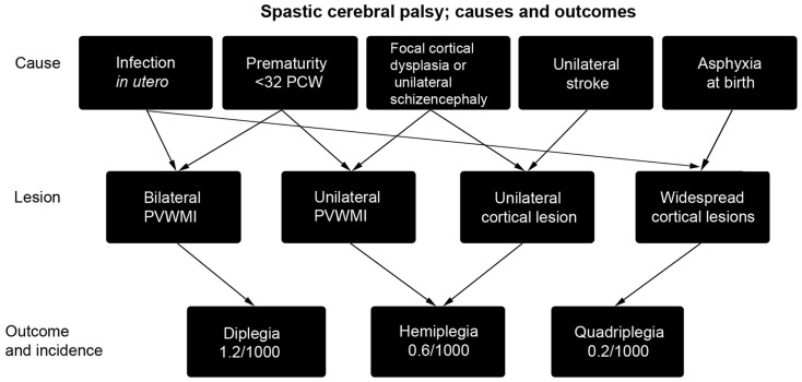
Interventions to treat cerebral palsy should be initiated as soon as possible in order to restore the nervous system to the correct developmental trajectory. One drawback to this approach is that interventions have to undergo exceptionally rigorous assessment for both safety and efficacy prior to use in infants. Part of this process should involve research using animals but how good are our animal models? Part of the problem is that cerebral palsy is an umbrella term that covers a number of conditions. There are also many causal pathways to cerebral palsy, such as periventricular white matter injury in premature babies, perinatal infarcts of the middle cerebral artery, or generalized anoxia at the time of birth, indeed multiple causes, including intra-uterine infection or a genetic predisposition to infarction, may need to interact to produce a clinically significant injury. In this review, we consider which animal models best reproduce certain aspects of the condition, and the extent to which the multifactorial nature of cerebral palsy has been modeled. The degree to which the corticospinal system of various animal models human corticospinal system function and development is also explored. Where attempts have already been made to test early intervention in animal models, the outcomes are evaluated in light of the suitability of the model.

Keywords: cerebral palsy; corticospinal tract; hypoxia/ischemia; perinatal stroke; periventricular white matter injury.


Figures

Figure 1
Figure 1
A summary of the causes of spastic cerebral palsy, and the particular outcomes they lead to [reproduced with permission from Ref. (11)]. Asphyxia at birth may arise from prolapsed cord, intrapartum hemorrhage, uterine rupture, or maternal cardiac arrest. As arrows indicate, multiple causes may combine to produce cerebral palsy (4) and may also interact with subtle genetic variations in individuals that cause predisposition to stroke (6). PCW, post-conceptional weeks; PVWMI, periventricular white matter injury.
Figure 2
Figure 2
A comparative timetable of oligodendrocyte development between rodent and human. The time of greatest vulnerability to hypoxia/ischemia (arrow) is at the pre-oligodendrocyte stage of development. Based on the information from Ref. (–36). E, Embryonic day; PCW, post-conceptional weeks.
Figure 3
Figure 3
This figure compares four stages of development of the corticospinal system in rodent and human. At stage 1 segmental circuits are connected, and local circuitry is also forming in the forebrain, but there is no connectivity between the two. Stage 2; thalamic afferents invade the subplate, and the corticospinal tract waits in the white matter to innervate the spinal cord gray matter. Stage 3; thalamic afferents innervate layer IV of the cortex at the same time as corticospinal fibers innervate the spinal cord, thus the spinal cord and sensorimotor cortex become reciprocally connected. Spindle bursts in response to spontaneous movement are recorded in somatosensory cortex. Stage 4; the subplate dissolves and corticospinal connections and muscle afferent projections are refined in the spinal cord and dorsal column nuclei. DCN, dorsal column nuclei; DH, dorsal horn; DRG, dorsal root ganglion; SP, subplate; VH, ventral horn; IV, V, VI, cortical layers. Arrows represent ingrowth of axons, dashed lines withdrawal of axon terminals. Axon projections colored gray have not changed at that stage in the figure. Based on information from Ref. (42, 50, 57, 75, 92, 100, 101).
Figure 4
Figure 4
The outcome measures employed in a sample of 36 rodent studies that modeled PVWMI or PIS, some of which involved experimental therapies. Blue columns depict the proportion of studies that studied lesion size in the short term (within a week) or in the longer term, either using MRI, or histology. Green shows studies of changes in molecular markers in response to lesions, e.g., markers of apoptosis, myelin, and gliosis. Red/orange shows behavioral testing in adolescent or adult animals following perinatal lesions. These are divided into tests of memory and cognition (e.g., mazes) sensorimotor (e.g., rotarod, reaching, and ladder walking) and anxiety (open field). The 36 studies sampled are those involving rodents cited in Sections “Models of Periventricular White Matter Injury” and “Models of Perinatal Ischemic Stroke.”

References

    1. O’Collins VE, Macleod MR, Donnan GA, Horky LL, van der Worp BH, Howells DW. 1,026 Experimental treatments in acute stroke. Ann Neurol (2006) 59:467–77. 10.1002/ana.20741 - DOI - PubMed
    1. Howells DW, Macleod MR. Evidence-based translational medicine. Stroke (2013) 44:1466–71 10.1161/STROKEAHA.113.000469 - DOI - PubMed
    1. Ouskoi M, Coutinho F, Dykeman J, Jette N, Pringsheim T. An update on the prevalence of cerebral palsy: a systematic review and meta-analysis. Dev Med Child Neurol (2013) 55:509–19. 10.1111/dmcn.12080 - DOI - PubMed
    1. Stanley F, Blair E, Alberman E. Cerebral Palsies: Epidemiology and Causal Pathways. Clinics in Developmental Medicine No. 151. London: Mac Keith Press; (2000).
    1. Cowan F, Rutherford M, Groenendaal F, Eken P, Mercuri E, Bydder GM, et al. Origin and timing of brain lesions in term infants with neonatal encephalopathy. Lancet (2003) 361:736–42. 10.1016/S0140-6736(03)12658-X - DOI - PubMed

LinkOut - more resources