

Fluoroscopic evaluation of oropharyngeal dysphagia: anatomic, technical, and common etiologic factors
- PMID: 25539237
- PMCID: PMC4331119
- DOI: 10.2214/AJR.13.12374
Fluoroscopic evaluation of oropharyngeal dysphagia: anatomic, technical, and common etiologic factors
Abstract
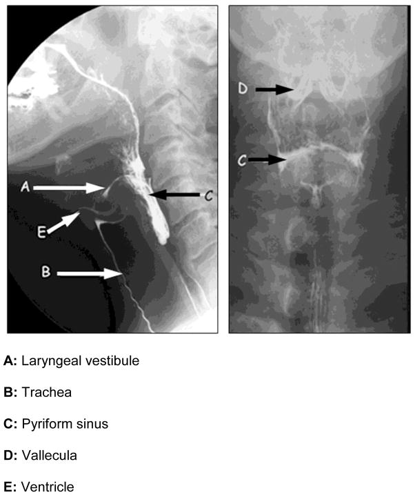
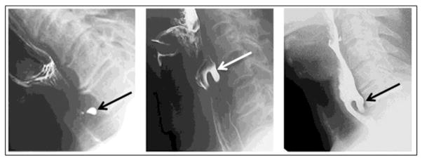
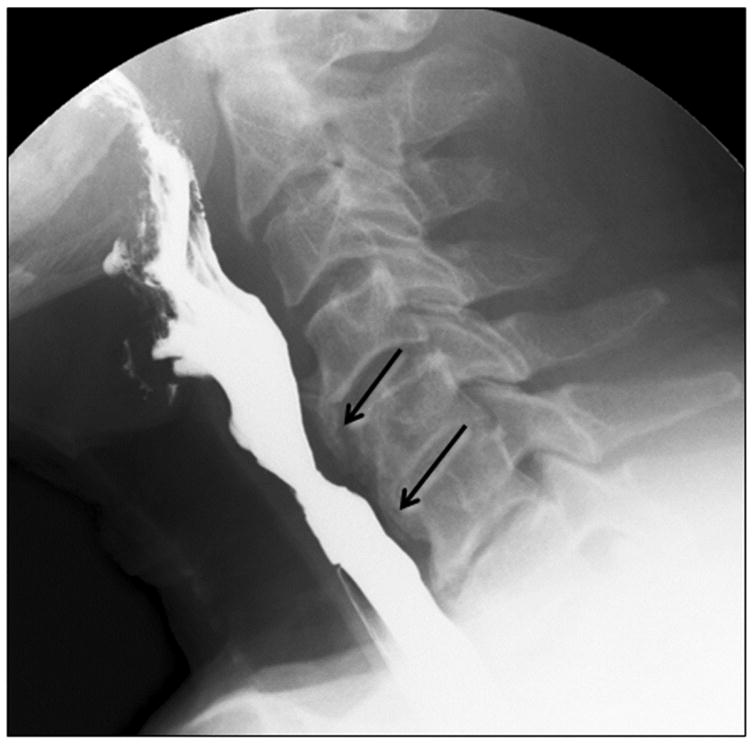
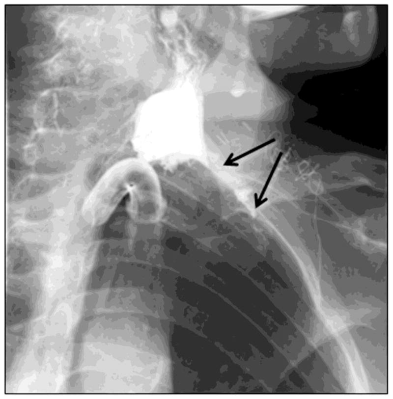
Objective: The purposes of this article are to review the anatomy of the upper gastrointestinal tract; review techniques and contrast agents used in the fluoroscopic examination of the oropharynx and hypopharynx; provide a pictorial review of some important causes of oropharyngeal dysphagia; and link these causes to key findings in the clinical history to assist in establishing a clinical diagnosis.
Conclusion: Many important causes and presentations of oropharyngeal dysphagia are sometimes overlooked during conventional upper gastrointestinal studies. Videofluoroscopic evaluation for assessment of both structural abnormalities and motility disorders of the oropharynx by use of various compositions of barium contrast medium is the standard of practice. Using best-practices radiographic techniques and having knowledge of swallowing mechanisms and various diseases are important for assessment of dysphagia. Dynamic fluoroscopic imaging remains an essential tool for assessing functional disorders of swallowing. Detailed videofluoroscopic assessment can guide treatment decisions with the goal of decreasing the secondary complications of dysphagia.
Keywords: fluoroscopy; oropharyngeal dysphagia.
Figures


Comment in
-
Fluoroscopic Evaluation of Oropharyngeal Dysphagia.AJR Am J Roentgenol. 2015 Aug;205(2):W227. doi: 10.2214/AJR.15.14453. AJR Am J Roentgenol. 2015. PMID: 26204314 No abstract available.
References
-
- Palmer JB, Drennan JC, Baba M. Evaluation and treatment of swallowing impairments. American Family Physician. 2000;61:2453–2462. - PubMed
-
- Prasse JE, Kikano GE. An overview of dysphagia in the elderly. Advanced Studies in Medicine. 2004;4(10):527–533.
-
- Steele CM, Greenwood C, Ens I, Robertson C, Seidman-Carlson R. Mealtime difficulties in a home for the aged: not just dysphagia. Dysphagia. 1997;12:43–50. - PubMed
-
- Logemann JA. Manual for the videofluorographic study of swallowing: Second edition. Austin, TX: Pro-Ed, Inc; 1993.
-
- Hiiemae KM, Palmer JB. Tongue movements in feeding and speech. Crit Rev Oral Biol Med. 2003;14:413–429. - PubMed
Publication types
MeSH terms
Grants and funding
LinkOut - more resources
Full Text Sources
Medical
