

Noise alters guinea pig's blood-labyrinth barrier ultrastructure and permeability along with a decrease of cochlear Claudin-5 and Occludin
- PMID: 25539640
- PMCID: PMC4299297
- DOI: 10.1186/s12868-014-0136-0
Noise alters guinea pig's blood-labyrinth barrier ultrastructure and permeability along with a decrease of cochlear Claudin-5 and Occludin
Abstract
Background: Noise exposure (NE) is a severe modern health hazard that induces hearing impairment. However, the noise-induced ultrastructural changes of blood-labyrinth barrier (BLB) and the potential involvements of tight junction proteins (TJP) remain inconclusive. We investigated the effects of NE on not only the ultrastructure of cochlea and permeability of BLB but also the expression of TJP within the guinea pig cochlea.
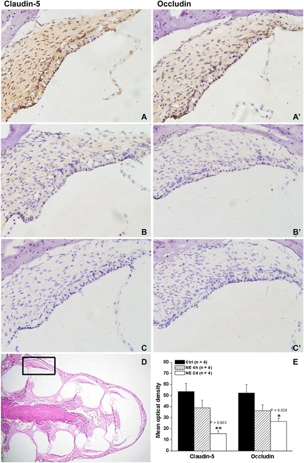
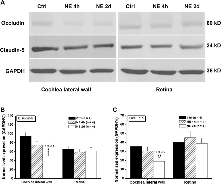
Results: Male albino guinea pigs were exposed to white noise for 4 h or 2 consecutive days (115 dB sound pressure level, 6 hours per day) and the hearing impairments and light microscopic change of BLB were evaluated with auditory brainstem responses (ABR) and the cochlear sensory epithelia surface preparation, respectively. The cochlear ultrastructure and BLB permeability after NE 2d were revealed with transmission electron microscope (TEM) and lanthanum nitrate-tracing techniques, respectively. The potential alterations of TJPs Claudin-5 and Occludin were quantified with immunohistochemistry and western blot. NE induced significant hearing impairment and NE 2d contributed to significant outer hair cell (OHC) loss that is most severe in the first row of outer hair cells. Furthermore, the loosen TJ and an obvious leakage of lanthanum nitrate particles beneath the basal lamina were revealed with TEM. Moreover, a dose-dependent decrease of Claudin-5 and Occludin was observed in the cochlea after NE.
Conclusions: All these findings suggest that both decrease of Claudin-5 and Occludin and increased BLB permeability are involved in the pathologic process of noise-induced hearing impairment; however, the causal relationship and underlying mechanisms should be further investigated.
Figures
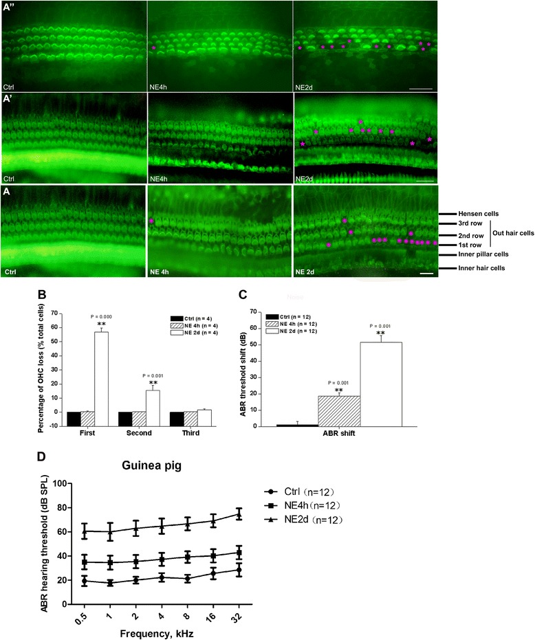
 and
and  indicate Hair Cell loss.
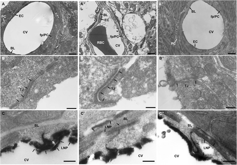
References
-
- Henderson DHB, Bielefeld EC. Patterns and mechanisms of noise induced cochlear pathology. In: Schacht JPA, Fay RR, editors. Auditory trauma, protection, and repair. New York: Springer; 2008. p. 22.
Publication types
MeSH terms
Substances
LinkOut - more resources
Full Text Sources
Other Literature Sources
Medical
Miscellaneous
