. 2015 Jan 29;33(5):726-33.
doi: 10.1016/j.vaccine.2014.11.035. Epub 2014 Dec 21.

Mucosal immunization with an attenuated Salmonella vaccine partially protects white-tailed deer from chronic wasting disease

Affiliations

Mucosal immunization with an attenuated Salmonella vaccine partially protects white-tailed deer from chronic wasting disease

Fernando Goñi et al. Vaccine. .

Abstract

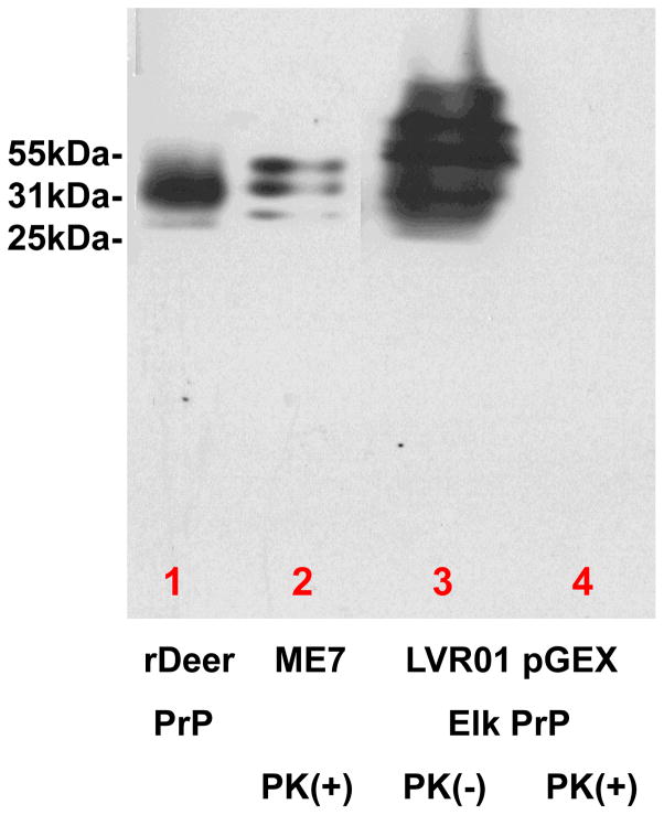
Prion disease is a unique category of illness, affecting both animals and humans, in which the underlying pathogenesis is related to a conformational change of a normal, self-protein called PrP(C) (C for cellular) to a pathological and infectious conformer known as PrP(Sc) (Sc for scrapie). Bovine spongiform encephalopathy (BSE), a prion disease believed to have arisen from feeding cattle with prion contaminated meat and bone meal products, crossed the species barrier to infect humans. Chronic wasting disease (CWD) infects large numbers of deer and elk, with the potential to infect humans. Currently no prionosis has an effective treatment. Previously, we have demonstrated we could prevent transmission of prions in a proportion of susceptible mice with a mucosal vaccine. In the current study, white-tailed deer were orally inoculated with attenuated Salmonella expressing PrP, while control deer were orally inoculated with vehicle attenuated Salmonella. Once a mucosal response was established, the vaccinated animals were boosted orally and locally by application of polymerized recombinant PrP onto the tonsils and rectal mucosa. The vaccinated and control animals were then challenged orally with CWD-infected brain homogenate. Three years post CWD oral challenge all control deer developed clinical CWD (median survival 602 days), while among the vaccinated there was a significant prolongation of the incubation period (median survival 909 days; p=0.012 by Weibull regression analysis) and one deer has remained CWD free both clinically and by RAMALT and tonsil biopsies. This negative vaccinate has the highest titers of IgA in saliva and systemic IgG against PrP. Western blots showed that immunoglobulins from this vaccinate react to PrP(CWD). We document the first partially successful vaccination for a prion disease in a species naturally at risk.

Keywords: Bovine spongiform encephalopathy; Chronic wasting disease; Immunization; Mucosal vaccination; Prion protein; Salmonella vaccine strain; White-tailed deer.


Conflict of interest statement

Conflict of Interest:

The authors have no conflicts of interest.

Figures

Figure 1
Figure 1
Analysis of the expression of cervid PrP in the Salmonella LVR01 vaccine strain, by Western blot using monoclonal anti-PrP 7D9/6D11 [18]. In lane one cervid rPrP was run. In lane 2 PrP strain ME7 was run following proteinase K treatment. Lane 3 shows the expression of cervid PrP by the Salmonella LVR01 and lane 4 the same preparation with proteinase K treatment. As can be seen in lane 4 the cervid PrP was completely digested, while, as expected, the ME7 PrPSc in lane 2 was PK resistant. This shows that our Salmonella LVR01 can express high levels of cervid PrP and also that this cervid PrP is fully PK sensitive.
Figure 2
Figure 2
2A): Titers of IgA in the cervid feces. The feces IgA were obtained using our published methods [15;18]. The titers illustrate the importance of mucosal application of a boost of oligomerized recombinant cervid PrP after a primary mucosal immune response is established. T0 refers to “time zero” or pre-immunization titers, while T5, T6 and T7 refer to the fifth and subsequent collections of cervid feces. The T5 titer of anti-PrP IgA in vaccinated deer was significantly higher than control T5 anti-PrP IgA (p<0.05), but was low. This titer improved in T6 and 7 following boosts of oligomerized recombinant cervid PrP (T6, p=0.0009 and T7, p<0.0001). 2B): Cross-reactive anti-deer PrP IgA titers (measured with anti-goat IgA) in saliva of vaccinated (red bars) and control deer (blue bars). For T5 titer the p=0.0036. T7 had a much higher titer (compared to T5) with the p=0.008. 2C): Anti-deer PrP IgG titers in plasma of vaccinated (red bars) and control deer (blue bars). The anti-PrP IgG titers in the plasma were low but significantly different from controls. For T5 titer the p=0.0067. For T6 the p=0.01. For T7 the p=0.016. 2D): Cross-reactive anti-deer PrP IgM titers (measured with anti-goat IgM) in plasma of vaccinated (red bars) and control deer (blue bars). The titer from T5 to T7 is increasing. For T5 titer the p=0.01. For T6 the p=0.002. For T7 the p=0.01.
Figure 3
Figure 3
In 3A purified deer Ig from plasma of deer 781 (with full protection) was used for Western blots and compared to purified deer Ig from a control deer (deer 786, figure 3B). All lanes were equally loaded with 4μg protein. Anti-deer IgG was used for detection of the Western blots in 3A and 3B. In lane 1 the lysate of vector LVR01 was run and a very strong reaction is seen both in the vaccinated and control deer to the highly immunogenic Salmonella, as expected. The control showed no reaction to any of the PrP antigens loaded in lanes 2–5 of Fig 3B (which was the case for all the control deer, data not shown). Deer 781 plasma IgG reacted strongly to polymerized deer recombinant PrP (lane 5, see red arrow) and faintly to aggregated deer recombinant PrP (lane 4, blue arrow). Deer 781 also detected a distinct band of polymerized sheep PrP (lane 3, black arrow), as well as faintly detecting aged sheep recombinant PrP (lane 2, see green arrow)
Figure 4
Figure 4
Western blots analysis of the reactivity of the plasma IgG and saliva IgA of vaccinated deer 781 versus CWD brain and scrapie preparations. In each gel (A–C) each lane was loaded with samples following PK digestion consisting of: lane 1, Elk brain homogenate; lane 2, 20% brain homogenate of a mule deer with CWD; lane 3, 20% brain homogenate of a 22L scrapie brain; lane 4, 1% brain homogenate of a CWD infected white-tailed deer; lane 5, 10% brain homogenate of a cervid PrP expressing Tg mouse infected with CWD; lane 6, 10% brain homogenate of a CWD infected white-tailed deer. In A, the primary antibody was anti-PrP monoclonal antibodies 7D9/6D11 at 1:8000. The secondary antibody was anti-mouse IgG-HRP at 1:2000. In B, ammonium sulphate precipitated Ig from the plasma of deer 781 was used at 1:200 as a primary antibody. The secondary antibody was anti-deer IgG-HRP (1:2000). In C, ammonium sulphate precipitated Ig from the saliva of deer 781 was used at 1:100 as a primary antibody. The secondary antibody was anti-goat IgA-HRP at 1:1250 (which was cross reactive with cervid IgA). As can been seen in B and C, the plasma IgG and saliva IgA from deer 781 is reactive to both CWD and scrapie preparations.
Figure 5
Figure 5
Anti-PrP immunostaining of biopsies in control vector only Salmonella treated deer (Figure 5A and C) compared to vaccinated deer 781 that remained negative for CWD (Figure 5B and D). In A and B tonsil biopsies are shown, while in C and D RAMALT biopsies are shown. The scale bars = 200μm. 5E): Western blot of brain homogenates from a deer negative for CWD in lanes 1 and 2 and a positive control deer with CWD in lanes 3 and 4 (neither deer is from our study). In lanes 5 and 6 brain homogenates were run from one of the LVR01 vector control deer from our study 120 days after challenge. The lanes alternate for proteinase K (PK) non-treated homogenate and treated homogenate. As can be seen, there is no PrPCWD in the assay negative control deer (lane 2), while PrPCWD is evident in the positive control deer (lane 4) and from our study control LVR01 vector exposed and CWD challenged deer.
Figure 5
Figure 5
Anti-PrP immunostaining of biopsies in control vector only Salmonella treated deer (Figure 5A and C) compared to vaccinated deer 781 that remained negative for CWD (Figure 5B and D). In A and B tonsil biopsies are shown, while in C and D RAMALT biopsies are shown. The scale bars = 200μm. 5E): Western blot of brain homogenates from a deer negative for CWD in lanes 1 and 2 and a positive control deer with CWD in lanes 3 and 4 (neither deer is from our study). In lanes 5 and 6 brain homogenates were run from one of the LVR01 vector control deer from our study 120 days after challenge. The lanes alternate for proteinase K (PK) non-treated homogenate and treated homogenate. As can be seen, there is no PrPCWD in the assay negative control deer (lane 2), while PrPCWD is evident in the positive control deer (lane 4) and from our study control LVR01 vector exposed and CWD challenged deer.
Figure 6
Figure 6
Current Kaplan-Meier survival curve of the control LVRO1 vector exposed (n=6) and vaccinated deer (n=5). The vaccinated deer had partial protection from CWD infection (p=0.012 by Weibull regression analysis). Deer 781 in the vaccinated group has remained free from clinical CWD (~1310 days after challenge) with negative tonsil and RAMALT biopsies, in spite of sharing a den with CWD infected deer for > 1yr. In this study two deer in each group were included with a Prnp polymorphism of G/S at codon 96 (other polymorphisms were matched and are associated with sensitivity to CWD). Codon 96 G/S versus G/G is the most common (25%) deer polymorphism (one of 5) that confers partial resistance to CWD infection [21;22]. Significantly deer 781 was fully sensitive to CWD (codon 96G/G). The one deer in the control group that had a prolonged incubation of 953 days (other controls incubations were: 477, 477, 538, 666, and 670 days) had codon 96G/S (which confers some resistance to CWD infection); hence, its longer incubation period was likely related to this polymorphism.

References

    1. Colby DW, Prusiner SB. Prions. Cold Spring Harb Perspect Biol. 2011 Jan;3(1):a006833. - PMC - PubMed
    1. Harman JL, Silva CJ. Bovine spongiform encephalopathy. J Am Vet Med Assoc. 2009 Jan 1;234(1):59–72. - PubMed
    1. Williams ES. Chronic wasting disease. Vet Pathol. 2005 Sep;42(5):530–49. - PubMed
    1. Saunders SE, Bartelt-Hunt SL, Bartz JC. Occurrence, transmission, and zoonotic potential of chronic wasting disease. Emerg Infect Dis. 2012 Mar;18(3):369–76. - PMC - PubMed
    1. Mitchell GB, Sigurdson CJ, O’Rourke KI, et al. Experimental oral transmission of chronic wasting disease to reindeer (Rangifer tarandus tarandus) PLoS ONE. 2012;7(6):e39055. - PMC - PubMed

Publication types

MeSH terms