. 2014 Dec 24:11:214.
doi: 10.1186/s12974-014-0214-3.

Molecular mechanisms involved in HIV-1 Tat-mediated induction of IL-6 and IL-8 in astrocytes

Affiliations

Molecular mechanisms involved in HIV-1 Tat-mediated induction of IL-6 and IL-8 in astrocytes

Anantha Ram Nookala et al. J Neuroinflammation. .

Abstract

Background: HIV-associated neurocognitive disorders (HAND) exist in approximately 50% of infected individuals even after the introduction of highly active antiretroviral therapy. HIV-1 Tat has been implicated in HIV-associated neurotoxicity mediated through production of pro-inflammatory cytokines like IL-6 and IL-8 by astrocytes among others as well as oxidative stress. However, the underlying mechanism(s) in the up-regulation of IL-6 and IL-8 are not clearly understood. The present study was designed to determine the mechanism(s) responsible for IL-6 and IL-8 up-regulation by HIV-1 Tat.

Methods: SVG astrocytes were transiently transfected with a plasmid encoding HIV-1 Tat. The HIV-1 Tat-mediated mRNA and protein expression levels of both IL-6 and IL-8 in SVG astrocytes were quantified using real time RT-PCR and multiplex cytokine assay respectively. We also employed immunocytochemistry for staining of IL-6 and IL-8. The underlying signaling mechanism(s) were identified using pharmacological inhibitors and siRNA for different intermediate steps involved in PI3K/Akt, p38 MAPK and JNK MAPK pathways. Appropriate controls were used in the experiments and the effect of pharmacological antagonists and siRNA were observed on both mRNA expression and protein levels.

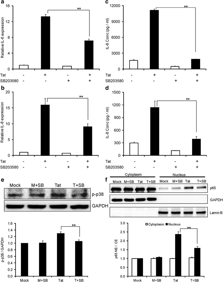
Results: Both IL-6/IL-8 mRNA and protein showed peak expressions at 6 hours and 96 hours post-transfection, respectively. Elevated levels of IL-6/IL-8 were also confirmed by immunocytochemistry. Our studies indicated that both NF-kB and AP-1 transcription factors were involved in IL-6 and IL-8 expression mediated by HIV-1 Tat; however, AP-1 was differentially activated for either cytokine. In the case of IL-6, p38δ activated AP-1 whereas JNK but not p38 MAPK was involved in AP-1 activation for IL-8 production. On the other hand both PI3K/Akt and p38 MAPK (β subunit) were found to be involved in activation of NF-κB that led to IL-6 and IL-8 production.

Conclusion: Our results demonstrate HIV-1 Tat-mediated induction of both IL-6 and IL-8 in a time-dependent manner in SVG astrocytes. Furthermore, we also showed the involvement of NF-κB and AP-1 transcription factors regulated by PI3/Akt, p38 MAPK and JNK MAPK upstream signaling molecules. These results present new therapeutic targets that could be used in management of HAND.


Figures

Figure 1
Figure 1
HIV-1 Tat induces time dependent expression of IL-6 and IL-8 in SVG astrocytes. Seven hundred thousand SVG astrocytes were transfected with 0.3 μg of HIV-1 Tat plasmid using Lipofectamine 2000. (a, d) Cells were harvested at 1, 3, 6, 12, 24, 48 and 72 hours and total RNA was isolated. The expression levels of IL-6 and IL-8 were determined by real time RT-PCR. The values represented are normalized to their mock-transfected controls. (b, e) The concentrations of IL-6 and IL-8 in cell culture supernatants were measured at 6, 12, 24, 48, 72 and 96 hours after transfection by multiplex cytokine assay. Open bars and closed bars represent protein concentrations of mock- and HIV-1 Tat- transfected cells, respectively. Each experiment was done at least in triplicate and each bar represents the ± SE of three individual experiments. (c, f) Primary astrocytes from 2 independent donors were treated with 200 ng/ml of Tat protein for 2 hours and total RNA was isolated. The expression levels of IL-6 and IL-8 were determined by real time RT-PCR. Statistical analyses was performed by Student’s t-test and ** denotes P-value of ≤ 0.01.
Figure 2
Figure 2
Immunocytochemistry of IL-6 and IL-8 mediated by HIV-1 Tat in astrocytes. (a-i, k-s) Five hundred thousand SVG astrocytes were grown on a cover slip and transfected with plasmid encoding HIV-1 Tat. The over expression of IL-6 and IL-8 by HIV-1 Tat (g-i, q-s) was compared with control cells (a-c, k-m) and mock-transfected cells (d-f, n-p). The cells were costained with a mixture of antibodies against GFAP (red) and either IL-6 or IL-8 (green). The nucleus was stained blue using 4′,6-diamidino-2-phenylindole (DAPI). The images for different fluorophores were obtained using an inverted confocal microscope. The quantification of IL-6 and IL-8 were done using imageJ software (j, t). Each experiment was done at least in triplicate and each bar represents the ± SE of three individual experiments. Statistical analyses was performed by Student’s t-test and ** denotes P-value of ≤ 0.01.
Figure 3
Figure 3
Involvement of NF-κB in HIV-Tat mediated up-regulation of IL-6 and IL-8. (a) SVG astrocytes were either mock-transfected or transfected with HIV-1 Tat plasmid and translocation of p65 was measured at 3, 6, 9 and 12 hours. Open bars and closed bars represent cytoplasm and nuclear fractions, respectively. (b) Primary astrocytes were treated with 200 ng/ml Tat protein and p-IκBα protein levels were measured from 0 minutes to 60 minutes. The bar graph represents the mean values obtained from two independent donors. (c-g) Astrocytes were pretreated with 10 μM concentration of NF-κB inhibitor (BAY11-7082) 1 hour prior to the transfection. The expressions of IL-6 and IL-8 were determined at 6 hours and 48 hours post transfection for mRNA (c, d) and protein (e, f), respectively. The values represented are normalized their mock-transfected controls. (g) Astrocytes were either mock-transfected or transfected with HIV-1 Tat plasmid for a duration of 6 hours and translocation of p65 was measured. (h-k) Astrocytes were transfected with siRNA (scrambled or p65 or p50) for 48 hours, followed by transfection with HIV-1 Tat plasmid. The expression of IL-6 and IL-8 was determined at 6 hours and 48 hours post transfection for mRNA (h, i) and protein (j, k), respectively. Each experiment was done at least in triplicate and each bar represents the ± SE of three individual experiments. Statistical analyses was performed by one-way ANOVA and ** denotes P-value of ≤ 0.01.
Figure 4
Figure 4
Inhibition of HIV-1 Tat-induced expression of IL-6 and IL-8 by inhibitors of p38 mitogen activated-protein kinase (MAPK). (a-f) Astrocytes were pretreated with 10 μM concentration of p38 MAPK inhibitor (SB203580) 1 hour prior to the transfection. (a, b) The expression levels of IL-6 and IL-8 at mRNA were determined by real time RT-PCR at 6 hours post transfection. The values represented are normalized to their mock-transfected controls. (c, d) The protein concentrations of IL-6 and IL-8 were determined in the cell culture supernatants at 48 hours post transfection by multiplex cytokine assay. (e) Astrocytes were either mock-transfected or transfected with HIV-1 Tat plasmid for a duration of 6 hours and p-p38 levels were measured in whole cell extracts. (f) Astrocytes were either mock-transfected or transfected with HIV-1 Tat plasmid for duration of 6 hours and translocation of p65 was measured. A representative Western blot is shown in panels (e) and (f). Each experiment was done at least in triplicate and each bar represents the ± SE of three individual experiments. Statistical analyses was performed by one-way ANOVA and ** denotes P-value of ≤ 0.01.
Figure 5
Figure 5
Involvement of p38 mitogen activated-protein kinase (MAPK) in the induction of IL-6 and IL-8 by HIV-1 Tat. (a-f) SVG astrocytes were transfected with siRNA (scrambled or p38α or p38β or p38γ or p38δ) for 48 hours. Then they are mock-transfected or transfected with plasmid encoding HIV-1 Tat. (a, b) The expression levels of IL-6 and IL-8 at mRNA were determined by real time RT-PCR at 6 hours post transfection. The values represented are normalized to their mock-transfected controls. (c, d) The protein concentrations of IL-6 and IL-8 were determined in the cell culture supernatants at 48 hours post transfection by multiplex cytokine assay. (e) Astrocytes were transfected with p38β siRNA for 48 hours prior to transfection with HIV-1 Tat plasmid. The translocation of p65 is measured 6 hours after transfection. Open bars and closed bars represent cytoplasm and nuclear fractions, respectively. (f) Astrocytes were transfected with p38δ siRNA for 48 hours, followed by transfection with HIV-1 Tat plasmid. The levels of p-c-jun were measured after 6 hours of transfection. A representative Western blot is shown in panels (e) and (f). Each experiment was done at least in triplicate and each bar represents the ± SE of three individual experiments. Statistical analyses was performed by one-way ANOVA and ** denotes P-value of ≤ 0.01.
Figure 6
Figure 6
HIV-1 Tat-mediated expression of IL-6 and IL-8 involves activator protein-1 (AP-1) transcription factor. (a-d) Astrocytes were transfected with either scrambled or C/EBPα or C/EBPγ or AP-1 siRNA for a duration of 48 hours, followed by either mock transfection or transfection with HIV-1 Tat plasmid. (a, b) The expression levels of IL-6 and IL-8 at mRNA were determined by real time RT-PCR at 6 hours post transfection. The values represented are normalized to their mock-transfected controls. (c, d) Multiplex cytokine assay was employed to measure the protein concentrations of IL-6 and IL-8 in the cell culture supernatants at 48 hours post transfection. Each experiment was done at least in triplicate and each bar represents the ± SE of three individual experiments. Statistical analyses was performed by one-way ANOVA and ** denotes P-value of ≤ 0.01.
Figure 7
Figure 7
Involvement of C-Jun N-terminal kinase/mitogen-activated protein kinase (JNK MAPK) in HIV-1 Tat-mediated expression of IL-8. (a-f) SVG astrocytes were pretreated with JNK MAPK inhibitor (SP600125) for 1 hour prior to transfection. (a, b) The expression levels of IL-6 and IL-8 at mRNA level were determined at 6 hours post transfection by real time RT-PCR. The values represented are normalized to their mock- transfected controls. (c, d) IL-6 and IL-8 protein concentrations in the cell culture supernatants at 48 hours post transfection were determined by multiplex cytokine assay. (e) Astrocytes were either mock-transfected or transfected with HIV-1 Tat plasmid for duration of 6 hours and p-JNK and JNK were measured in whole cell extracts. (f) The levels of p-c-jun were measured in whole cell extracts 6 hours after the transfection. (g) Astrocytes were transfected with JNK1 siRNA for 48 hours, followed by transfection with HIV-1 Tat plasmid. The levels of p-c-jun were measured after 6 hours of transfection. A representative Western blot is shown in the panels (e), (f) and (g). Each experiment was done at least in triplicate and each bar represents the ± SE of three individual experiments. Statistical analyses was performed by one-way ANOVA and ** denotes P-value of ≤ 0.01.
Figure 8
Figure 8
HIV-1 Tat-mediated expression of IL-6 and IL-8 involves PI3K/Akt pathway. (a-f) Astrocytes were pretreated with a specific PI3K inhibitor (LY294002) for 1 hour prior to transfection. (a, b) The expression levels of IL-6 and IL-8 at the level of mRNA were determined at 6 hours post transfection by real time RT-PCR. The values represented are normalized to their mock-transfected controls. (c, d) IL-6 and IL-8 protein concentrations in the cell culture supernatants at 48 hours post transfection were determined by multiplex cytokine assay. (e) Astrocytes were either mock-transfected or transfected with HIV-1 Tat plasmid for duration of 6 hours and p-Akt levels were measured in whole cell extracts. (f) Astrocytes were transfected for a duration of 6 hours and translocation of p65 was measured. Glyceraldehyde 3-phosphate dehydrogenase (GAPDH) and LaminB were used as internal loading controls for cytoplasmic and nuclear protein fractions, respectively. A representative Western blot is shown in figures (e) and (f). (g-j) Astrocytes were transfected with either scrambled or Akt1 or Akt2 or Akt3 siRNA for a duration of 48 hours, followed by either mock transfection or transfection with HIV-1 Tat plasmid. (g, h) The levels of IL-6 and IL-8 at mRNA level were determined by real time RT-PCR at 6 hours post transfection. (i, j) The protein concentrations of IL-6 and IL-8 in cell culture supernatants at 48 hours post transfection were determined by multiplex cytokine assay. Each experiment was done at least in triplicate and each bar represents the ± SE of three individual experiments. Statistical analyses was performed by one-way ANOVA and ** denotes P-value of ≤ 0.01 and * denotes P-value of ≤ 0.05.
Figure 9
Figure 9
Schematic of signaling pathways involved in HIV-1 Tat-mediated up-regulation of IL-6 and IL-8 in astrocytes. The major signaling pathways involved in HIV-1 Tat-mediated up-regulation of IL-6 are PI3K/Akt and p38 MAPK which lead to the activation of NF-κB and AP-1 (solid line). Induction of IL-8 by HIV-1 Tat involves PI3K/Akt, p38 and C-Jun N-terminal kinase/mitogen activated-protein kinase (JNK MAPK) signaling pathways, leading to the activation of NF-κB and AP-1 transcription factors (broken line). The siRNAs used to target various isoforms are indicated by a green color whereas drug targets are indicated by a blue color. The involvement and absence of a particular isoform or a signaling molecule is indicated by a dark color and a pale color, respectively. Specific inhibitors used for targeting signaling molecules are indicated by a red color.

References

    1. Forloni G, Mangiarotti F, Angeretti N, Lucca E, De Simoni MG. Beta-amyloid fragment potentiates IL-6 and TNF-alpha secretion by LPS in astrocytes but not in microglia. Cytokine. 1997;9:759–762. doi: 10.1006/cyto.1997.0232. - DOI - PubMed
    1. Nagatsu T, Sawada M. Inflammatory process in Parkinson’s disease: role for cytokines. Curr Pharm Des. 2005;11:999–1016. doi: 10.2174/1381612053381620. - DOI - PubMed
    1. Maimone D, Guazzi GC, Annunziata P. IL-6 detection in multiple sclerosis brain. J Neurol Sci. 1997;146:59–65. doi: 10.1016/S0022-510X(96)00283-3. - DOI - PubMed
    1. Kaul M, Garden GA, Lipton SA. Pathways to neuronal injury and apoptosis in HIV-associated dementia. Nature. 2001;410:988–994. doi: 10.1038/35073667. - DOI - PubMed
    1. Yuan L, Qiao L, Wei F, Yin J, Liu L, Ji Y, Smith D, Li N, Chen D. Cytokines in CSF correlate with HIV-associated neurocognitive disorders in the post-HAART era in China. J Neurovirol. 2013;19:144–149. doi: 10.1007/s13365-013-0150-5. - DOI - PMC - PubMed

Publication types

MeSH terms

Substances

LinkOut - more resources