. 2015 Jan;43(2):875-92.
doi: 10.1093/nar/gku1307. Epub 2014 Dec 24.

DNA ligase III acts as a DNA strand break sensor in the cellular orchestration of DNA strand break repair

Affiliations

DNA ligase III acts as a DNA strand break sensor in the cellular orchestration of DNA strand break repair

Ismail Abdou et al. Nucleic Acids Res. 2015 Jan.

Abstract

In the current model of DNA SSBR, PARP1 is regarded as the sensor of single-strand breaks (SSBs). However, biochemical studies have implicated LIG3 as another possible SSB sensor. Using a laser micro-irradiation protocol that predominantly generates SSBs, we were able to demonstrate that PARP1 is dispensable for the accumulation of different single-strand break repair (SSBR) proteins at sites of DNA damage in live cells. Furthermore, we show in live cells for the first time that LIG3 plays a role in mediating the accumulation of the SSBR proteins XRCC1 and PNKP at sites of DNA damage. Importantly, the accumulation of LIG3 at sites of DNA damage did not require the BRCT domain-mediated interaction with XRCC1. We were able to show that the N-terminal ZnF domain of LIG3 plays a key role in the enzyme's SSB sensing function. Finally, we provide cellular evidence that LIG3 and not PARP1 acts as the sensor for DNA damage caused by the topoisomerase I inhibitor, irinotecan. Our results support the existence of a second damage-sensing mechanism in SSBR involving the detection of nicks in the genome by LIG3.


Figures

Figure 1.
Figure 1.
Comparative induction of base damage and strand breaks by different laser micro-IR conditions. Laser micro-irradiation was performed on HeLa cells using either 750-nm multi-photon excitation or a 405-nm laser diode. The production of base damage was gauged on the basis of production of 8-oxo-dG, while XRCC1 recruitment was used as a marker of strand break induction.
Figure 2.
Figure 2.
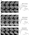
Recruitment of OGG1 and XRCC1 under different laser conditions. Recruitment kinetics of (A) the BER protein OGG1, (B) BER/SSBR scaffold protein XRCC1 and (C) XRCC1 mutant L360D were compared following irradiation of HeLa cells expressing pEGFP-OGG1 or wild type or mutant XRCC1-mGFP with either 750-nm multi-photon excitation or 405-nm laser excitation. The recruitment of the XRCC1 mutant, L360D, was tested in cells co-expressing WT XRCC1-mRFP (shown in Supplementary Figure S2). Error bars represent SEM from three independent experiments each analyzing 12 cells (i.e. n = 36).
Figure 3.
Figure 3.
Recruitment and retention of SSBR proteins following multi-photon 750-nm laser micro-irradiation. EGFP-PARP1, XRCC1-mGFP and EGFP-LIG3 show near instantaneous recruitment to sites of DNA damage, and PNKP-mGFP is also rapidly recruited. Laser micro-irradiation using multi-photon 750 nm was carried out as outlined in the Materials and Methods section using HeLa cells expressing fluorescently tagged versions of indicated proteins. Recruitment curves show quantification of signals over the observed time scale starting at the time when the damage is introduced by the laser (t = 0). Error bars represent SEM from three independent experiments for a total of 36 individual cells.
Figure 4.
Figure 4.
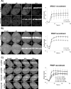
Recruitment of XRCC1 and PNKP in PARP1 WT and KO cells. The recruitment of SSBR proteins was monitored in PARP+/+ and PARP−/− MEFS expressing (A) XRCC1-mGFP and (B) PNKP-mGFP subjected to 750-nm multiphoton micro-irradiation. Error bars represent SEM from three independent experiments for a total of 36 individual cells.
Figure 5.
Figure 5.
PARP1 inhibition and the recruitment of SSBR proteins to sites of DNA damage. HeLa cells expressing fluorescently tagged SSBR proteins were treated with the PARP inhibitor, AG14361, and then subjected to laser micro-irradiation. PARP inhibition only affected early recruitment events of (A) XRCC1, (B) PNKP and (C) LIG3 with almost no effect on the late events of accumulation of all the proteins at sites of DNA damage. For recruitment curves, error bars represent SEM from three independent experiments for a total of 36 individual cells.
Figure 6.
Figure 6.
LIG3 knockdown and the recruitment of XRCC1 and PNKP to sites of DNA damage. Laser micro-irradiation was performed on HeLa cells expressing reduced levels of LIG3 (see Supplementary Figure S5) and (A) XRCC1-mRFP or (B) PNKP-mRFP. Reduced background levels of LIG3 lead to decreased overall recruitment of XRCC1 and PNKP to sites of DNA damage. (C) Simultaneous inhibition of PARP1 (using AG14361) and knockdown of LIG3 showed an additive effect on the reduction of the amount of PNKP recruited to sites of DNA damage. For recruitment curves, error bars represent SEM from three independent experiments for a total of 36 individual cells. Note that the mRFP photobleaches during laser micro-irradiation resulting in an initial loss of fluorescence at the damage sites.
Figure 7.
Figure 7.
Comparison between the recruitment of full-length LIG3 and LIG3 lacking the zinc finger to sites of DNA damage. (A) HeLa cells or (B) EM9 cells expressing full-length (FL) LIG3 or mutant ΔZnF LIG3 were subjected to laser micro-irradiation. In HeLa cells, FL LIG3 was robustly recruited to sites of DNA damage while ΔZnF LIG3 was recruited less efficiently. Furthermore, FL-LIG3 was recruited to sites of DNA damage even in the absence of XRCC1 (EM9 cells) while ΔZnF-LIG3 could not. For recruitment curves, error bars represent SEM; n = 36. Both cell lines were tested with mRFP- and EGFP-tagged proteins and the tags were shown not to influence the result.
Figure 8.
Figure 8.
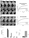
The ZnF domain is required for the damage sensing function of LIG3. (A) Comparison of the recruitment of fluorescently tagged full-length (FL) LIG3 and the ZnF and ZnF-DBD domains of LIG3 to micro-irradiated DNA in HeLa cells. (B) Comparison of recruitment of wild type (WT) and the DNA binding mutant of ZnF-R31I to micro-irradiated DNA in HeLa cells. For recruitment curves shown in (A) and (B), bars represent SEM; n = 36. (C) Expression of the ZnF domain of LIG3 retards single-strand break repair. HeLa cells expressing either the GFP-ZnF or GFP alone (control) were treated with 100 μM hydrogen peroxide for 40 min on ice and then strand break repair was monitored by the alkaline comet assay and quantification of tail moments at the indicated time points as described in the Materials and Methods section. Expanding the ordinate (plot on the right-hand side) showed that even in the absence of the hydrogen peroxide the ZnF expressing cells exhibit a slightly higher background level of damage.
Figure 9.
Figure 9.
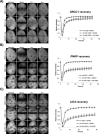
PARP1 inhibition and the retention of SSBR proteins at sites of DNA damage. FRAP analysis on HeLa cells expressing GFP-tagged (A) XRCC1, (B) PNKP and (C) LIG3, respectively, before and after DNA damage with 10 mM hydrogen peroxide in the absence and presence of 2 μM AG14361 as described in the Materials and Methods section. ‘Prebleach’ indicates no photobleaching and ‘Bleach’ is the 0-s time point. For recovery curves, error bars represent SEM; n = 24.
Figure 10.
Figure 10.
LIG3 is an in vivo nick sensor for irinotecan-induced DNA damage. FRAP analysis showing differences in binding kinetics after 5 mM irinotecan (IRI) treatment in HeLa cells expressing PARP1 and LIG3. ‘Prebleach’ indicates no photobleaching and ‘Bleach’ is the 0-s time point. Error bars represent SEM; n = 24.
Figure 11.
Figure 11.
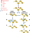
Two pathways exist for the short patch repair of SSBs. In the canonical pathway (PARP1 dependent) (A) PARP1 senses DNA damage and rapidly catalyzes the formation of PAR residues that allow for (B) chromatin expansion which in turn facilitates (C) the recruitment of downstream repair proteins. In the second pathway (PARP1 independent) (D) XRCC1–LIG3 complex continuously scans the DNA, upon sensing an interruption (via LIG3), (E) the complex is capable of causing a localized nucleosomal disruption (dependent on LIG3) (42), and the scaffold XRCC1 is capable of (F) loading downstream repair factors, PNKP and Polβ, and then repair continues as previously described (G).

Similar articles

Cited by

References

    1. Hoeijmakers J.H. Genome maintenance mechanisms for preventing cancer. Nature. 2001;411:366–374. - PubMed
    1. Caldecott K. Single-strand break repair and genetic disease. Nat. Rev. Genet. 2008;9:619–631. - PubMed
    1. Rouleau M., Patel A., Hendzel M.J., Kaufmann S.H., Poirier G.G. PARP inhibition: PARP1 and beyond. Nat. Rev. Cancer. 2010;10:293–301. - PMC - PubMed
    1. Weinfeld M., Mani R.S., Abdou I., Aceytuno R.D., Glover J.N.M. Tidying up loose ends: the role of polynucleotide kinase/phosphatase in DNA strand break repair. Trends Biochem. Sci. 2011;36:262–271. - PMC - PubMed
    1. Wilson S.H., Beard W.A, Shock D.D., Batra V.K., Cavanaugh N.A, Prasad R., Hou E.W., Liu Y., Asagoshi K., Horton J.K., et al. Base excision repair and design of small molecule inhibitors of human DNA polymerase β. Cell. Mol. Life Sci. 2010;67:3633–3647. - PMC - PubMed

Publication types

MeSH terms