. 2014 Dec 24:12:78.
doi: 10.1186/s12964-014-0078-2.

Succinate causes pathological cardiomyocyte hypertrophy through GPR91 activation

Affiliations

Succinate causes pathological cardiomyocyte hypertrophy through GPR91 activation

Carla J Aguiar et al. Cell Commun Signal. .

Abstract

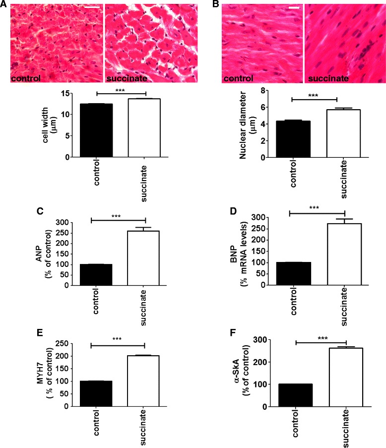
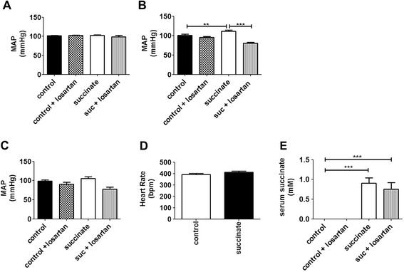
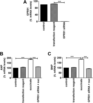
Background: Succinate is an intermediate of the citric acid cycle as well as an extracellular circulating molecule, whose receptor, G protein-coupled receptor-91 (GPR91), was recently identified and characterized in several tissues, including heart. Because some pathological conditions such as ischemia increase succinate blood levels, we investigated the role of this metabolite during a heart ischemic event, using human and rodent models.

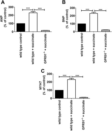
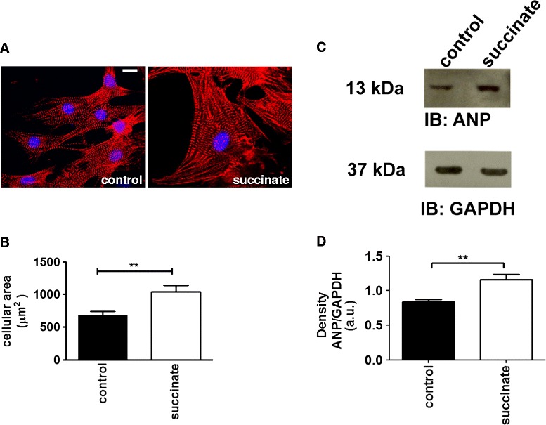
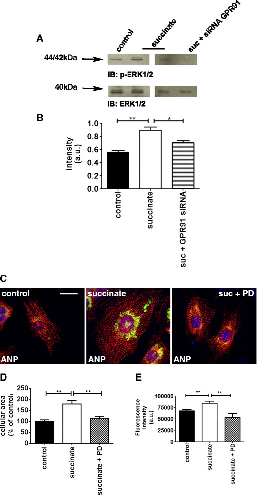
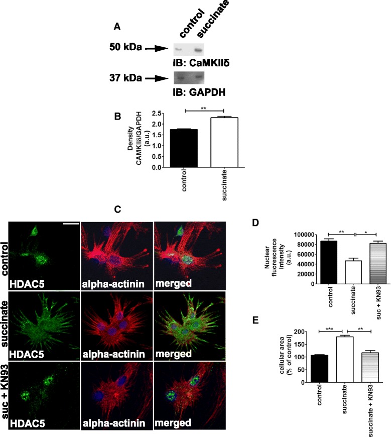
Results: We found that succinate causes cardiac hypertrophy in a GPR91 dependent manner. GPR91 activation triggers the phosphorylation of extracellular signal-regulated kinase 1/2 (ERK1/2), the expression of calcium/calmodulin dependent protein kinase IIδ (CaMKIIδ) and the translocation of histone deacetylase 5 (HDAC5) into the cytoplasm, which are hypertrophic-signaling events. Furthermore, we found that serum levels of succinate are increased in patients with cardiac hypertrophy associated with acute and chronic ischemic diseases.

Conclusions: These results show for the first time that succinate plays an important role in cardiomyocyte hypertrophy through GPR91 activation, and extend our understanding of how ischemia can induce hypertrophic cardiomyopathy.


Figures

Figure 1
Figure 1
Succinate induces pathologic cardiac hypertrophy. A. Cardiac morphometric analysis after 5 days of consecutive succinate administration. Upper panels show representative images of cell width. The specimen was stained with hematoxylin and eosin (original magnification; x 100, scale bars; 50 μm). The bar graph shows quantitative analysis of cardiomyocyte diameter (n = 50, ***p < 0.001). B. The upper panels show representative images of the nuclear diameter. The bar graph summarizes data from the nuclear diameter. (n = 5, ***p < 0.001). Scale bar 6 μm. C. ANP mRNA levels in freshly isolated adult cardiomyocytes from control and succinate treated rats. D. BNP mRNA levels in freshly isolated adult cardiomyocytes from control and succinate treated rats. E. MYH7 mRNA levels in freshly isolated adult cardiomyocytes from control and succinate treated rats. F. α-SkA mRNA levels in freshly isolated adult cardiomyocytes from control and succinate treated rats. These results represent the mean ± S.E. of three separate experiments (***p < 0.001).
Figure 2
Figure 2
Sustained levels of serum succinate cause increase in blood pressure on day 4 of treatment, but do not affect heart rate throughout the experiment. Medium Arterial Pressure levels in control animals and succinate treated animals, with or without IV injection of losartan on the 3rd day (A), 4th day (B) and 5th day of treatment (C) (**p < 0.01, ***p < 0.001). D. Mean heart rate of control and succinate treated rats during the experiment. E. Serum succinate concentration measured on the last day of experiment.
Figure 3
Figure 3
High blood levels of succinate induce cardiac hypertrophy through GPR91. A-C. ANP, BNP and BNP mRNA levels in cardiomyocytes from control (WT) and knockout mice (GPR91−/−) subjected to intravenous administration of succinate for 5 days (n = 6, ***p < 0.001).
Figure 4
Figure 4
Succinate induces hypertrophy in neonatal cardiomyocytes. A. Representative confocal images of neonatal cardiomyocytes double-labeled with DAPI (blue), and anti-α-actinin (red). Left panel shows control cells and right panel shows cells treated with succinate. B. Summary of cellular area, indicating that succinate induces hypertrophy. (*p < 0.05, n = 35 cells). C. Representative immunoblot of whole-cell protein lysates from neonatal cardiomyocytes probed with anti-ANP and anti-GAPDH. D. Bar graph shows that succinate significantly increases ANP levels. These results represent the mean ± S.E. of three separate experiments (**p < 0.01).
Figure 5
Figure 5
Hypertrophic effect of succinate in cardiomyocytes requires GPR91 expression. A. Real time PCR showed significant knockdown of GPR91 mRNA levels in cells transfected with siRNA against GPR91 (n = 3 independent experiments, ***p < 0.001). B-C. Knockdown of GPR91 decreased ANP and BNP mRNA levels. (***p < 0.001).
Figure 6
Figure 6
Succinate activates the ERK1/2 hypertrophic signaling pathway. A. Immunoblot of whole cell lysates showing increased phosphorylation of ERK1/2 and absence of phosphorylated ERK1/2 when GPR91 is silenced with siRNA. B. Bar graph shows that succinate significantly increases phosphorylation of ERK1/2 levels and fails to increase ERK1/2 phosphorylation when GPR91 is efficiently silenced. These results represent the mean ± S.E. of three separate experiments (**p < 0.01). C. Cells were treated with succinate and ERK1/2 inhibitor PD 098059. Immunofluorescence staining with DAPI (blue), anti-α-actinin (red) and ANP (green). D-E. Summary of cellular area and fluorescence intensity indicating that inhibition of ERK1/2 signaling pathway reverses the hypertrophic effect of succinate. (**p < 0.01, n = 50 cells).
Figure 7
Figure 7
Succinate activates the CaMKIIδ hypertrophic signaling pathway. A. Immunoblot for CaMKIIδ of whole cell lysates from primary cultures of neonatal cardiomyocytes. B. Bar graph shows that succinate significantly increases CAMKIIδ levels. These results represent the mean ± S.E. of three separate experiments. (**p < 0.01). C. Representative images of cardiomyocytes immunostained with antibodies against HDAC5 (green), α-actinin (red) and DAPI (blue). Succinate decreased HDAC5 nuclear export. Scale bar represents 10 μm. KN93 is a selective CaMKIIδ inhibitor. Nuclear export of HDAC5 induced by succinate is dependent on CaMKIIδ. D. Quantification of the nuclear fluorescence for HDAC5 (*p < 0.05, **p < 0.01). E. Summary of cellular area (**p < 0.01, ***p < 0.001), 45 cells).

References

    1. Heineke J, Molkentin JD. Regulation of cardiac hypertrophy by intracellular signalling pathways. Nat Rev Mol Cell Biol. 2006;7:589–600. doi: 10.1038/nrm1983. - DOI - PubMed
    1. Wilkins BJ, Molkentin JD. Calcineurin and cardiac hypertrophy: where have we been? Where are we going? J Physiol. 2002;541:1–8. doi: 10.1113/jphysiol.2002.017129. - DOI - PMC - PubMed
    1. van Rooij E, Doevendans PA, de Theije CC, Babiker FA, Molkentin JD, de Windt LJ. Requirement of nuclear factor of activated T-cells in calcineurin-mediated cardiomyocyte hypertrophy. J Biol Chem. 2002;277:48617–48626. doi: 10.1074/jbc.M206532200. - DOI - PubMed
    1. Berenji K, Drazner MH, Rothermel BA, Hill JA. Does load-induced ventricular hypertrophy progress to systolic heart failure? Am J Physiol Heart Circ Physiol. 2005;289:H8–H16. doi: 10.1152/ajpheart.01303.2004. - DOI - PubMed
    1. Bers DM. Altered cardiac myocyte Ca regulation in heart failure. Physiology (Bethesda) 2006;21:380–387. doi: 10.1152/physiol.00019.2006. - DOI - PubMed

Publication types

MeSH terms