Review
. 2015 Jan;95(1):83-123.
doi: 10.1152/physrev.00025.2013.

The organic anion transporter (OAT) family: a systems biology perspective

Affiliations
Review

The organic anion transporter (OAT) family: a systems biology perspective

Sanjay K Nigam et al. Physiol Rev. 2015 Jan.

Abstract

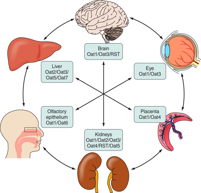
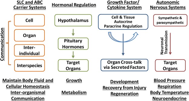
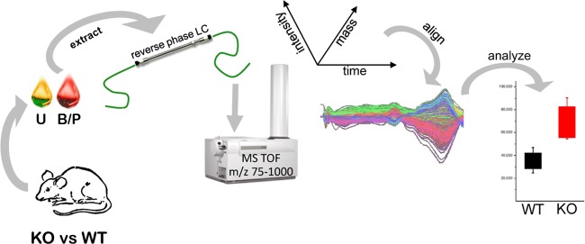
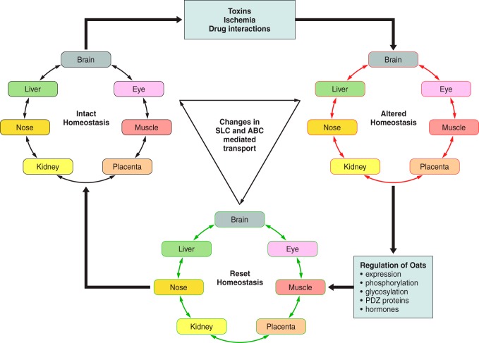
The organic anion transporter (OAT) subfamily, which constitutes roughly half of the SLC22 (solute carrier 22) transporter family, has received a great deal of attention because of its role in handling of common drugs (antibiotics, antivirals, diuretics, nonsteroidal anti-inflammatory drugs), toxins (mercury, aristolochic acid), and nutrients (vitamins, flavonoids). Oats are expressed in many tissues, including kidney, liver, choroid plexus, olfactory mucosa, brain, retina, and placenta. Recent metabolomics and microarray data from Oat1 [Slc22a6, originally identified as NKT (novel kidney transporter)] and Oat3 (Slc22a8) knockouts, as well as systems biology studies, indicate that this pathway plays a central role in the metabolism and handling of gut microbiome metabolites as well as putative uremic toxins of kidney disease. Nuclear receptors and other transcription factors, such as Hnf4α and Hnf1α, appear to regulate the expression of certain Oats in conjunction with phase I and phase II drug metabolizing enzymes. Some Oats have a strong selectivity for particular signaling molecules, including cyclic nucleotides, conjugated sex steroids, odorants, uric acid, and prostaglandins and/or their metabolites. According to the "Remote Sensing and Signaling Hypothesis," which is elaborated in detail here, Oats may function in remote interorgan communication by regulating levels of signaling molecules and key metabolites in tissues and body fluids. Oats may also play a major role in interorganismal communication (via movement of small molecules across the intestine, placental barrier, into breast milk, and volatile odorants into the urine). The role of various Oat isoforms in systems physiology appears quite complex, and their ramifications are discussed in the context of remote sensing and signaling.


Figures

FIGURE 1.
FIGURE 1.
OAT structure and the mechanism of OAT-mediated uptake and transport of organic anions. A: illustration of the predicted topology of organic anion transporters. Two pairs of 6-transmembrane domains are connected by a large intracellular loop and both NH2 and COOH termini are intracellular (G, glycosylation sites; P, PKC phosphorylation sites). B: a renal proximal tubule cell is depicted as a prototypical epithelial cell to illustrate the Oat-mediated uptake and transcellular movement of organic anionic substrates (OA) from the blood to the urine. Oat1 and Oat3 (A), localized to the basolateral membrane of the proximal tubule cell, transport OA across the basolateral membrane and into the cell through the exchange of dicarboxylates (DC). As a secondary active membrane transporter system (76), the Oat-mediated entry of OA is linked to the transmembrane electrochemical potential of dicarboxylates generated by their movement against a concentration gradient and intracellular accumulation maintained through the action of the Na+/dicarboxylate cotransporter (B). Thus the energy driving this ”tertiary“ mechanism is the ATP consumed by the Na+-K+-ATPase in generating the sodium gradient (C). OA exit into the urinary luminal space (D) is via transporters found on the apical membrane. [Modified from Eraly et al. (69), with permission from ASPET.]
FIGURE 2.
FIGURE 2.
Interorgan communication mediated by organic anion transporters. Organic anion transporters (Oats) have been localized to most barrier epithelia. In these tissues, the Oats represent rate-limiting steps involved in the uptake and transcellular movement of small molecule anionic substrates (including metabolites, toxins, and drugs) between body fluid compartments. These various substrates, including many with informational content (e.g., signaling molecules, hormones, and growth factors, as well as toxins and xenobiotics), are ”sensed" by the other organs via their own set of variably expressed transporters and information is shared between tissues and organs. [Modified from Ahn and Bhatnagar (2), with permission from Wolters Kluwer Health.]
FIGURE 3.
FIGURE 3.
Transporter-mediated remote sensing and signaling. The Oats, members of the SLC family of solute carriers, are believed to function along with members of the ATP-binding cassette (ABC) transport system, to maintain body fluid and cellular homeostasis. The movement of the small molecule substrates handled by these transport systems is postulated to provide a means of communication between cells, as well as between tissues/organs. This intraorganismal process can be viewed as being analogous and working with regulatory mechanisms of the autonomic nervous system, growth factor/cytokine system, and neuroendocrine system. However, through their secretion (e.g., milk) or excretion (e.g., urine), these substrates are also postulated to allow for interorganismal communication between individuals of the same species (e.g., mother/neonate) or of different species (e.g., predator/prey). [Modified from Wu et al. (284), with permission from ASPET.]
FIGURE 4.
FIGURE 4.
Strategy utilized for the en masse identification of endogenous OAT substrates. An untargeted metabolomics analysis strategy used to identify endogenous substrates of the Oats is depicted (279, 285). Samples of body fluids [e.g., blood/plasma (B/P; red) and urine (U; yellow)] were collected from wild-type (WT) and Oat-deficient (KO) mice, and extracts of these specimens were subjected to reverse-phase liquid chromatography (LC) followed by time-of-flight mass spectrophotometry (MS TOF) (279). LC-MS features were statistically ranked and aligned based on their mass, time of flight, and intensity (224). Metabolites with significant differences between WT and Oat-KO mice were identified by searching available metabolomics databases. The ability of some of the identified metabolites to interact with Oats was then validated in wet-lab functional assays (279, 285). [From Wikoff et al. (279). Copyright 2011 American Chemical Society.]
FIGURE 5.
FIGURE 5.
Oat6 is expressed in olfactory epithelium. Oat6, initially identified using an in silico homology-based analysis of the Ensembl mouse genome database (160), was localized to the olfactory mucosa by in situ hybridization in coronal sections of nasal mucosa [A–C; anti-sense Oat6 (A), sense Oat6 (B), olfactory marker protein control (C); scale bar = 100 μm]. D–F: RT-PCR analysis of mouse nasal olfactory organs reveals epithelial expression of Oat6. D: Oat6 is expressed in whole main olfactory epithelium (MOE) (lane 7) and whole vomeronasal organ (VNO) (lane 8), but not in MOE sensory neurons (lanes 1–3) or VNO sensory neurons (lanes 4–6). (Lane 9, no template control.) E: β-tubulin control. F: olfactory marker protein control. G: dose-dependent inhibition of the uptake of labeled estrone sulfate by various odorant molecules in Xenopus oocytes microinjected with either Oat6 (solid line) or Oat1 (dashed line). [Modified from Kaler et al. (113), with permission from Elsevier.]
FIGURE 6.
FIGURE 6.
Chromosomal clustering of members of the SLC22 family of genes. The discovery of the organic anion transporters allowed for the chromosomal mapping of their genes, and many of the Oats were found to exist in pairs and/or clusters (70), which was also found to be true for Octs and Oatps (70, 205). For example, OAT1 and OAT3 were found to exist as a tandem repeat with no other genes between them on human chromosome 11, as well as on mouse chromosome 19. Subsequent sequence analysis of adjacent areas of these chromosomes identified additional transporters clustered together with OAT1 (SLC22A6) and OAT3 (SLC22A8) on both the human and mouse chromosomes (283). The figure depicts the organization of this SLC22 organic anion transporter-containing cluster on human chromosome 11 (top) and the corresponding region on mouse chromosome 19 (bottom). The significance of this genomic clustering remains to be clarified. [Modified from Wu et al. (283).]
FIGURE 7.
FIGURE 7.
Hnf4α and Hnf1α, regulate drug transporter expression in the developing kidney. A: bar graph demonstrating the changes in the expression of phase I and phase II DMEs, as well as phase III transporters in whole embryonic rat kidneys cultured in the presence of a small molecule antagonist of Hnf4α (120). B, top: viral transduction of mouse embryonic fibroblasts (MEFs) with both Hnf1α and Hnf4α leads to the formation of cells with a proximal tubule-like character. B, bottom: qPCR analysis of MEFs virally transduced with Hnf1α, Hnf4α, or both revealed highest expression of transporter genes when both Hnf1α and Hnf4α are present. C: screenshots of p300 ChIP-seq in adult kidney cortex. P300 binding sites are highly enriched in Oat1 and Oat3, as well as in in the Hnf1α locus. D: two of the most highly enriched transcription factor binding motifs were Hnf4α and Hnf1α. [Modified from Martovetsky et al. (149), with permission from ASPET.]
FIGURE 8.
FIGURE 8.
A: molecular dynamic simulation of Oat1-mediated transport. Ribbon diagram of superimposed Oat1 structural conformers obtained at 40 ns (brown) and 94 ns (green) of a ∼100-ns molecular dynamic transport simulation (253). The transport simulation was performed on a homology-based computational model of Oat1, and the superimposition of the structural conformers allowed for visualization of Oat1 movements during the intial stages of substrate transport. Alterations in the distances between amino acid residues suggested that the early stages of Oat1-mediated transport were characterized by opening of the extracellular portion of the transporter allowing substrates access to a transporter channel. [Modified from Tsigelny et al. (253), with permission from Springer Science and Business Media.] B: pharmacophore modeling of Oat1 substrates. A pharmacophore model based on the 3-dimensional chemical structures common to certain Oat1 substrates. Colored spheres represent various structural features of the pharmacophore [e.g., hydrophobic (green), aromatic (orange), and hydrogen-bond acceptor (red)]. Such a model can be used to virtually screen chemical libraries for potential novel substrates which can then be validated in transport assays. [Modified from Wikoff et al. (279) Copyright 2011 American Chemical Society.]
FIGURE 9.
FIGURE 9.
Phylogeny of human and mouse Oats and Usts. Slc22a6 (Oat1) and Slc22a8 (Oat3), both of which exist in mouse and human, appear to have arisen from tandem duplication of an Oat ancestral gene. Multiple rounds of duplication of the Ust (unknown substrate transporter) ancestral gene are proposed to have led to several Ust-like genes by the time of human and mouse divergence. After this, certain mouse Usts are proposed to have undergone further duplication to generate other mouse-specific Ust genes. Some Usts exist in human while others exist in mouse, but no orthologous Usts exist in both species. [Modified from Wu et al. (283).]
FIGURE 10.
FIGURE 10.
The expression of the SLC22 family of transporters during kidney development. A: microarray analyses of developing embryonic [early embryonic (e13, e14, e15, e16 days of gestation), intermediate embryonic (e17, e18), late embryonic (e19, e20, e21,e22)], postnatal (birth and 1 wk), and mature (week 4 and adult) rodent kidneys demonstrate major increases in the expression of Slc22 transporters during kidney development and postnatal maturation. The grouping of the different stages of kidney development was based on a cluster analysis of gene expression data across the time course of kidney development (252). B: bar graph showing the clearance of the prototypical Oat substrate, PAH, in postnatal mice at 1, 2, and 3 wk of age. The rate of clearance increases during maturation of the postnatal kidney. C: comparison of Slc22 gene expression between consecutive stages of kidney development indicates that major changes in expression occur between the late embryo (late emb)/postnatal and postnatal/mature. [Modified from Sweeney et al. (230), with permission from ASPET.]
FIGURE 11.
FIGURE 11.
Oat-mediated transport in kidney organ cultures. A and B: uptake of a fluorescent Oat substrate (6-carboxyfluorscein, 6CF; green) in cultured embryonic rodent kidneys. A: low-magnification examination of Oat-mediated transport of 6CF (green) in an embryonic rodent kidney cultured for 4 days. Red staining indicates the collecting ducts labeled specifically with fluorescently labeled Dolichos biflorus lectin. 6CF accumulates in tubular structures distinct from the collecting duct. B: higher magnification view of kidney from A. The green fluorescent probe accumulates in nascent proximal tubular structures; the green staining does not overlap with the developing collecting ducts which are indicated by red staining. CD, collecting duct; CF, carboxyfluorescein. [Modified from Truong et al. (250). Copyright the American Society for Biochemistry and Molecular Biology.]
FIGURE 12.
FIGURE 12.
Network-based approach for linking Oat1-mediated transport to metabolism. A, inset: an integrative computational approach was used to analyze both gene expression data from the kidneys of Oat1 knockout mouse and metabolomics data derived from the plasma and urine of Oat1 knockout animals. Certain predictions based on the comparison of these two separate network analyses are supported by wet-lab data. B: the integrative network-based analyses of a comparison of the wild-type and Oat1-knockout mouse suggested alterations in many metabolic reactions in the Oat1 knockout. Implicated pathways included those involved in nucleotide, fatty acid, carnitine, chondroitin sulfate, inositol phosphate, and biotin metabolism. [From Ahn et al. (4). Copyright the American Society for Biochemistry and Molecular Biology.]
FIGURE 13.
FIGURE 13.
Potential role of Oats and other transporters in normal and perturbed homeostasis. Members of the SLC and ABC transporter families are involved in the handling of a wide variety of endogenous and exogenous substrates, including nutrients, metabolites, signaling molecules, toxins, and drugs. As described in the text, it is hypothesized that these transporters are essential for maintaining normal homeostasis and play key roles in resetting the system after homeostasis is altered. Transporter-mediated interorgan communication is disrupted after insults to the system (e.g., toxins, ischemia, or competitive inhibition by other substrates), leading to perturbed substrate clearance. Resetting and eventual reestablishment of intact homeostatsis likely occurs through changes in expression and/or function of the transporters (e.g., transcriptional, translational, or posttranslational modifications) in either the injured tissues or in other tissues participating in the larger remote-sensing network involving SLC and ABC transporters. [Modified from Ahn and Nigam (5), with permission from ASPET.]

References

    1. Adelibieke Y, Shimizu H, Muteliefu G, Bolati D, Niwa T. Indoxyl sulfate induces endothelial cell senescence by increasing reactive oxygen species production and p53 activity. J Ren Nutr 22: 86–89, 2012. - PubMed
    1. Ahn SY, Bhatnagar V. Update on the molecular physiology of organic anion transporters. Curr Opin Nephrol Hypertens 17: 499–505, 2008. - PubMed
    1. Ahn SY, Eraly SA, Tsigelny I, Nigam SK. Interaction of organic cations with organic anion transporters. J Biol Chem 284: 31422–31430, 2009. - PMC - PubMed
    1. Ahn SY, Jamshidi N, Mo ML, Wu W, Eraly SA, Dnyanmote A, Bush KT, Gallegos TF, Sweet DH, Palsson BO, Nigam SK. Linkage of organic anion transporter-1 to metabolic pathways through integrated ”omics“-driven network and functional analysis. J Biol Chem 286: 31522–31531, 2011. - PMC - PubMed
    1. Ahn SY, Nigam SK. Toward a systems level understanding of organic anion and other multispecific drug transporters: a remote sensing and signaling hypothesis. Mol Pharmacol 76: 481–490, 2009. - PMC - PubMed

Publication types

MeSH terms

Substances