. 2015 Mar 15;113(6):1941-51.
doi: 10.1152/jn.00555.2014. Epub 2014 Dec 24.

Decomposition of surface EMG signals from cyclic dynamic contractions

Affiliations

Decomposition of surface EMG signals from cyclic dynamic contractions

Carlo J De Luca et al. J Neurophysiol. .

Abstract

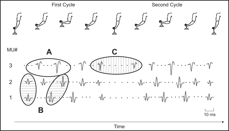
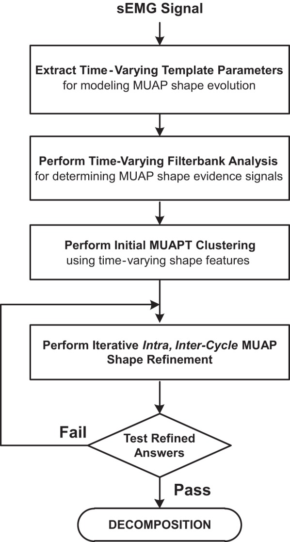
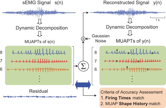
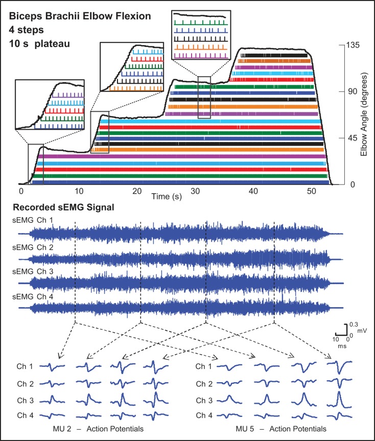
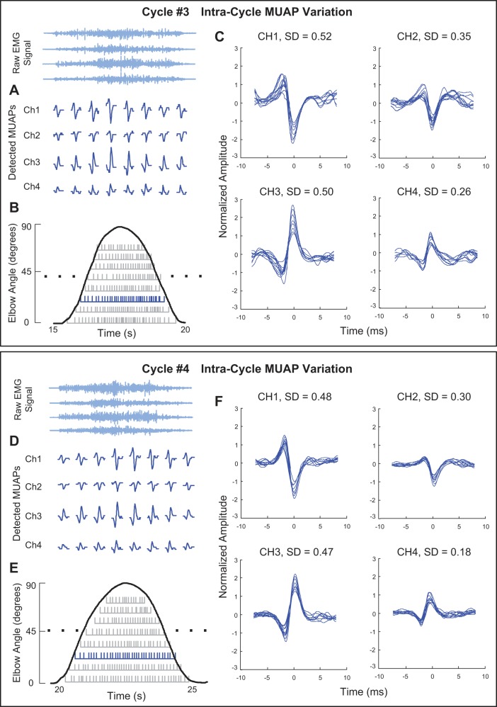
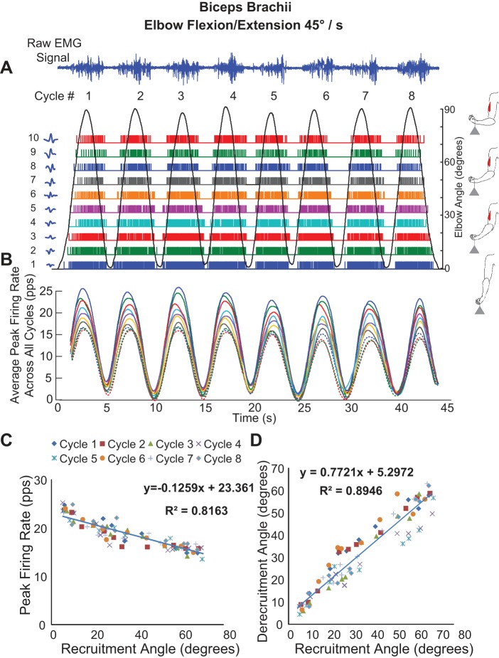
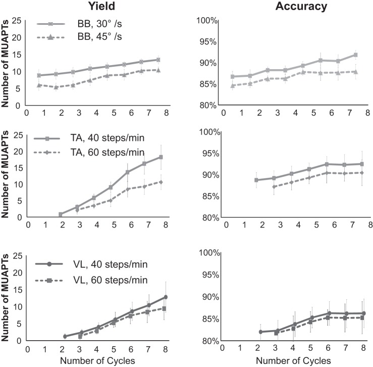
Over the past 3 decades, various algorithms used to decompose the electromyographic (EMG) signal into its constituent motor unit action potentials (MUAPs) have been reported. All are limited to decomposing EMG signals from isometric contraction. In this report, we describe a successful approach to decomposing the surface EMG (sEMG) signal collected from cyclic (repeated concentric and eccentric) dynamic contractions during flexion/extension of the elbow and during gait. The increased signal complexity introduced by the changing shapes of the MUAPs due to relative movement of the electrodes and the lengthening/shortening of muscle fibers was managed by an incremental approach to enhancing our established algorithm for decomposing sEMG signals obtained from isometric contractions. We used machine-learning algorithms and time-varying MUAP shape discrimination to decompose the sEMG signal from an increasingly challenging sequence of pseudostatic and dynamic contractions. The accuracy of the decomposition results was assessed by two verification methods that have been independently evaluated. The firing instances of the motor units had an accuracy of ∼90% with a MUAP train yield as high as 25. Preliminary observations from the performance of motor units during cyclic contractions indicate that during repetitive dynamic contractions, the control of motor units is governed by the same rules as those evidenced during isometric contractions. Modifications in the control properties of motoneuron firings reported by previous studies were not confirmed. Instead, our data demonstrate that the common drive and hierarchical recruitment of motor units are preserved during concentric and eccentric contractions.

Keywords: dynamic contractions; firing rate; gait; motor units.


Figures

Fig. 1.
Fig. 1.
Anticipated challenges that are likely present when decomposing surface electromyographic (sEMG) data acquired during cyclic dynamic contractions. Real motor unit (MU) action potential (MUAP) data for 3 different MUs are shown from the biceps brachii muscle of a subject recorded during 2 consecutive 5-s elbow flexion/extension contraction cycles while holding an 8-lb weight. Three potential challenges are identified by the oval outlines: A, intracycle shape change (no shading); B, intracycle shape similarity (horizontal bar shading); and C, intercycle shape change (vertical bar shading).
Fig. 2.
Fig. 2.
Flow diagram of the different functional components of the algorithm developed for decomposing sEMG signals acquired from cyclic dynamic contractions.
Fig. 3.
Fig. 3.
A diagram illustrating the decompose-synthesize-decompose-compare (DSDC) method used to assess the accuracy of the decomposition algorithm. A recorded sEMG signal s(n) is decomposed to identify its MUAP trains (MUAPTs). A reconstructed signal y(n) is synthesized by summing together the real MUAPTs from s(n) and time-varying Gaussian noise for which the variance is set equal to that of the residual signal from the decomposition. The reconstituted signal y(n) is then decomposed and compared with the MUAPTs within the synthesized signal. The Xs indicate discrepancies between the MUAPTs of y(n) and s(n), which are designated as errors based on a comparison of the MU firing times and MUAP shapes.
Fig. 4.
Fig. 4.
Decomposition results from a staircase trajectory contraction from 1 subject acquired from the biceps brachii muscle during elbow flexion/extension with an 8-lb weight. The staircase trajectory ranged from 0 to 135° of elbow flexion with 4 increments or “steps” of 10-s duration each. The firing instances of different MUs are identified by differently colored bars. Typical MUAPs identified in each of the 4 channels (Ch) of the recorded sEMG signal are shown for 2 example MUs at different joint angles throughout the contraction. The callout boxes identify the recruitment and firing patterns of MUs during the transition period between steps. The solid black lines represent the elbow angle measured from an electrogoniometer. Assessed accuracy was 93.6%.
Fig. 5.
Fig. 5.
Decomposition results for data acquired from the biceps brachii muscle during the 3rd (Cycle #3, top plot) and 4th (Cycle #4, bottom plot) cyclic contractions of multicycle elbow flexion and extension (0–90°). The plots include an example of typical changes in the MUAP shape from MUAPT 3 for the 4 channels of data acquired from the sensor (A and D), the firing instances of 8 MUAPTs (gray bars) derived from the decomposition algorithm for each of the cyclic contractions (B and E), and superimposed MUAP waveforms for each of the 4 channels (C and F). The standard deviation (SD) of the MUAP amplitude, normalized as a percentage of the smallest action potential of the MUAPT, is presented for each channel in C and F.
Fig. 6.
Fig. 6.
Decomposition of sEMG data recorded from 1 subject during a multicycle contraction of the biceps brachii muscle (8 cycles) during repetitive elbow flexion/extension at an angular velocity of 45°/s. A: the sEMG signals and firing instances of MUAPTs derived from decomposing these signals are shown for changes in elbow angle (black sinusoidal line). The MUAP waveform templates obtained at recruitment are shown adjacent to the y-axis for each train. B: the mean firing rate values of the firing instances shown in A (MUAPTs 17 are shown as solid colored lines, and MUAPTs 810 are shown as dotted colored lines). C: the peak firing rate values are plotted vs. the angle at which the MUAPT was recruited and derecruited. The results of a linear regression analysis of the data are shown. D: the angle at which each MU is recruited is plotted vs. the angle at which it is derecruited. The results of a linear regression analysis of the data are shown. pps, Pulses per second.
Fig. 7.
Fig. 7.
Decomposition of sEMG data recorded from the tibialis anterior muscle from 1 subject during 8 gait cycles for a walking speed of 60 steps per minute. A: the sEMG signals and firing instances of MUAPTs derived from decomposing these signals are shown for changes in ankle angle (black line). The MUAP waveform templates at recruitment are shown adjacent to the y-axis for each train. B: the mean firing rate values of the firing instances shown in A (MUAPTs 1-7 are shown as solid colored lines, and MUAPTs 8-10 are shown as dotted colored lines). C: the peak firing rate values are plotted vs. the angle at which the MUAPT was recruited and derecruited. The results of a linear regression analysis of the data are shown. D: the angle at which each MU is recruited is plotted vs. the angle at which it is derecruited. The results of a linear regression analysis of the data are shown.
Fig. 8.
Fig. 8.
Decomposition algorithm performance is shown as a function of contraction cycle for the 3 muscles studied (BB, biceps brachii; TA, tibialis anterior; and VL, vastus lateralis). Data for each contraction cycle is plotted as mean values with SD bars for all subjects tested for all contractions. Data are plotted separately for the 2 different elbow contraction speeds (top plots) and walking speeds (2 bottom plots). Accuracy is calculated using the DSDC method.

References

    1. Bertram MF, Nishida T, Minieka MM, Janssen I, Levy CE. Effects of temperature on motor unit action potentials during isometric contraction. Muscle Nerve 18: 1443–1446, 1995. - PubMed
    1. De Luca CJ. Myoelectrical manifestations of localized muscular fatigue in humans. Crit Rev Biomed Eng 11: 251–279, 1984. - PubMed
    1. De Luca CJ, Adam A, Wotiz R, Gilmore LD, Nawab SH. Decomposition of surface EMG signals. J Neurophysiol 96: 1646–1657, 2006. - PubMed
    1. De Luca CJ, Contessa P. Hierarchical control of motor units in voluntary contractions. J Neurophysiol 107: 178–195, 2012. - PMC - PubMed
    1. De Luca CJ, Erim Z. Common drive of motor units in regulation of muscle force. Trends Neurosci 17: 299–305, 1994. - PubMed

Publication types