. 2015 Feb;167(2):351-66.
doi: 10.1104/pp.114.250365. Epub 2014 Dec 24.

Two activities of long-chain acyl-coenzyme A synthetase are involved in lipid trafficking between the endoplasmic reticulum and the plastid in Arabidopsis

Affiliations

Two activities of long-chain acyl-coenzyme A synthetase are involved in lipid trafficking between the endoplasmic reticulum and the plastid in Arabidopsis

Dirk Jessen et al. Plant Physiol. 2015 Feb.

Abstract

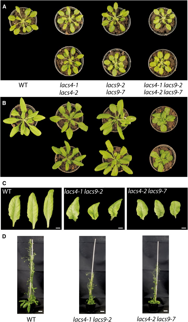
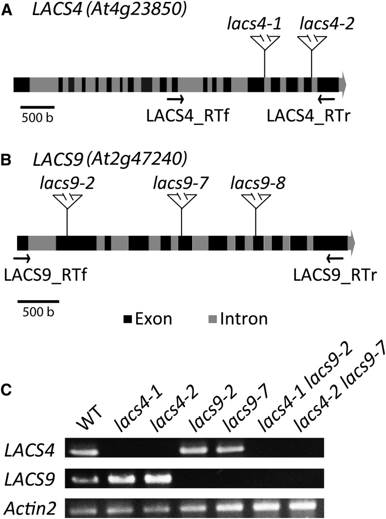
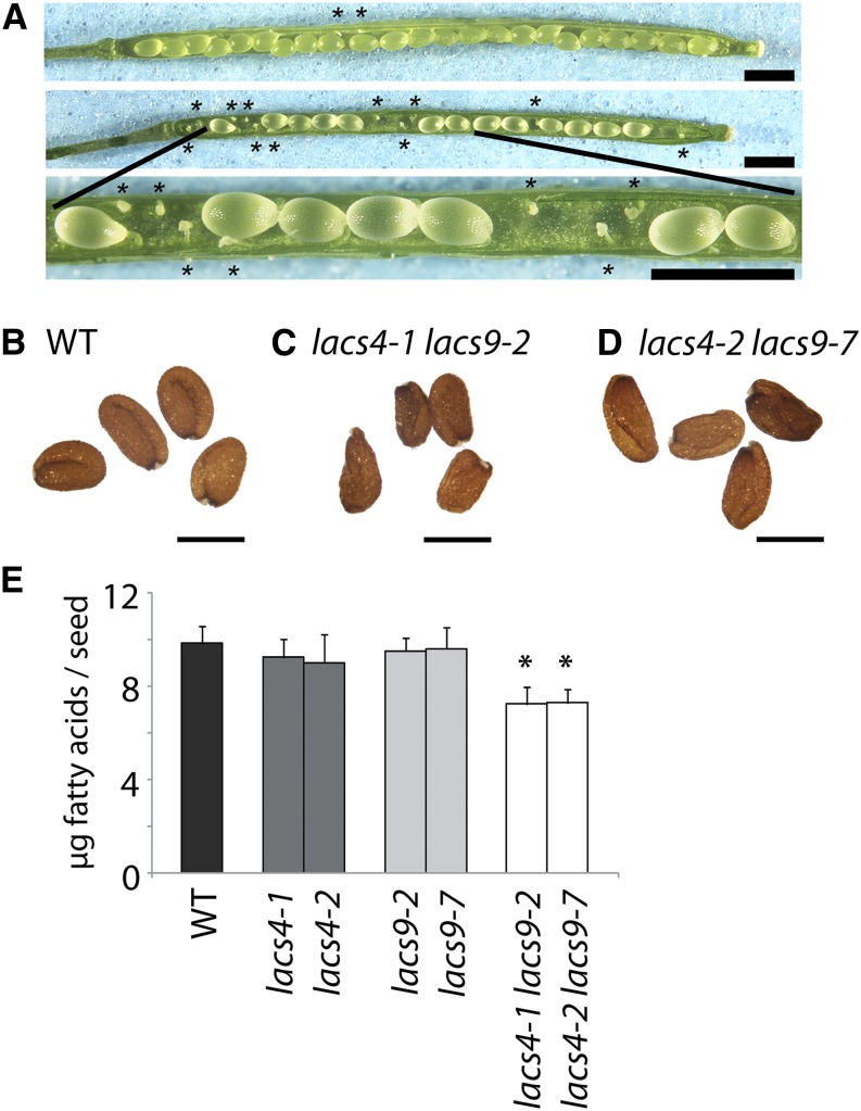
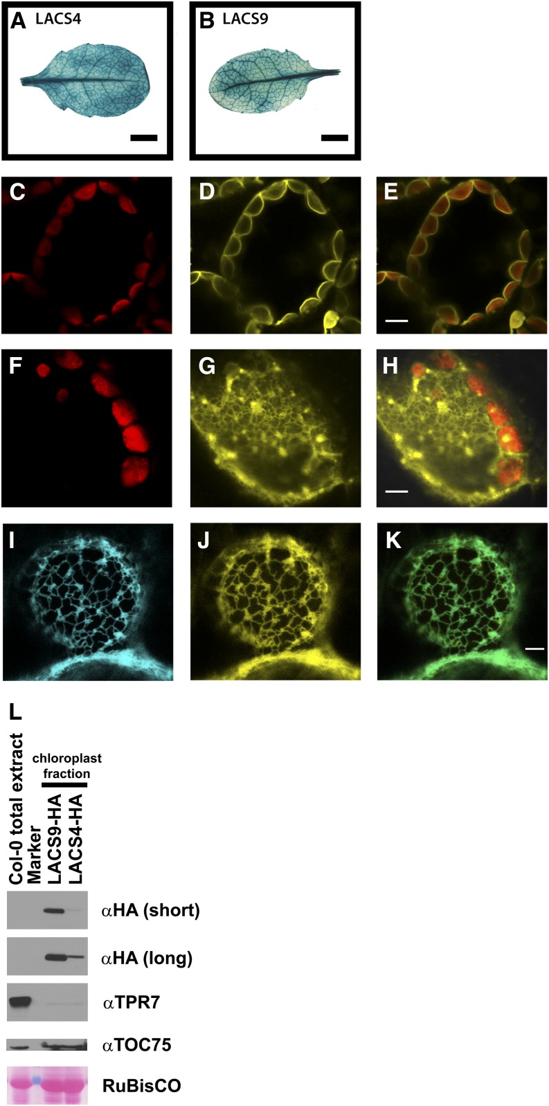
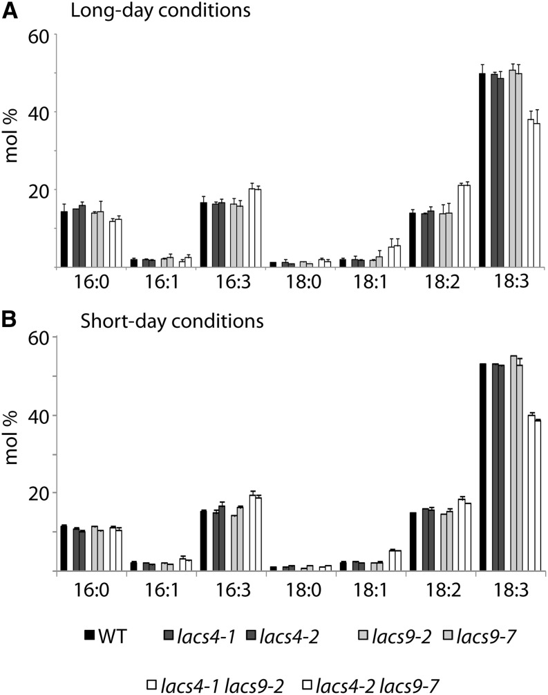
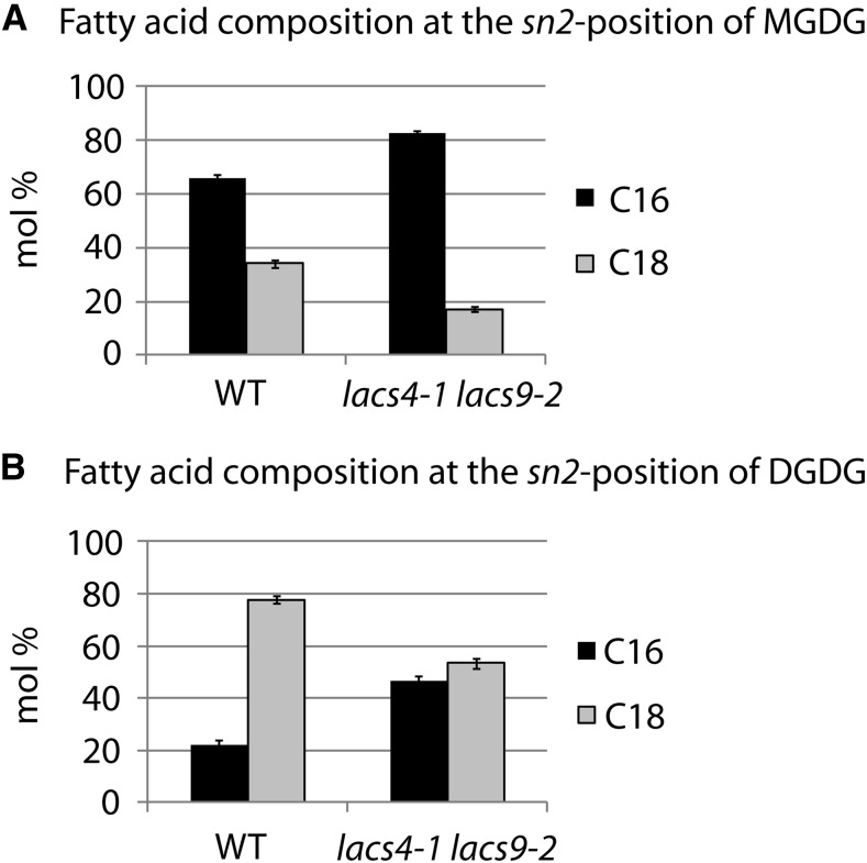
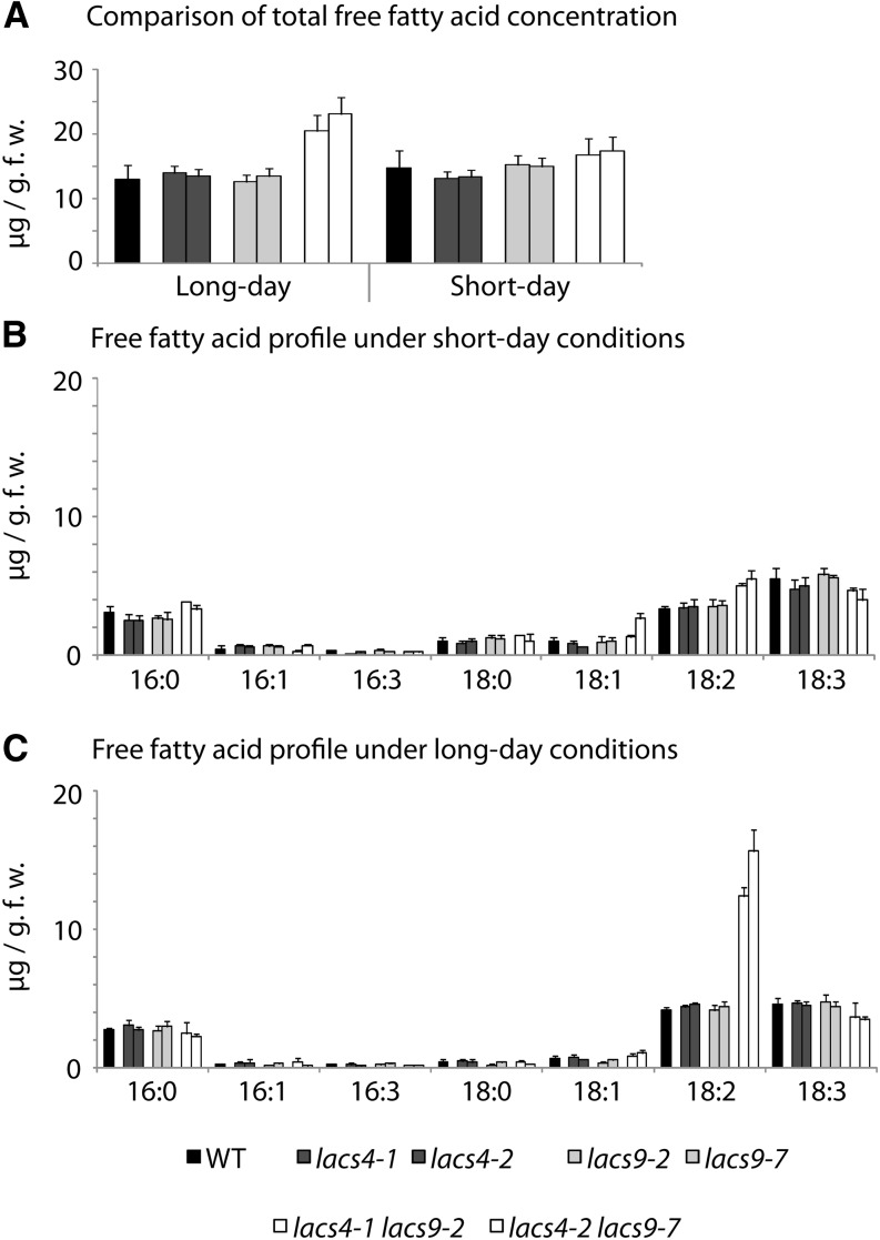
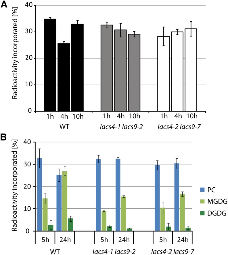
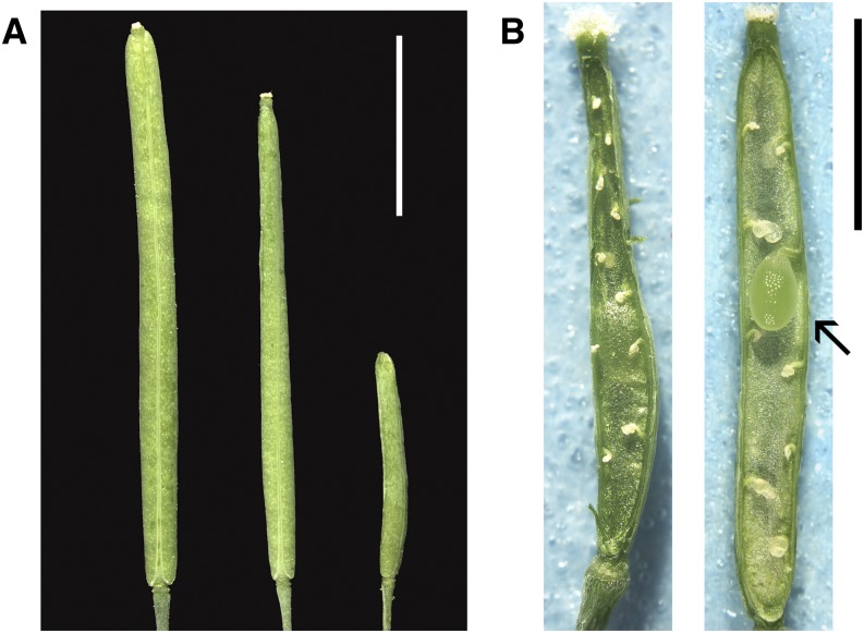
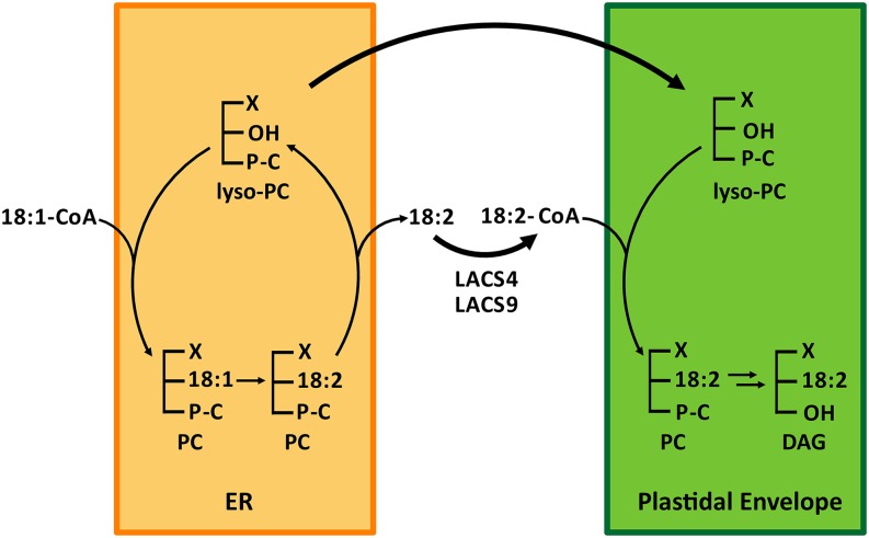
In plants, fatty acids are synthesized within the plastid and need to be distributed to the different sites of lipid biosynthesis within the cell. Free fatty acids released from the plastid need to be converted to their corresponding coenzyme A thioesters to become metabolically available. This activation is mediated by long-chain acyl-coenzyme A synthetases (LACSs), which are encoded by a family of nine genes in Arabidopsis (Arabidopsis thaliana). So far, it has remained unclear which of the individual LACS activities are involved in making plastid-derived fatty acids available to cytoplasmic glycerolipid biosynthesis. Because of its unique localization at the outer envelope of plastids, LACS9 was regarded as a candidate for linking plastidial fatty export and cytoplasmic use. However, data presented in this study show that LACS9 is involved in fatty acid import into the plastid. The analyses of mutant lines revealed strongly overlapping functions of LACS4 and LACS9 in lipid trafficking from the endoplasmic reticulum to the plastid. In vivo labeling experiments with lacs4 lacs9 double mutants suggest strongly reduced synthesis of endoplasmic reticulum-derived lipid precursors, which are required for the biosynthesis of glycolipids in the plastids. In conjunction with this defect, double-mutant plants accumulate significant amounts of linoleic acid in leaf tissue.


Figures

Figure 1.
Figure 1.
Phenotype of the lacs4 lacs9 double mutants. A and B, Six-week-old wild type (WT) and mutant plants grown under either short- (A) or long-day (B) conditions. C, Close-up views of rosette leaves from 6-week-old plants of the wild type and both lacs4 lacs9 double-mutant lines grown under long-day conditions. Bars = 0.3 cm. D, Morphology of 8-week-old plants of the wild type and both lacs4 lacs9 double-mutant lines grown under long-day conditions. Bars = 2 cm.
Figure 2.
Figure 2.
Identification of lacs4 and lacs9 mutant lines. A and B, Schematic gene structures of LACS4 (A) and LACS9 (B) depicting exons in dark gray and introns in light gray. For both genes, the independent T-DNA insertion sites are indicated. Arrows indicate positions of primers used for RT-PCR. C, RT-PCR analyses showing the absence of full-length transcripts of LACS4 in both lacs4 mutant alleles and LACS9 in both lacs9 mutant alleles. The two lacs4 lacs9 double-mutant lines are deficient in both transcripts. WT, Wild type.
Figure 3.
Figure 3.
Morphology and lipid phenotype of seeds. A, Seed development in two randomly chosen siliques of lacs4-1 lacs9-2 showing highly variable numbers of abortive ovules (indicated by asterisks). Bars = 1 mm. B to D, Photographs of randomly chosen seeds of the wild type (B) and both lacs4 lacs9 double-mutant lines (C and D). Bars = 500 µm. E, Total fatty acid content of mature seeds of the wild type and all mutant lines under investigation. *, Significantly different values between the wild type (WT) and the respective mutant line (P ≤ 0.05).
Figure 4.
Figure 4.
LACS4 and LACS9 are expressed in leaf tissue but show different subcellular localization. A and B, Analysis of gene expression in leaves using promoter:GUS reporter constructs for LACS4 (A) and LACS9 (B). Bars = 0.5 cm. C to E, Subcellular localization of LACS9 in leaf mesophyll cells of stable transgenic Arabidopsis plants was investigated by confocal fluorescence microscopy. Images show chloroplast autofluorescence (C), the signal of LACS9 fused to enhanced yellow fluorescent protein (EYFP) under control of the endogenous promoter (D), and the merged image (E). Bar = 5 µm. F to H, Subcellular localization of LACS4 in leaf mesophyll cells of stable transgenic Arabidopsis plants. Confocal images show chloroplast autofluorescence (F), the signal of LACS4 fused to EYFP under control of the endogenous promoter (G), and the merged image (H). Bar = 5 µm. I to K, Colocalization of LACS4 with an ER marker in leaf mesophyll cells upon transient expression in N. benthamiana. Confocal images show the signal of an ER marker fused to enhanced cyan fluorescent protein (ECFP; I), the signal of LACS4 fused to EYFP under control of the 35S promoter (J), and the merged image (K). Bar = 5 µm. L, Immunoblot analysis of LACS proteins in chloroplast fractions of transgenic plants expressing human influenza hemagglutinin (HA)-tagged LACS4 or LACS9 under control of the respective native promoter. Upon short exposure, only LACS9-HA is detected in chloroplast preparations (αHA [short]). Small amounts of HA-tagged LACS4 in purified chloroplasts can only be detected after longer exposure time (αHA [long]). TOC75 (for translocon at the outer envelope membrane of chloroplasts, 75 kD) was used as the chloroplast outer envelope marker, and tetratricopeptide repeat protein7 (TPR7) was used as an ER marker. Total leaf extract of wild-type plants (Col-0 total extract) was used as the positive control for TPR7 and TOC75 immunoblots and the negative control for the HA immunoblot. Ponceau S staining of Rubisco was used as the loading control.
Figure 5.
Figure 5.
Alterations of leaf fatty acid composition in lacs4 lacs9 double mutants are not affected by light regime. The fatty acid profiles of leaf total lipids of the wild type (WT) and all mutants under investigation were analyzed from plants grown under long- (A) or short-day (B) conditions. Each value represents the mean of three independent replicates, with error bars indicating sd.
Figure 6.
Figure 6.
Fatty acid composition at the sn-2 position of MGDG (A) and DGDG (B). The lipid classes were purified by TLC and digested by Rhizopus sp. lipase. The fatty acid compositions of the resulting lysoderivatives were determined by gas chromatography analysis. Black bars represent the sum of 16-carbon fatty acids, and gray bars represent the sum of 18-carbon fatty acids. The values represent the mean of three independent replicates, with error bars indicating sd. WT, Wild type.
Figure 7.
Figure 7.
The combined inactivation of LACS4 and LACS9 results in elevated levels of free fatty acids in plants grown under long-day conditions. A, Concentration of total free fatty acids in leaf tissue of the wild type (WT) and all mutant lines under investigation. Values are given in micrograms per gram of fresh weight (g.f.w.). B and C, Profiles of free fatty acids in the wild type and mutant lines grown under short- (B) or long-day (C) conditions. Each value represents the mean of five independent replicates, with error bars indicating sd.
Figure 8.
Figure 8.
Compromised lipid transfer from the ER to plastids in both lacs4 lacs9 double mutants. A, In vivo pulse-chase acetate labeling of MGDG. Detached leaves of the wild type (WT) and both lacs4 lacs9 double-mutant lines were floated on buffer containing [14C]acetate for 1 h before they were chased for the times indicated. Leaf material was collected, and lipid extracts were prepared and separated by two-dimensional TLC. The developed TLC plate was exposed to imaging plates, and the radiolabeled MGDG was quantified using a phosphorimager. B, In vivo pulse-chase labeling of lipids using [14C]oleic acid. The experimental setup was similar to that described for the labeling by acetate. The incorporation of radiolabeled oleic acid into the lipid classes PC, MGDG, and DGDG is shown for the wild type compared with both lacs4 lacs9 double-mutant lines. Each value represents the mean of three independent replicates. Error bars indicate sd.
Figure 9.
Figure 9.
The reproductive development of the lacs4 lacs8 lacs9 triple mutant. A, Siliques (from left to right) of the wild type, lacs4-1 lacs9-2, and lacs4-1 LACS8/lacs8-1 lacs9-2, respectively. Bar = 5 mm. B, Seed development in siliques of lacs4-1 LACS8/lacs8-1 lacs9-2. The majority of the siliques contained only abortive ovules and only very rarely set seed (indicated by an arrow). Bar = 2 mm.
Figure 10.
Figure 10.
Working model describing the LACS-mediated transfer of fatty acids from the ER to the chloroplast envelope. By the Lands cycle at the ER, mainly de novo synthesized oleic acid is incorporated into the sn-2 position of PC. The oleic acid is converted by the desaturase FAD2 to linoleic acid (18:2), and PC molecules with this signature are the preferred substrate for lipid transfer toward the plastid. We propose that 18:2 of remodeled PC is released through deacylation by phospholipase A2 to establish a pool of lyso-PC and a pool of free linoleic acid, both destined for lipid transfer to the plastid. As shown earlier (Bessoule et al., 1995), the lyso-PC is well suited to migrate between membranes and is able to move from the ER to the plastid. In parallel, LACS4 and LACS9 are both involved in converting the free linoleic acid into a tightly coupled pool of linoleoyl-CoA, which is used by plastidial acyl-CoA-lyso-PC acyltransferase to resynthesize PC in the plastidial envelope. Parts of the resynthesized PC stay in the envelope, whereas another portion is converted via PA into DAG, which is the immediate substrate for glycolipid biosynthesis. X, C16, or C18 fatty acid.

Similar articles

Cited by

References

    1. Alonso JM, Stepanova AN, Leisse TJ, Kim CJ, Chen H, Shinn P, Stevenson DK, Zimmerman J, Barajas P, Cheuk R, et al. (2003) Genome-wide insertional mutagenesis of Arabidopsis thaliana. Science 301: 653–657 - PubMed
    1. Andersson MX, Goksör M, Sandelius AS (2007) Optical manipulation reveals strong attracting forces at membrane contact sites between endoplasmic reticulum and chloroplasts. J Biol Chem 282: 1170–1174 - PubMed
    1. Andrews J, Keegstra K (1983) Acyl-CoA synthetase is located in the outer membrane and acyl-CoA thioesterase in the inner membrane of pea chloroplast envelopes. Plant Physiol 72: 735–740 - PMC - PubMed
    1. Aronsson H, Jarvis P (2002) A simple method for isolating import-competent Arabidopsis chloroplasts. FEBS Lett 529: 215–220 - PubMed
    1. Awai K, Xu C, Tamot B, Benning C (2006) A phosphatidic acid-binding protein of the chloroplast inner envelope membrane involved in lipid trafficking. Proc Natl Acad Sci USA 103: 10817–10822 - PMC - PubMed

Publication types