. 2015 Mar;89(5):2792-800.
doi: 10.1128/JVI.02693-14. Epub 2014 Dec 24.

Intracellular expression of camelid single-domain antibodies specific for influenza virus nucleoprotein uncovers distinct features of its nuclear localization

Affiliations

Intracellular expression of camelid single-domain antibodies specific for influenza virus nucleoprotein uncovers distinct features of its nuclear localization

Joseph Ashour et al. J Virol. 2015 Mar.

Abstract

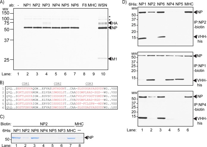
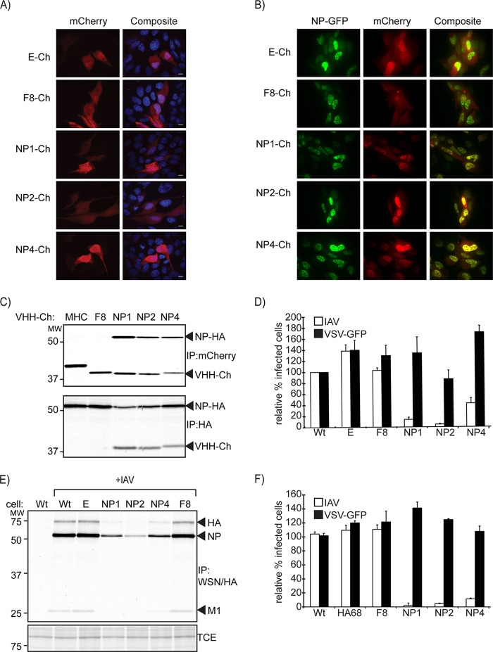
Perturbation of protein-protein interactions relies mostly on genetic approaches or on chemical inhibition. Small RNA viruses, such as influenza A virus, do not easily lend themselves to the former approach, while chemical inhibition requires that the target protein be druggable. A lack of tools thus constrains the functional analysis of influenza virus-encoded proteins. We generated a panel of camelid-derived single-domain antibody fragments (VHHs) against influenza virus nucleoprotein (NP), a viral protein essential for nuclear trafficking and packaging of the influenza virus genome. We show that these VHHs can target NP in living cells and perturb NP's function during infection. Cytosolic expression of NP-specific VHHs (αNP-VHHs) disrupts virus replication at an early stage of the life cycle. Based on their specificity, these VHHs fall into two distinct groups. Both prevent nuclear import of the viral ribonucleoprotein (vRNP) complex without disrupting nuclear import of NP alone. Different stages of the virus life cycle thus rely on distinct nuclear localization motifs of NP. Their molecular characterization may afford new means of intervention in the virus life cycle.

Importance: Many proteins encoded by RNA viruses are refractory to manipulation due to their essential role in replication. Thus, studying their function and determining how to disrupt said function through pharmaceutical intervention are difficult. We present a novel method based on single-domain-antibody technology that permits specific targeting and disruption of an essential influenza virus protein in the absence of genetic manipulation of influenza virus itself. Characterization of such interactions may help identify new targets for pharmaceutical intervention. This approach can be extended to study proteins encoded by other viral pathogens.


Figures

FIG 1
FIG 1
VHHs isolated from the influenza virus VHH library are specific for NP. (A) Lysate from 35S-labeled MDCK cells infected with influenza virus were subjected to immunoprecipitation with purified VHH containing a C-terminal 6×His tag immobilized on Ni-NTA beads. Samples were analyzed using SDS-PAGE and autoradiography. Asterisks denote expected mobility for PB2 (upper asterisk) and PB2/PA (lower asterisk). (B) ClustalW-based alignment of αNP-VHH CDR1, CDR2, and CDR3 amino acid sequences (in red). (C) His-tagged NP was incubated briefly with His-tagged versions of the given VHHs. Biotinylated αNP-VHH2 immobilized on streptavidin beads was then used to precipitate available NP-His. Precipitated NP-His was run on SDS-PAGE and visualized by Coomassie staining. (D) His-tagged NP was incubated briefly with His-tagged versions of the given VHHs. Biotinylated αNP-VHH2 (top), VHH4 (middle), and VHH1 (bottom) immobilized on streptavidin beads were then used to pull down NP-His. Precipitated protein was run on SDS-PAGE and detected via immunoblotting with αHis-HRP antibody.
FIG 2
FIG 2
αNP-VHH-Ch and NP-GFP colocalize in the nucleus of transfected MDCK cells. (A) Plasmids expressing αNP-VHH-Ch were transfected into MDCK cells. At 24 posttransfection, cells were fixed and permeabilized with saponin. DNA was stained with Hoechst, and cells were imaged via confocal microscopy. (B) Plasmids expressing αNP-VHH-Ch and NP-GFP were cotransfected into MDCK cells. At 24 h posttransfection, cells were imaged using live confocal microscopy. (C) NP and αNP-VHH-Ch interact in the cytosol. 293T cells were transfected with expression plasmids for αNP-VHH-Ch and NP-HA. At 24 posttransfection, cells were metabolically labeled with [35S]cysteine/methionine for 20 min and subsequently lysed in NP-40-containing lysis buffer. The lysate was split into two samples and subjected to immunoprecipitation using antibodies against HA or mCherry. Samples were analyzed via SDS-PAGE and autoradiography. (D) αNP-VHH-Ch expression in the cytosol specifically inhibits influenza virus replication. MDCK cells constitutively expressing αNP-VHH-Ch were challenged with influenza virus or VSV-GFP. At 6 h postinfection, cells were fixed, and IAV-infected cells were stained with Alexa Fluor (AF) 647-coupled αNP-VHH1 under permeabilizing conditions. Levels of infection were quantified by flow cytometry and normalized to that in the WT sample. Means and standard errors of the means (SEM) from three independent experiments are shown. (E) Expression of αNP-VHH-Ch in MDCK cells results in decreased influenza virus expression upon virus challenge. MDCK cells stably expressing VHH-Ch were challenged with influenza virus (MOI, 1.0). Two hours postinfection, cells were metabolically labeled with [35S]methionine for 20 min. Cells were lysed in NP-40-containing lysis buffer, and the lysate was subsequently probed with WSN serum and FluB1 antibody. Samples were analyzed by SDS-PAGE and autoradiography. (F) αNP-VHH-mediated inhibition is independent of cell type or mCherry fusion. A549 cells inducibly expressing αNP-VHH-HA were treated with 1 μg/ml doxycycline for 24 h or left untreated. Cells were subsequently challenged with influenza virus or VSV-GFP. At 6 h postinfection, cells were fixed, and IAV-infected cells were stained with AF 647-coupled α-NP-VHH1. Levels of infection were quantified by flow cytometry, and infections obtained in induced samples were normalized to their uninduced counterpart. Means and SEM from three independent experiments are shown.
FIG 3
FIG 3
αNP-VHH2 and -4 do not influence NP activity in a mini-genome assay. 293T cells were transfected with plasmids expressing influenza virus PB1, PB2, PA, NP, and VHH-Ch and a plasmid transcribing a model genome segment encoding neuraminidase with a sortag motif inserted at the C terminus (NAsrt) (under the control of the pPol1 promoter). At 24 h posttransfection, cells were lysed in NP-40-containing lysis buffer. Samples were analyzed via Western blotting using antibody against the HA epitope, NP, mCherry, and GAPDH.
FIG 4
FIG 4
αNP-VHH-Ch inhibits nuclear import of the incoming vRNP complex. MDCK cells stably expressing VHH-Ch were infected with influenza virus (MOI, 500) in the presence of cycloheximide. Four hours after infection, cells were fixed and permeabilized with 4% paraformaldehyde and 0.5% NP-40. Staining was performed with Hoechst (blue) and anti-NP (green) before visualization by confocal microscopy.

Similar articles

Cited by

References

    1. Gottschalk A, Fezekas De St Groth S. 1960. On the relationship between the indicator profile and prosthetic group of mucoproteins inhibitory for influenza virus haemagglutinin. J Gen Microbiol 22:690–697. doi:10.1099/00221287-22-3-690. - DOI - PubMed
    1. Graham ER, Gottschalk A. 1960. Studies on mucoproteins. I. The structure of the prosthetic group of ovine submaxillary gland mucoprotein. Biochim Biophys Acta 38:513–524. - PubMed
    1. Hirst GK. 1948. The nature of the virus receptors of red cells; evidence on the chemical nature of the virus receptors of red cells and of the existence of a closely analogous substance in normal serum. J Exp Med 87:301–314. doi:10.1084/jem.87.4.301. - DOI - PMC - PubMed
    1. Matlin KS, Reggio H, Helenius A, Simons K. 1981. Infectious entry pathway of influenza virus in a canine kidney cell line. J Cell Biol 91:601–613. doi:10.1083/jcb.91.3.601. - DOI - PMC - PubMed
    1. Doms RW, Helenius A, White J. 1985. Membrane fusion activity of the influenza virus hemagglutinin. The low pH-induced conformational change. J Biol Chem 260:2973–2981. - PubMed