. 2015 Mar;89(5):2866-74.
doi: 10.1128/JVI.02901-14. Epub 2014 Dec 24.

Alpha-defensin HD5 inhibits furin cleavage of human papillomavirus 16 L2 to block infection

Affiliations

Alpha-defensin HD5 inhibits furin cleavage of human papillomavirus 16 L2 to block infection

Mayim E Wiens et al. J Virol. 2015 Mar.

Abstract

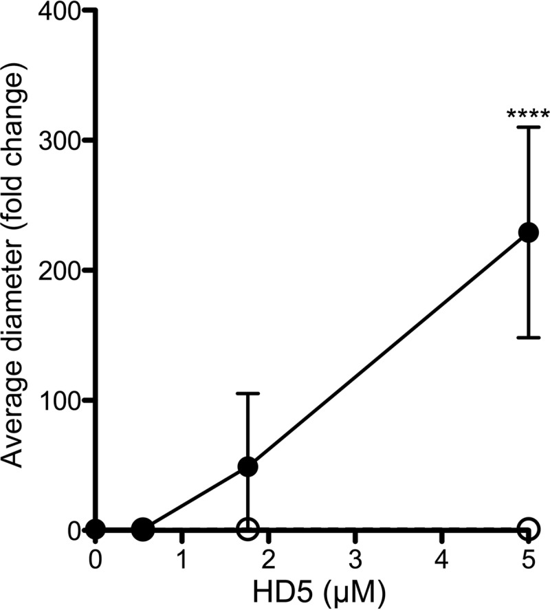
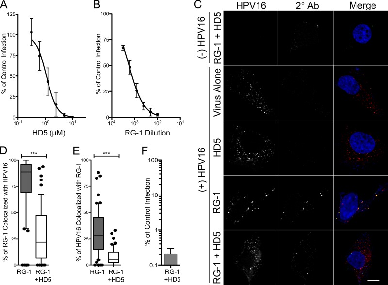
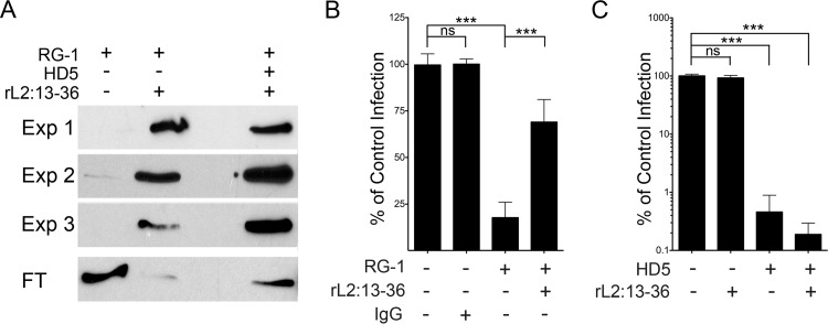
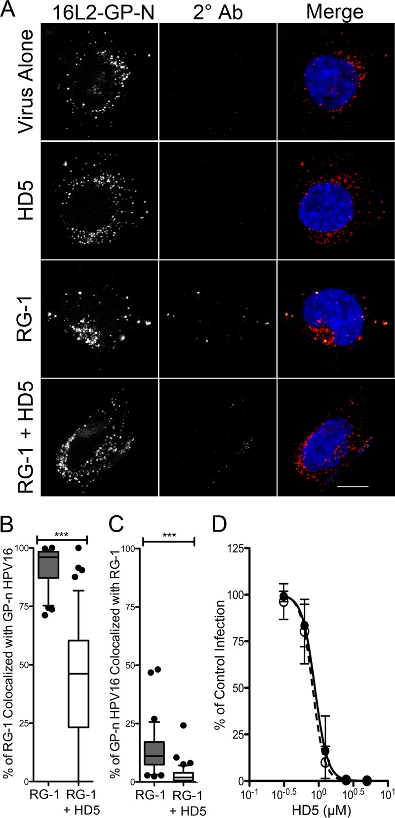
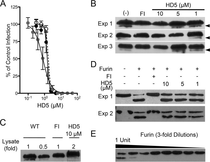
Human papillomavirus (HPV) is a significant oncogenic virus, but the innate immune response to HPV is poorly understood. Human α-defensin 5 (HD5) is an innate immune effector peptide secreted by epithelial cells in the genitourinary tract. HD5 is broadly antimicrobial, exhibiting potent antiviral activity against HPV at physiologic concentrations; however, the specific mechanism of HD5-mediated inhibition against HPV is unknown. During infection, the HPV capsid undergoes several critical cell-mediated viral protein processing steps, including unfolding and cleavage of the minor capsid protein L2 by host cyclophilin B and furin. Using HPV16 pseudovirus, we show that HD5 interacts directly with the virus and inhibits the furin-mediated cleavage of L2 at the cell surface during infection at a step downstream of the cyclophilin B-mediated unfolding of L2. Importantly, HD5 does not affect the enzymatic activity of furin directly. Thus, our data support a model in which HD5 prevents furin from accessing L2 by occluding the furin cleavage site via direct binding to the viral capsid.

Importance: Our study elucidates a new antiviral action for α-defensins against nonenveloped viruses in which HD5 directly interferes with a critical host-mediated viral processing step, furin cleavage of L2, at the cell surface. Blocking this key event has deleterious effects on the intracellular steps of virus infection. Thus, in addition to informing the antiviral mechanisms of α-defensins, our studies highlight the critical role of furin cleavage in HPV entry. Innate immune control, mediated in part by α-defensins expressed in the genital mucosa, may influence susceptibility to HPV infections that lead to cervical cancer. Moreover, understanding the mechanism of these natural antivirals may inform the design of therapeutics to limit HPV infection.


Figures

FIG 1
FIG 1
HD5 binding aggregates HPV16 PsV. The mean diameter of HPV15 PsV was measured upon incubation with increasing concentrations of wild-type HD5 (black circles) or HD5 Abu (open circles). Data are the average fold increases in diameter for each condition compared to untreated controls from 4 independent experiments ± standard deviations (SD). ****, P < 0.0001.
FIG 2
FIG 2
(A) HD5 neutralizes AF555-HPV16 PsV in complete medium. HeLa cells were infected with AF555-HPV16 PsV incubated with increasing concentrations of HD5 in complete medium. Data are from three independent experiments normalized to control infection in the absence of inhibitor ± SD. IC50 = 1.1 μM, 95% CI = 0.93 to 1.32 μM. (B) RG-1 antibody neutralizes AF555-HPV16 PsV. HeLa cells were infected with AF555-HPV16 PsV incubated with increasing concentrations of RG-1 antibody in complete medium. Data are from three independent experiments normalized to control infection in the absence of inhibitor ± SD. IC50 = 1,759-fold dilution, 95% CI = 1,428- to 2,167-fold dilution. (C) The presence of HD5 prevents binding of the RG-1 antibody to HPV16 during cell entry. Images of HaCaT cells 12 h p.i. with [(+) HPV16] or without [(−) HPV16] AF555-HPV16 PsV in the presence of no inhibitor (virus alone), 5 μM HD5 (HD5), 5.4 μg/ml RG-1 antibody equivalent to a 200-fold dilution (RG-1), or 5 μM HD5 and 5.4 μg/ml RG-1 together (RG-1 + HD5). Individual panels depict maximum intensity z-projections of signal above the threshold for images in the z-stack that are coplanar with the nucleus for HPV16 (red) and RG-1 (2° Ab, green). In the merged images, the nucleus is blue. Scale bar is 10 μm. Manders coefficient values M1 (D) and M2 (E) are plotted as percentages of RG-1 colocalized with HPV16 and percentages of HPV16 colocalized with RG-1, respectively, for 50 to 60 cells for each condition. Whiskers are 5 to 95%, the horizontal line is the median, and outliers are depicted as individual points. ***, P < 0.0001. (F) HPV16 PsV treated with a combination of 5.4 μg/ml RG-1 and 5 μM HD5 is neutralized. Data are the means ± SD from 3 independent experiments normalized to infection in the absence of inhibitor.
FIG 3
FIG 3
HD5 does not block RG-1-L2 epitope binding. (A) RG-1 was immunoprecipitated by rL2:13–36 in the presence or absence of 5 μM HD5. Shown are bound antibody from three independent experiments (Exp 1 to 3) and a representative unbound fraction (FT) from one experiment visualized by immunoblotting. (B) Excess rL2:13–36 rescues HPV16 from RG-1 neutralization. Infection of HeLa cells by HPV16 PsV incubated with RG-1 alone or in competition with a 500-fold molar excess of rL2:13–36 was quantified relative to infection in the absence of inhibitor. BSA was used to normalize protein levels in all samples, and mouse IgG1 was used as an isotype control for RG-1. Data are means ± SD from three independent experiments. ***, P < 0.0001. (C) rL2:13–36 does not rescue HPV16 infection from HD5 neutralization. Infection of HeLa cells by HPV16 PsV incubated with 5 μM HD5 alone or in competition with a 500-fold molar excess of rL2:13–36 was quantified relative to infection in the absence of inhibitor. BSA was used to normalize protein levels in all samples. Data are means ± SD from three independent experiments. ***, P < 0.0001.
FIG 4
FIG 4
The cyclophilin B-independent HPV16 mutant (16L2-GP-N) remains sensitive to HD5 neutralization. (A) Images of HaCaT cells 12 h p.i. with AF555-16L2-GP-N PsV in the presence of no inhibitor (virus alone), 5 μM HD5 (HD5), 5.4 μg/ml RG-1 antibody (RG-1), or 5 μM HD5 and 5.4 μg/ml RG-1 together (RG-1 + HD5). Individual panels depict maximum intensity z-projections of signal above the threshold for images in the z-stack that are coplanar with the nucleus for 16L2-GP-N (red) and RG-1 (2° Ab; green). In the merged images, the nucleus is blue. Scale bar is 10 μm. Manders coefficient values M1 (B) and M2 (C) are plotted as percentages of RG-1 colocalized with 16L2-GP-N and percentages of 16L2-GP-N colocalized with RG-1, respectively, for 45 to 60 cells for each condition. Whiskers are 5 to 95%, the horizontal line is the median, and outliers are depicted as individual points. ***, P < 0.0001. (D) HD5 neutralizes 16L2-GP-N. HeLa cells were infected with WT HPV16 PsV (black circles, IC50 = 0.88 μM, 95% CI = 0.78 to 0.99 μM) or 16L2-GP-N PsV (open circles, IC50 = 0.89 μM, 95% CI = 0.72 to 0.92 μM) incubated with increasing concentrations of HD5. Data are the means ± SD from three independent experiments compared to control infection in the absence of inhibitor.
FIG 5
FIG 5
(A) Myc-L2-HA HPV16 PsV is sensitive to HD5. HeLa cells were infected with WT HPV16 PsV in SFM (black circles, IC50 = 1.47 μM, 95% CI = 1.35 to 1.6 μM), Myc-L2-HA PsV in SFM (open circles, IC50 = 1.71 μM, 95% CI = 1.6 to 1.8 μM), or 10 times as much Myc-L2-HA PsV in complete medium (gray circles, IC50 = 0.79 μM, 95% CI = 0.69 to 0.9 μM) incubated with increasing concentrations of HD5. Data are means ± SD from three independent experiments normalized to infection in the absence of inhibitor. (B) HD5 inhibits furin cleavage of L2. HeLa cells were infected with Myc-16L2-HA HPV16 PsV in the presence of 40 μM furin inhibitor (FI), 1 to 10 μM HD5, or no inhibitor (−). L2 cleavage was assessed by immunoblotting of cell lysates 16 h p.i. using an anti-HA antibody. Cleaved L2 (arrow) is visible as a faster-migrating band below uncleaved L2. Shown are three independent experiments. (C) Analysis of a greater amount of lysate confirms the inhibition of furin cleavage. Different amounts of HD5-treated HPV16 PsV lysate, indicated by fold change relative to the amounts loaded in panel B, were assessed by immunoblotting with anti-HA antibody. (D) HD5 does not directly affect the enzymatic activity of furin. A total of 1.8 ng rL2:1–160 was digested with 1 U of furin in the presence or absence of the indicated inhibitors for 1 h at 30°C. Samples were immunoblotted using an anti-His antibody. Cleaved rL2:1–160 is the faster-migrating band. Shown are two independent experiments. (E) Titration of furin required for rL2:1–160 cleavage. rL2:1–160 was digested with a 3-fold dilution series of furin, starting at 1 U of total furin. Samples were resolved on a reducing gel and immunoblotted using anti-His antibody. Cleaved rL2:1–160 is the faster-migrating band.

References

    1. Hazlett L, Wu M. 2011. Defensins in innate immunity. Cell Tissue Res 343:175–188. doi:10.1007/s00441-010-1022-4. - DOI - PubMed
    1. Lehrer RI, Lu W. 2012. Alpha-defensins in human innate immunity. Immunol Rev 245:84–112. doi:10.1111/j.1600-065X.2011.01082.x. - DOI - PubMed
    1. Fan SR, Liu XP, Liao QP. 2008. Human defensins and cytokines in vaginal lavage fluid of women with bacterial vaginosis. Int J Gynaecol Obstet 103:50–54. doi:10.1016/j.ijgo.2008.05.020. - DOI - PubMed
    1. Com E, Bourgeon F, Evrard B, Ganz T, Colleu D, Jegou B, Pineau C. 2003. Expression of antimicrobial defensins in the male reproductive tract of rats, mice, and humans. Biol Reprod 68:95–104. doi:10.1095/biolreprod.102.005389. - DOI - PubMed
    1. Spencer JD, Hains DS, Porter E, Bevins CL, DiRosario J, Becknell B, Wang H, Schwaderer AL. 2012. Human alpha defensin 5 expression in the human kidney and urinary tract. PLoS One 7:e31712. doi:10.1371/journal.pone.0031712. - DOI - PMC - PubMed

Publication types

MeSH terms