. 2015 Mar;89(5):2494-506.
doi: 10.1128/JVI.03421-14. Epub 2014 Dec 24.

Chicken and duck myotubes are highly susceptible and permissive to influenza virus infection

Affiliations

Chicken and duck myotubes are highly susceptible and permissive to influenza virus infection

Belinda Baquero-Perez et al. J Virol. 2015 Mar.

Abstract

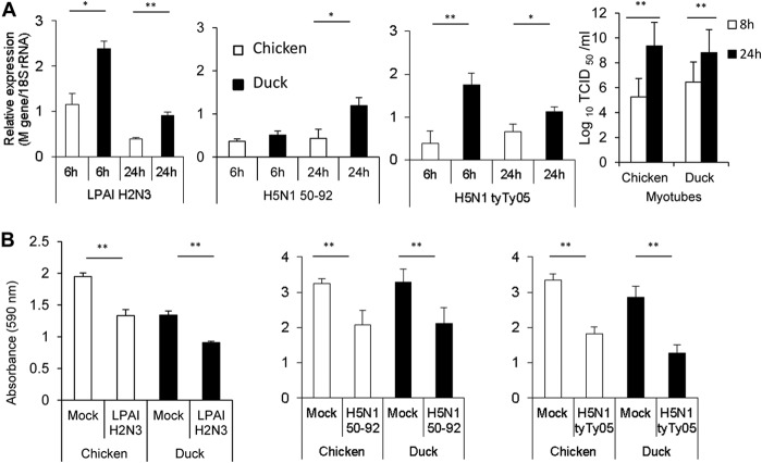
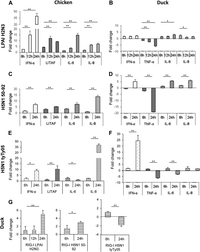
Skeletal muscle, at 30 to 40% of body mass, is the most abundant soft tissue in the body. Besides its primary function in movement and posture, skeletal muscle is a significant innate immune organ with the capacity to produce cytokines and chemokines and respond to proinflammatory cytokines. Little is known about the role of skeletal muscle during systemic influenza A virus infection in any host and particularly avian species. Here we used primary chicken and duck multinucleated myotubes to examine their susceptibility and innate immune response to influenza virus infections. Both chicken and duck myotubes expressed avian and human sialic acid receptors and were readily susceptible to low-pathogenicity (H2N3 A/mallard duck/England/7277/06) and high-pathogenicity (H5N1 A/turkey/England/50-92/91 and H5N1 A/turkey/Turkey/1/05) avian and human H1N1 (A/USSR/77) influenza viruses. Both avian host species produced comparable levels of progeny H5N1 A/turkey/Turkey/1/05 virus. Notably, the rapid accumulation of viral nucleoprotein and matrix (M) gene RNA in chicken and duck myotubes was accompanied by extensive cytopathic damage with marked myotube apoptosis (widespread microscopic blebs, caspase 3/7 activation, and annexin V binding at the plasma membrane). Infected chicken myotubes produced significantly higher levels of proinflammatory cytokines than did the corresponding duck cells. Additionally, in chicken myotubes infected with H5N1 viruses, the induction of interferon beta (IFN-β) and IFN-inducible genes, including the melanoma differentiation-associated protein 5 (MDA-5) gene, was relatively weak compared to infection with the corresponding H2N3 virus. Our findings highlight that avian skeletal muscle fibers are capable of productive influenza virus replication and are a potential tissue source of infection.

Importance: Infection with high-pathogenicity H5N1 viruses in ducks is often asymptomatic, and skeletal muscle from such birds could be a source of infection of humans and animals. Little is known about the ability of influenza A viruses to replicate in avian skeletal muscle fibers. We show here that cultured chicken and duck myotubes were highly susceptible to infection with both low- and high-pathogenicity avian influenza viruses. Infected myotubes of both avian species displayed rapid virus accumulation, apoptosis, and extensive cellular damage. Our results indicate that avian skeletal muscle fibers of chicken and duck could be significant contributors to progeny production of highly pathogenic H5N1 viruses.


Figures

FIG 1
FIG 1
Infected chicken and duck myotubes but not myoblasts show progressive accumulation of viral NP. (A to A″ and D to D′) At 6 h p.i. with LPAI H2N3 virus at a MOI of 0.1, almost all chicken and duck myotubes but only a few myoblasts were immunopositive for viral NP (brown). (B, B′, E, and E′) At 12 h p.i., more viral NP accumulated in chicken and duck myotubes, as evident by the intensity of NP detection. (C, C′, F, and F′) By 24 h p.i., the remaining attached chicken and duck myotubes showed even more intense NP expression, while few myoblasts were infected (C′, arrows). All cells were immunolabeled at the same time; differences in the intensity of labeling indicate different amounts of intracellular NP. Cells were counterstained with Harris' hematoxylin to visualize nuclei. (G and H) A similar NP expression outcome with human H1N1 (A/USSR/77) virus at a MOI of 0.1 was found for chicken and duck myotubes (data from 6 h p.i. are shown). (I and J) At a higher MOI of 1.0 with LPAI H2N3 virus, chicken and duck myotubes and myoblasts (arrows) showed comparable viral NP expression levels (data from 6 h p.i. are shown).
FIG 2
FIG 2
Infected duck myotubes show higher levels of accumulation of virus M gene RNA than do the corresponding myoblasts or MDCK cells. (A) Duck myotube, duck myoblast, and MDCK cell cultures were infected with LPAI H2N3 virus at a MOI of 0.1. Duck myotubes had significantly (P < 0.005) higher levels of intracellular viral M gene RNA (normalized to the 18S RNA gene) than did myoblasts and MDCK cells at 6 h, 12 h, and 24 h of infection. (B) Duck myotubes also accumulated the most viral M gene RNA in culture supernatants by 24 h of infection. Results show the means of data from three biological replicates, with error bars indicating standard deviations. One-way analysis of variance followed by Tukey's multiple-comparison test was used (**, P < 0.005). (C) Chicken and duck muscle cells (myotubes and myoblasts) and MDCK cells coexpressed avian and human sialic acid receptor types. The human α-2,6-linked sialic acid receptor (green) and avian α-2,3-linked sialic acid receptor (red) were detected with Sambucus nigra agglutinin (SNA) and Maackia amurensis agglutinin II (MAA II) lectins, respectively. Nuclei were counterstained by using DAPI (blue). Merged and individual fluorescent images show extensive expression of both receptors in all three cell types.
FIG 3
FIG 3
Avian influenza virus-infected chicken and duck myotubes show comparable progeny virus outputs and similar reductions in cell viability. (A) One LPAI H2N3 and two HPAI H5N1 viruses at a MOI of 1.0 conferred higher levels of accumulation of M gene RNA in duck than in chicken myotubes (*, P < 0.05; **, P < 0.005 [determined by an unpaired t test]); however, comparable increasing outputs of progeny H5N1 tyTy05 virus were detected for both avian species, based on TCID50 virus assays using infected supernatants on MDCK cells (*, P < 0.05 [determined by a two-sample t test]). (B) Chicken and duck myotubes infected at a MOI of 1.0 for 24 h displayed a significant reduction in cell viability based on MTT assays (**, P < 0.005 [determined by an unpaired t test]). There was no significant difference in reduced viability between infected chicken and infected duck myotubes. Data points are the means of data from four wells of a 96-well plate, with error bars indicating standard deviations.
FIG 4
FIG 4
Influenza virus-infected chicken and duck myotubes show extensive cytopathic damage. Chicken and duck myotubes were infected with LPAI H2N3 virus at a MOI of 1.0 for 24 h. (A and E) Uninfected control chicken and duck myotubes show extensive expression of the muscle-specific intermediate filament desmin (brown). (B and F) Cytopathic damage of chicken and duck myotubes is seen as widespread rounding and detachment of cells (arrows). (C, C′, G, and G′) Whole chicken and duck myotubes appear to have degenerated into numerous small membrane-lined blebs (apoptotic bodies) (myotube boundaries are delineated). (D, D′, H, and H′) Commonly, sections of chicken and duck myotubes contained aggregates of apoptotic blebs with concentrated viral NP, as evident by immunocytochemical NP detection. Morphologically intact myonuclei were found in the sarcoplasm of the duck myotube (H′, arrows). (I and K) Phase-contrast microscopy of mock-infected chicken (I) and duck (K) myotube cultures show typical myotubes. (J and L) In contrast, similar myotube detachment and rounding (arrows) were evident in HPAI H5N1 50-92 virus-infected (MOI of 1.0) chicken (J) and duck (L) myotubes at 24 h p.i. (M to P) Translocation of PS to the outer membrane was detected (green fluorescence) by annexin V-EGFP binding to chicken and duck muscle cells (myotubes and myoblasts) infected with LPAI H2N3 virus (MOI of 1.0) for 24 h. Infected chicken (M) and duck (N) myotubes were extensively positive for PS translocation; some cells had lost membrane integrity, as evident by the nuclear uptake of PI (red fluorescence). Myonuclei labeled with PI displayed chromatin condensation and/or fragmentation in chicken and duck myotubes, whose boundaries are outlined (M and N, respectively). A morphologically intact nucleus with condensed and/or fragmented nuclear contents is highlighted (M, arrows). Chromatin condensation and fragmentation were also seen in chicken (O) and duck (P) myoblasts. (Q) Chicken and duck myotube cultures infected with LPAI H2N3 virus at a MOI of 1.0 significantly activated the effector caspases 3 and 7 at 24 h and 48 h p.i. (*, P < 0.05; **, P < 0.01 [determined by an unpaired t test]) (RLU/s, relative light units per second). (Q′) Results are also represented as fold changes. There was, however, no significant difference in the activation of caspases 3 and 7 between the two avian species. Data points are the means of data from three wells from a 96-well plate, with error bars indicating standard deviations.
FIG 5
FIG 5
Chicken myotubes display a more vigorous cytokine response to avian influenza virus infection than do duck myotubes. (A and B) Chicken myotubes infected with a LPAI H2N3 virus showed a strong induction of IFN-α (40-fold induction) and a moderate induction of proinflammatory cytokines (LITAF, IL-6, and IL-8) (A); the corresponding duck muscle cells showed weaker induction, with progressive downregulation of the TNF-α response (B). (C and E) With HPAI H5N1 50-92 (C) or HPAI H5N1 tyTy05 (E) virus, the level of IFN-α mRNA induction was not >10-fold, while IL-8 mRNA was more strongly induced in chicken than in duck muscle cells. (D and F) In duck cells, TNF-α gene transcription was downregulated by both HPAI H5N1 viruses, unlike LITAF gene induction in the corresponding chicken cells (C and E). Duck IFN-α mRNA induction was greater with HPAI H5N1 tyTy05 virus (F) than with HPAI H5N1 50-92 virus (D). (G) Duck myotubes infected with LPAI H2N3 and HPAI H5N1 50-92 viruses showed modest upregulation of the viral RNA sensor RIG-I, but with HPAI H5N1 tyTy05 virus, RIG-I expression was downregulated. mRNA levels were normalized to the 18S rRNA gene and are expressed as fold changes in relation to uninfected controls at each p.i. time point. The fold change for each gene is the mean of data from three biological replicates, with error bars indicating standard deviations. A significant increase or decrease in mRNA levels between 6 h p.i. and later times of infection was determined by a two-sample unpaired t test (*, P < 0.05; **, P < 0.01).
FIG 6
FIG 6
LPAI H2N3 virus induces a more vigorous antiviral response than do HPAI H5N1 viruses in chicken myotubes. (A) Chicken myotubes infected with LPAI H2N3 virus at a MOI of 1.0 induced a strong upregulation of IFN-β, which correlated with the upregulation of the viral RNA sensor MDA-5 as well as the IFN-inducible Mx1, 2′,5′-OAS, and PKR genes. (B and C) However, with HPAI H5N1 50-92 (B) and HPAI H5N1 tyTy05 (C) viruses, both at a MOI of 1.0, the level of IFN-β mRNA induction was <20-fold, considerably lower than that for the corresponding LPAI H2N3 virus infection (A). In contrast to LPAI H2N3 virus infection (A), HPAI H5N1 50-92 (B) and HPAI H5N1 tyTy05 (C) viruses at 24 h p.i. downregulated the expression of the MDA-5 and PKR genes and elicited much weaker induction of Mx1 and 2′,5′-OAS gene expression. mRNA levels were normalized to the 18S rRNA gene and are expressed as fold changes in relation to uninfected controls at each p.i. time point. The fold change for each gene is the mean of data from three biological replicates, with error bars indicating standard deviations. A significant increase or decrease in mRNA levels between 6 h p.i. and later times of infection was calculated by a two-sample unpaired t test (*, P < 0.05; **, P < 0.01).

Similar articles

Cited by

References

    1. Chan PKS. 2002. Outbreak of avian influenza A(H5N1) virus infection in Hong Kong in 1997. Clin Infect Dis 34:S58–S64. doi:10.1086/338820. - DOI - PubMed
    1. Guan Y, Peiris JSM, Lipatov AS, Ellis TM, Dyrting KC, Krauss S, Zhang LJ, Webster RG, Shortridge KF. 2002. Emergence of multiple genotypes of H5N1 avian influenza viruses in Hong Kong SAR. Proc Natl Acad Sci U S A 99:8950–8955. doi:10.1073/pnas.132268999. - DOI - PMC - PubMed
    1. Peiris JSM, de Jong MD, Guan Y. 2007. Avian influenza virus (H5N1): a threat to human health. Clin Microbiol Rev 20:243–267. doi:10.1128/CMR.00037-06. - DOI - PMC - PubMed
    1. Guan Y, Smith GJD, Webby R, Webster RG. 2009. Molecular epidemiology of H5N1 avian influenza. Rev Sci Tech 28:39–47. - PubMed
    1. Sims LS, Brown IH. 2008. Multi-continental epizootics of H5N1 highly pathogenic avian influenza 1996–2007, p 251–286 InSwayne DE. (ed), Avian influenza. Blackwell Publishing, Oxford, United Kingdom.

Publication types

MeSH terms