. 2015 Feb 15;26(4):726-39.
doi: 10.1091/mbc.E14-08-1303. Epub 2014 Dec 24.

Seipin performs dissectible functions in promoting lipid droplet biogenesis and regulating droplet morphology

Affiliations

Seipin performs dissectible functions in promoting lipid droplet biogenesis and regulating droplet morphology

Bethany R Cartwright et al. Mol Biol Cell. .

Abstract

Seipin is necessary for both adipogenesis and lipid droplet (LD) organization in nonadipose tissues; however, its molecular function is incompletely understood. Phenotypes in the seipin-null mutant of Saccharomyces cerevisiae include aberrant droplet morphology (endoplasmic reticulum-droplet clusters and size heterogeneity) and sensitivity of droplet size to changes in phospholipid synthesis. It has not been clear, however, whether seipin acts in initiation of droplet synthesis or at a later step. Here we utilize a system of de novo droplet formation to show that the absence of seipin results in a delay in droplet appearance with concomitant accumulation of neutral lipid in membranes. We also demonstrate that seipin is required for vectorial budding of droplets toward the cytoplasm. Furthermore, we find that the normal rate of droplet initiation depends on 14 amino acids at the amino terminus of seipin, deletion of which results in fewer, larger droplets that are consistent with a delay in initiation but are otherwise normal in morphology. Importantly, other functions of seipin, namely vectorial budding and resistance to inositol, are retained in this mutant. We conclude that seipin has dissectible roles in both promoting early LD initiation and in regulating LD morphology, supporting its importance in LD biogenesis.


Figures

FIGURE 1:
FIGURE 1:
Seipin knockout drastically impedes de novo droplet formation. A galactose-inducible promoter was integrated into the genomic DGA1 locus in a strain lacking the other three NL acyltransferases (3KO(GALDGA1)) and in a strain with an additional seipin knockout (3KO(GALDGA1)fld1∆). Cells were introduced to rich galactose medium at t = 0 to induce droplet formation and stained with BODIPY at the indicated time points. (A) Representative fluorescence microscopy projection images at indicated time points after galactose induction, overlaid onto bright-field images. Scale bar: 5 μm. (B) Image of typical cells from each strain at 3 h. Scale bar: 2 μm. Intensity settings were kept constant between the two images to compare membrane brightness. (C) Percent of cells containing at least one FB. Error bars signify range from two experiments. (D) Number of distinct FBs per cells that have at least one FB. Error bars represent SDs from two experiments. (E) TG levels determined by TLC of lipid extracts from whole-cell lysates, normalized to cell pellet wet weight. Error bars represent range from 2 independent experiments. (F) TG synthesis activity of isolated membranes. Error bars signify SDs from two experiments. (G) Growth of cultures.  Starting OD was 0.3. Error bars, SEM from two experiments, each performed in duplicate.
FIGURE 2:
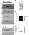
FIGURE 2:
In the absence of seipin, TG accumulates in the ER during de novo LD formation. 3KO(GALDGA1) and 3KO(GALDGA1)fld1∆ cells were induced in rich galactose medium for 6 h, lysed, and fractionated. (A and C) Enriched ER membranes were centrifuged through a sucrose gradient, and fractions were blotted with α-Dpm1p (ER) or α-porin antibody. “Both” indicates a mixture of isolated membranes (4:1 3KO(GALDGA1) to 3KO(GALDGA1)fld1∆ membranes by protein) from both strains; “wt” indicates the parental W303-1A strain. Representative blots for seipin from one of three independent experiments shown for 3KO(GALDGA1) and 3KO(GALDGA1)fld1∆ cells, and one of two independent experiments for porin. (B) Droplets and membranes from cells expressing Erg6-mRed and Sec63-GFP were separated and probed with α-Dpm1p or α-GFP; identical percents of LD and membrane fraction were analyzed. Parallel samples from cells expressing Erg6-DsRed were probed with α-DsRed. (Sec63-GFP interfered with the α-Erg6p signal.) Dpm1p is exclusively in the membrane fraction. (D) TG levels in the PNS fractions are compared in the strains indicated. (E) TG levels in the 3KO(GALDGA1) strain are compared with the 3KO(GALDGA1)fld1∆ strain in isolated droplets and enriched ER. The results of two experiments are shown in which organelles from the two strains were prepared in parallel. “100%” signifies the ratio of TG in the LD or ER fraction relative to TG in the PNS in the 3KO(GALDGA1) strain. Absolute values of TG (expressed as micrograms of trioleoyl glycerol equivalents) in the PNS and purified LDs are 697 and 88.9, respectively, in one experiment, and 263 and 52.4, respectively, for the other. Absolute values in the PNS and purified ER are 377 and 19.2, respectively, for one experiment, and 288 and 25.2, respectively, for the other. Comparison of these values with those from the 3KO(GALDGA1)fld1∆ strains reveals 36.5 and 40.2% (in the two experiments) as much TG in LDs from the seipin-minus strain, and 144 and 143% as much TAG in ER from the seipin-minus strain.
FIGURE 3:
FIGURE 3:
Deletion of 14 amino acids from the seipin N-terminus results in an SLD phenotype. Seipin knockout cells (fld1∆) were complemented with plasmids overexpressing FLD1, fld1∆Nterm, or empty vector and cultured in rich oleate medium. (A) Representative bright-field or fluorescence microscopy projection images after staining with BODIPY to visualize LDs. Scale bar: 5 μm. (B) Number of FBs per total number of cells. (C) Percent of cells displaying one or more SLDs (defined as >1 μm diameter). For B and C, error bars represent SEMs from four independent experiments; for each, n = 100 cells from at least three fields. (D–F) Analysis of electron micrographs (sample images in Figure 4A); error bars represent SEMs from 100 cells. (D) Number of LDs per total number of cells. (E) Number of SLDs. (F) Number of LD clusters (defined as >5 adjacent droplets). (G) Phospholipid to NL ratios of isolated LD fractions, analyzed by TLC. (H) Phospholipid levels of whole-cell lysates by TLC, normalized to cell pellet wet weight. (I) Neutral lipid levels of whole-cell lysates as in H. (G–I) Error bars represent SEMs from four independent experiments. *, p < 0.05; **, p < 0.01; ***, p < 0.001; ****, p < 0.0001 by one-way analysis of variance (ANOVA) with correction for multiple comparisons.
FIGURE 4:
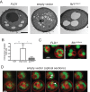
FIGURE 4:
Nuclear LDs in the absence of seipin. Cells lines and culture conditions as in Figure 3. (A) Representative electron micrographs. Scale bar: 1 µm. (B) Number of intranuclear droplets (defined as within an observable, intact nuclear envelope). Error bars represent SEMs from ∼30 cells sectioned at the level of a visible, intact nuclear envelope. (C and D) Cell lines also contained overexpressed CFP-HDEL to visualize the ER (false-colored red) and stained with BODIPY (green). (C) Fluorescence microscopy of fld1∆ cells containing FLD1 or fld1ΔNterm as indicated. (D) Fluorescence microscopy of consecutive 0.3-μm z-sections of fld1∆ cells with empty vector. Arrowheads, intranuclear LDs.
FIGURE 5:
FIGURE 5:
The N-terminal seipin-deletion phenotype is resistant to manipulation by inositol. Seipin knockout cells (fld1∆) complemented with plasmids overexpressing empty vector, FLD1, or fld1∆Nterm, and the ER marker CFP-HDEL were grown in minimal glucose medium with or without the indicated PL precursor supplements. (A) Representative fluorescence microscopy projection images after staining with BODIPY. Scale bar: 5 μm. CFP-HDEL is false-colored red in the merged images. Arrowhead indicates an LD–ER tangle, defined as an irregular BODIPY body colocalized with a similarly shaped ER density. (B and D) Percent of cells displaying SLDs. (C and E) Percent of cells displaying LD–ER tangles. Error bars for B–E represent SEMs from three independent experiments; for each, n = 100 cells from at least three fields. *, p < 0.05; **, p < 0.01; ***, p < 0.001; ****, p < 0.0001 by two-way ANOVA with correction for multiple comparisons.
FIGURE 6:
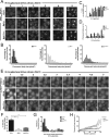
FIGURE 6:
N-terminal seipin deletion results in an initiation defect during de novo LD formation. Genomic knock-ins were generated at the seipin locus in the 3KO(GALDGA1) background for FLD1, fld1∆, and fld1∆Nterm. Cells were switched from raffinose to galactose media at t = 0 to induce droplet formation and stained with BODIPY. (A) Representative fluorescence microscopy projection images at indicated time points after galactose induction. Scale bars: 5 µm. (B) Histograms of FB size, given as area in pixels on a maximum-intensity projection image. (C) Percent of cells containing at least one FB. (D) Number of distinct FBs per cells that have at least one. Error bars in C and D represent SEMs from three independent experiments, each N = 100 cells from at least three fields. *, p < 0.05; #, p < 0.01; formula image, p < 0.0001 by one-way ANOVA with correction for multiple comparisons. (E) Time-lapse fluorescence microscopy of cells embedded into agar after 1 h of galactose induction in liquid culture at 27–29°C. Images taken in 10-min increments (see Supplemental Videos S1–S3); representative projections of 30-min increment montages are shown. Note that cells were precultured at high density to prevent division during imaging. (F) Percent of cells that displayed at least one FB over the course of the time lapse. Error bars represent SEMs from three independent experiments. ***, p < 0.001 by one-way ANOVA with correction for multiple comparisons. (G) Histogram of time to first appearance for each droplet. (H) Average intensity curves for FBs during the time lapse. Time 0 defined as the frame before first appearance of the droplet. Error bars represent SEMs from average droplet intensity values per time point over three independent experiments. Absolute intensity values are likely underestimated due to bleaching. Scale bars: 5 µm

References

    1. Adeyo O, Horn PJ, Lee S, Binns DD, Chandrahas A, Chapman KD, Goodman JM. The yeast lipin orthologue Pah1p is important for biogenesis of lipid droplets. J Cell Biol. 2011;192:1043–1055. - PMC - PubMed
    1. Agarwal AK, Garg A. Genetic basis of lipodystrophies and management of metabolic complications. Annu Rev Med. 2006;57:297–311. - PubMed
    1. Bi J, Wang W, Liu Z, Huang X, Jiang Q, Liu G, Wang Y, Huang X. Seipin Promotes adipose tissue fat storage through the ER Ca2+-ATPase SERCA. Cell Metab. 2014;19:861–871. - PubMed
    1. Binns D, Januszewski T, Chen Y, Hill J, Markin VS, Zhao Y, Gilpin C, Chapman KD, Anderson RG, Goodman JM. An intimate collaboration between peroxisomes and lipid bodies. J Cell Biol. 2006;173:719–731. - PMC - PubMed
    1. Binns D, Lee S, Hilton CL, Jiang QX, Goodman JM. Seipin is a discrete homooligomer. Biochemistry. 2010;49:10747–10755. - PMC - PubMed

Publication types

MeSH terms

Substances