Histological Analysis of γδ T Lymphocytes Infiltrating Human Triple-Negative Breast Carcinomas
- PMID: 25540645
- PMCID: PMC4261817
- DOI: 10.3389/fimmu.2014.00632
Histological Analysis of γδ T Lymphocytes Infiltrating Human Triple-Negative Breast Carcinomas
Abstract
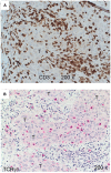
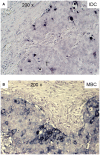
Breast cancer is the leading cause of cancer death in women and the second most common cancer worldwide after lung cancer. The remarkable heterogeneity of breast cancers influences numerous diagnostic, therapeutic, and prognostic factors. Triple-negative breast carcinomas (TNBCs) lack expression of HER2 and the estrogen and progesterone receptors and often contain lymphocytic infiltrates. Most of TNBCs are invasive ductal carcinomas (IDCs) with poor prognosis, whereas prognostically more favorable subtypes such as medullary breast carcinomas (MBCs) are somewhat less frequent. Infiltrating T-cells have been associated with an improved clinical outcome in TNBCs. The prognostic role of γδ T-cells within CD3(+) tumor-infiltrating T lymphocytes remains unclear. We analyzed 26 TNBCs, 14 IDCs, and 12 MBCs, using immunohistochemistry for the quantity and patterns of γδ T-cell infiltrates within the tumor microenvironment. In both types of TNBCs, we found higher numbers of γδ T-cells in comparison with normal breast tissues and fibroadenomas. The numbers of infiltrating γδ T-cells were higher in MBCs than in IDCs. γδ T-cells in MBCs were frequently located in direct contact with tumor cells, within the tumor and at its invasive border. In contrast, most γδ T-cells in IDCs were found in clusters within the tumor stroma. These findings could be associated with the fact that the patient's prognosis in MBCs is better than that in IDCs. Further studies to characterize these γδ T-cells at the molecular and functional level are in progress.
Keywords: breast cancer; histology; paraffin material; triple-negative breast cancer; γδ T-cells.
Figures


References
-
- Ferlay J, Soerjomataram I, Ervik M, Dikshit R, Eser S, Mathers C, et al. GLOBOCAN 2012 v1.0, Cancer Incidence and Mortality Worldwide: IARC Cancer Base No. 11 [Internet]. Lyon: International Agency for Research on Cancer; (2013). Available from: http://globocan.iarc.fr
-
- Loi S, Michiels S, Salgado R, Sirtaine N, Jose V, Fumagalli D, et al. Tumor infiltrating lymphocytes are prognostic in triple negative breast cancer and predictive for trastuzumab benefit in early breast cancer: results from the FinHER trial. Ann Oncol (2014) 25:1544–50.10.1093/annonc/mdu112 - DOI - PubMed
-
- Adams S, Gray R, Demaria S, Goldstein LJ, Perez EA, Shulman LN, et al. Prognostic value of tumor-infiltrating lymphocytes in triple-negative breast cancers from two phase III randomized adjuvant breast cancer trials: ECOG 2197 and ECOG 1199. J Clin Oncol (2014) 32:2959–66.10.1200/JCO.2013.55.0491 - DOI - PMC - PubMed
LinkOut - more resources
Full Text Sources
Other Literature Sources
Research Materials
Miscellaneous

