

Prevalence of tumor-infiltrating lymphocytes and PD-L1 expression in the soft tissue sarcoma microenvironment
- PMID: 25540867
- PMCID: PMC5505649
- DOI: 10.1016/j.humpath.2014.11.001
Prevalence of tumor-infiltrating lymphocytes and PD-L1 expression in the soft tissue sarcoma microenvironment
Abstract
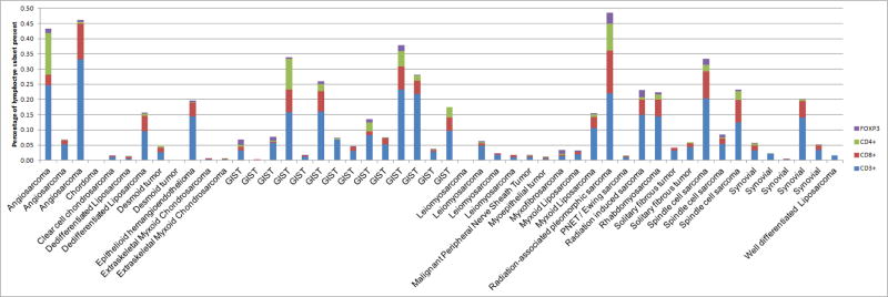
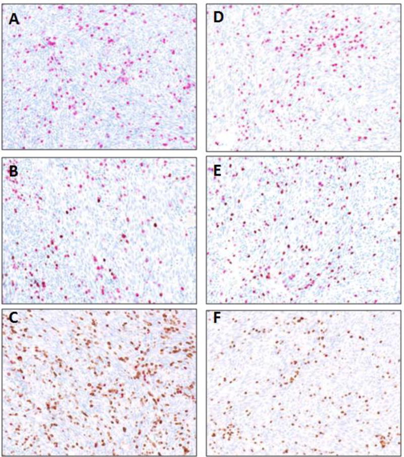
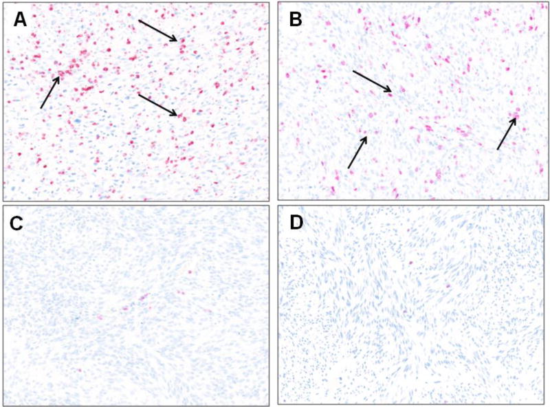
The prognostic and predictive implications of programmed death-ligand 1 (PD-L1) is unknown in sarcoma. We sought to examine the immune milieu in sarcoma specimens. We evaluated PD-L1 expression by immunohistochemistry in sarcoma specimens and quantified tumor-infiltrating lymphocytes (TIL). We correlated expression with clinical parameters and outcomes. Fifty sarcoma patients treated at Memorial Sloan Kettering Cancer Center were selected. Using the DAKO PD-L1 immunohistochemistry assay and archival formalin-fixed paraffin-embedded tissue specimens; PD-L1 expression was examined. Macrophage and lymphocyte PD-L1 status was determined qualitatively. TIL was quantified. Associations between PD-L1 expression in tumor, macrophages and lymphocytes, TIL and clinical-pathological characteristics were performed. The median age was 46 years (range, 22-76), and 66% of patients were men. Tumor, lymphocyte and macrophage PD-L1 expression was noted in 12%, 30% and 58%, respectively, with the highest prevalence in gastrointestinal stromal tumors (29%). Lymphocyte and macrophage infiltration was present in 98% and 90%, respectively. There was no association between clinical features, overall survival and PD-L1 expression in tumor or immune infiltrates. Lymphocyte and macrophage infiltration is common in sarcoma, but PD-L1 tumor expression is uncommon in sarcoma with the highest frequency observed in gastrointestinal stromal tumors. There was no association between PD-L1 expression, TIL and clinicopathological features and overall survival; however, this is limited by the heterogenous patient sample and minimal death events in the studied cohort.
Keywords: Immunotherapy; PD-1; PD-L1; Sarcoma; Tumor infiltrating lymphocyte CD3+, CD4+, CD8+, FOXP3+.
Copyright © 2015 Elsevier Inc. All rights reserved.
Figures
References
-
- Brennan MFAC, Maki RG. Management of Soft Tissue Sarcoma. New York: Springer; 2012.
-
- Rusakiewicz S, Semeraro M, Sarabi M, et al. Immune infiltrates are prognostic factors in localized gastrointestinal stromal tumors. Cancer Res. 2013;73:3499–3510. - PubMed
-
- Berghuis D, Santos SJ, Baelde HJ, et al. Pro-inflammatory chemokine-chemokine receptor interactions within the Ewing sarcoma microenvironment determine CD8(+) T-lymphocyte infiltration and affect tumour progression. J Pathol. 2011;223:347–357. - PubMed
-
- Zhang L, Zhao Y. The regulation of Foxp3 expression in regulatory CD4(+)CD25(+)T cells: multiple pathways on the road. J Cell Physiol. 2007;211:590–597. - PubMed
Publication types
MeSH terms
Substances
Grants and funding
LinkOut - more resources
Full Text Sources
Other Literature Sources
Medical
Research Materials
