Review
. 2015 Feb;36(1):65-91.
doi: 10.1210/er.2014-1079. Epub 2014 Dec 26.

Stem cells, progenitor cells, and lineage decisions in the ovary

Affiliations
Review

Stem cells, progenitor cells, and lineage decisions in the ovary

Katja Hummitzsch et al. Endocr Rev. 2015 Feb.

Abstract

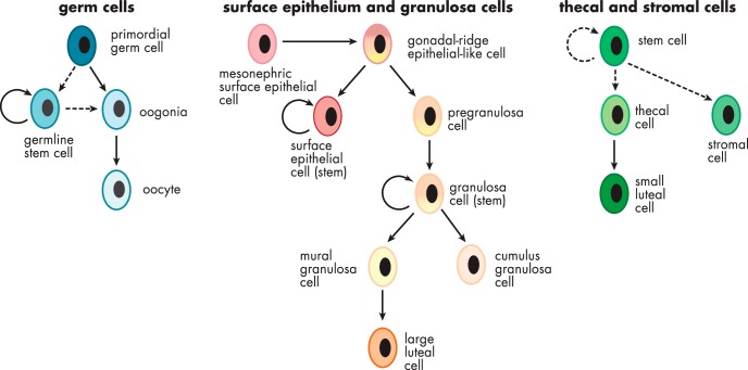
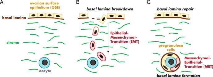
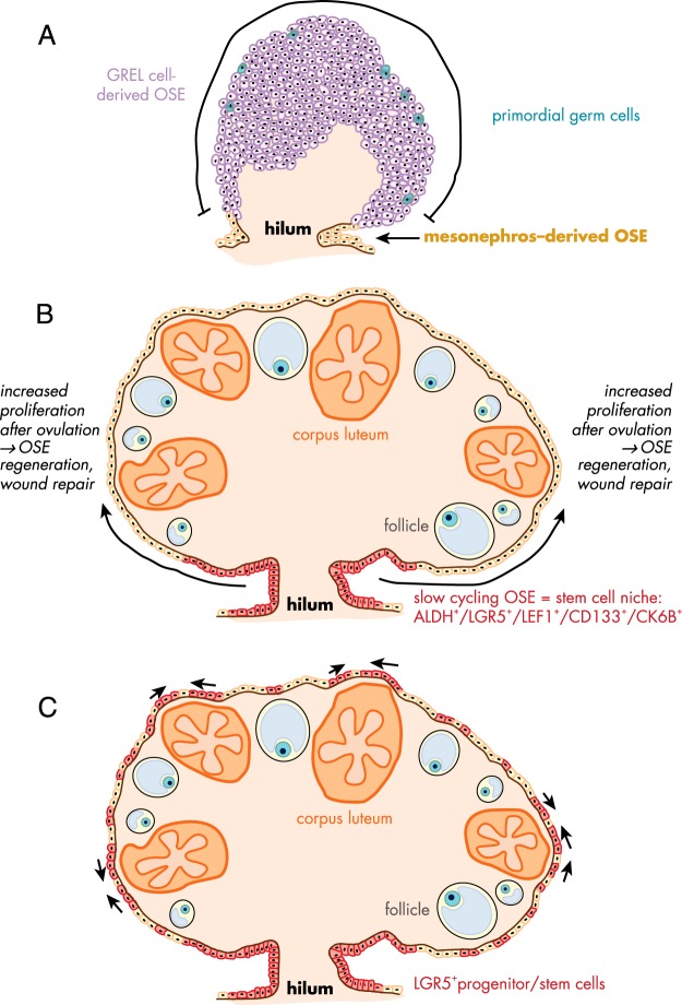
Exploring stem cells in the mammalian ovary has unleashed a Pandora's box of new insights and questions. Recent evidence supports the existence of stem cells of a number of the different cell types within the ovary. The evidence for a stem cell model producing mural granulosa cells and cumulus cells is strong, despite a limited number of reports. The recent identification of a precursor granulosa cell, the gonadal ridge epithelial-like cell, is exciting and novel. The identification of female germline (oogonial) stem cells is still very new and is currently limited to just a few species. Their origins and physiological roles, if any, are unknown, and their potential to produce oocytes and contribute to follicle formation in vivo lacks robust evidence. The precursor of thecal cells remains elusive, and more compelling data are needed. Similarly, claims of very small embryonic-like cells are also preliminary. Surface epithelial cells originating from gonadal ridge epithelial-like cells and from the mesonephric epithelium at the hilum of the ovary have also been proposed. Another important issue is the role of the stroma in guiding the formation of the ovary, ovigerous cords, follicles, and surface epithelium. Immune cells may also play key roles in developmental patterning, given their critical roles in corpora lutea formation and regression. Thus, while the cellular biology of the ovary is extremely important for its major endocrine and fertility roles, there is much still to be discovered. This review draws together the current evidence and perspectives on this topic.


Figures

Figure 1.
Figure 1.
Schematic diagram illustrating the potential and known cell lineages of the ovary.
Figure 2.
Figure 2.
Localization of extracellular matrix components in the stroma of fetal and adult bovine ovaries. A and B, Localization of fibrillin 3 (green) with the stromal cell marker COUP-TFII/NR2F2 (red) in fetal ovaries at gestational days 96 (A) and 182 (B). C, Fibrillin 1 expression (red) and components of laminin 111 (green) in fetal ovary at gestational day 79. D, Fibrillin 1 (green) localization in adult ovary. E, Localization of decorin fibers (green) and components of laminin 111 (red) in fetal ovary at gestational day 79. F, Localization of decorin fibers (green) in adult ovary. Nuclei are counterstained with DAPI (4',6-diamidino-2-phenylindole; blue). Scale bars, A and D, 25 μm; B, C, E, and F, 50 μm. [Panels A and B were reproduced from K. Hummitzsch et al: A new model of development of the mammalian ovary and follicles. PloS One. 2013;8:e55578 (31), with permission, and staining in panels C, E, and F was conducted as reported previously in the same article. Panel D was reproduced from M. J. Prodoehl et al: Fibrillins and latent TGFβ binding proteins in bovine ovaries of offspring following high or low protein diets during pregnancy of dams. Mol Cell Endocrinol. 2009;307:133–141 (98), with permission. Elsevier.]
Figure 3.
Figure 3.
Illustration of the conceptual processes needed to derive granulosa cells from the ovarian surface epithelium. There has been no discussion of these processes in the literature or any evidence to identify that they occur. Surface epithelial cells with a basal lamina and stromal interface (A) would first need to undergo an epithelial-mesenchymal transition (B) to break through the surface epithelial basal lamina and to become migratory and migrate to the oogonium (B). They would then need to undergo a mesenchymal-epithelial transition to form epithelial granulosa cells of follicles all enclosed by the follicular basal lamina (C).
Figure 4.
Figure 4.
Schematic diagram of ovarian development. [Reproduced from K. Hummitzsch et al: A new model of development of the mammalian ovary and follicles. PloS One. 2013;8:e55578 (31), with permission.]. Abbreviations: CK19, cytokeratin 19; DAPI, 4',6-diamidino-2-phenylindole.
Figure 5.
Figure 5.
Schematic diagram of the proposed development and repair of ovarian surface epithelium. A, In early fetal ovarian development, the ovarian surface epithelium (OSE) in the hilum region is derived from the mesonephros, whereas the remaining surface of ovary is covered by GREL cells, which later differentiate to surface epithelial cells or granulosa cells. [Adapted from K. Hummitzsch et al: A new model of development of the mammalian ovary and follicles. PloS One. 2013;8:e55578 (31).] B, Flesken-Nikitin et al identified an ALDH-, LGR5-, LEF-1-, CD133-, and CK6B-expressing stem cell niche in the hilum region of adult mice ovaries, which is responsible for the OSE repair after ovulation and is susceptible to malignant transformation. [Adapted from A. Flesken-Nikitin et al: Ovarian surface epithelium at the junction area contains a cancer-prone stem cell niche. Nature. 2013;495:241–245 (136).] C, A recent study in adult mice identified LGR5-positive OSE stem cells not only in the hilum region but also along the remaining ovarian surface as small clusters, mainly near ovulating follicles and on the apical side of corpora lutea. [Adapted from A. Ng et al: Lgr5 marks stem/progenitor cells in ovary and tubal epithelia. Nat Cell Biol. 2014;16:745–757 (137).] Abbreviations: ALDH, aldehyde dehydrogenase; LEF1, lymphoid enhancer-binding factor 1; CD133, cluster of differentiation 133; CK6B, cytokeratin 6B.
Figure 6.
Figure 6.
Morphological changes of the granulosa cells and the granulosa layer during follicle development. [Adapted from data derived from P. Da Silva-Buttkus et al: Effect of cell shape and packing density on granulosa cell proliferation and formation of multiple layers during early follicle development in the ovary. J Cell Sci. 2008;121:3890–3900 (271) and from R. J. Rodgers and H. F. Irving-Rodgers: Morphological classification of bovine ovarian follicles. Reproduction. 2010;139:309–318 (272).]

References

    1. Hsueh AJ, Kawamura K, Cheng Y, Fauser BC. Intraovarian control of early folliculogenesis [published online September 9, 2014]. Endocr Rev. doi.org/10.1210/er.2014–1020. - DOI - PMC - PubMed
    1. Matsuda F, Inoue N, Manabe N, Ohkura S. Follicular growth and atresia in mammalian ovaries: regulation by survival and death of granulosa cells. J Reprod Dev. 2012;58:44–50. - PubMed
    1. Scaramuzzi RJ, Baird DT, Campbell BK, et al. Regulation of folliculogenesis and the determination of ovulation rate in ruminants. Reprod Fertil Dev. 2011;23:444–467. - PubMed
    1. Chaves RN, de Matos MH, Buratini J, Jr, de Figueiredo JR. The fibroblast growth factor family: involvement in the regulation of folliculogenesis. Reprod Fertil Dev. 2012;24:905–915. - PubMed
    1. Chaves RN, Alves AM, Lima LF, Matos HM, Rodrigues AP, Figueiredo JR. Role of nerve growth factor (NGF) and its receptors in folliculogenesis. Zygote. 2013;21:187–197. - PubMed

Publication types