. 2014 Dec 26;9(12):e115963.
doi: 10.1371/journal.pone.0115963. eCollection 2014.

Impact of aging on the regenerative properties of bone marrow-, muscle-, and adipose-derived mesenchymal stem/stromal cells

Affiliations

Impact of aging on the regenerative properties of bone marrow-, muscle-, and adipose-derived mesenchymal stem/stromal cells

Olivia S Beane et al. PLoS One. .

Abstract

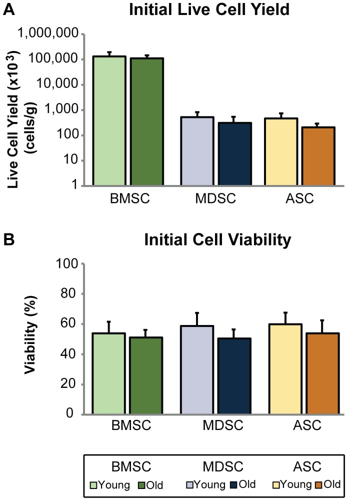
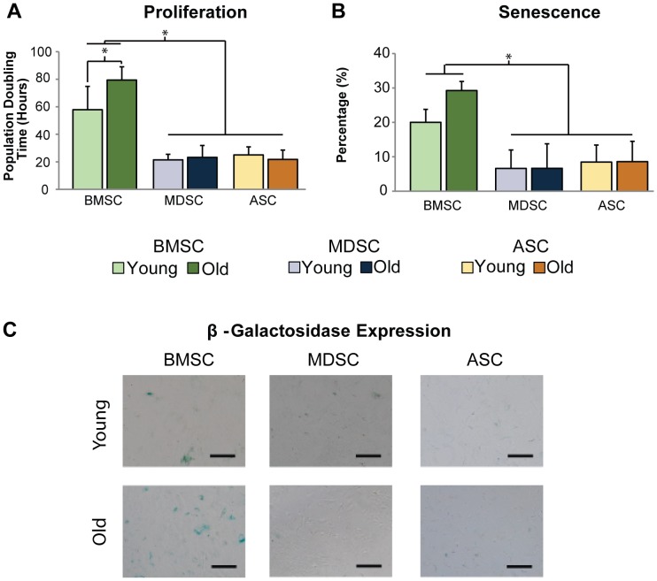
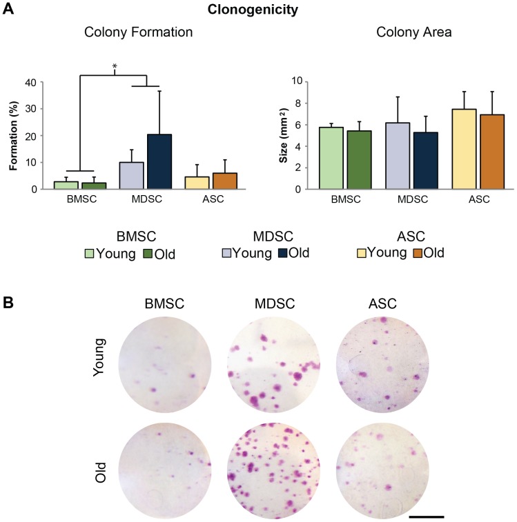
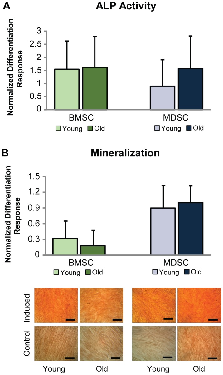
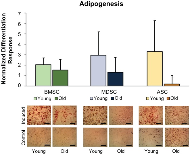
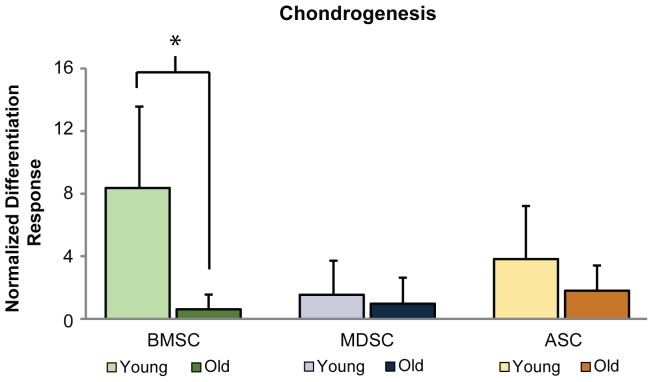
Mesenchymal stem/stromal cells (MSCs) are promising cell sources for regenerative therapies due to their multipotency and ready availability, but their application can be complicated by patient-specific factors like age or illness. MSCs have been investigated for the treatment of many musculoskeletal disorders, including osteoarthritis and osteoporosis. Due to the prevalence of these diseases in older populations, researchers have studied how aging affects MSC properties and have found that proliferation and differentiation potential are impaired. However, these effects have never been compared among MSCs isolated from multiple tissue sources in the same, healthy donor. Revealing differences in how MSCs are affected by age could help identify an optimal cell source for musculoskeletal therapies targeting older patients. MSCs were isolated from young and old rabbit bone marrow, muscle, and adipose tissue. Cell yield and viability were quantified after isolation procedures, and expansion properties were assessed using assays for proliferation, senescence, and colony formation. Multipotency was also examined using lineage-specific stains and spectrophotometry of metabolites. Results were compared between age groups and among MSC sources. Results showed that MSCs are differentially influenced by aging, with bone marrow-derived stem cells having impaired proliferation, senescence, and chondrogenic response, whereas muscle-derived stem cells and adipose-derived stem cells exhibited no negative effects. While age reduced overall cell yield and adipogenic potential of all MSC populations, osteogenesis and clonogenicity remained unchanged. These findings indicate the importance of age as a factor when designing cell-based therapies for older patients.


Conflict of interest statement

Competing Interests: The authors have declared that no competing interests exist.

Figures

Figure 1
Figure 1. MSC yield and viability following isolation.
Age adversely affected initial cell yield and viability of MSCs. After cell isolation, initial cell yield and viability were quantified. (A) Live cell counts were normalized to harvested tissue mass, with results indicating that cell yield was significantly reduced based on age as a factor (p<0.05). However, individual comparisons within tissue source showed no significant differences between age groups. BMSC yields were significantly larger than MDSC and ASC yields (p<0.001). (B) While viability was also reduced across MSC types, this trend did not quite reach significance (p = 0.051). Error bars depict standard deviations.
Figure 2
Figure 2. Cell expansion properties.
Characterization of MSC expansion properties revealed differences in age-related changes across MSC populations. (A) PDTs of old BMSCs were significantly longer than young BMSC PDTs (p<0.005). However, no differences were observed between age groups for MDSCs or ASCs. Additionally, BMSC PDTs were significantly longer than MDSCs and ASCs (p<0.001). (B) Quantitative and (C) qualitative assessment of β-galactosidase staining showed that senescence was greater in old BMSC populations than young, although this trend did not reach significance (p = 0.095). MDSC and ASC senescence was not affected by age. Statistical analysis also determined that senescence was significantly greater in BMSC populations than MDSCs and ASCs (p<0.001). Error bars depict standard deviations. (Scale bars  = 100 µm).
Figure 3
Figure 3. Clonogenicity of MSC populations.
Quantification of colony formation and area indicated that age had no universal effect on MSCs for either parameter. However, MDSC colony formation was significantly higher than BMSC formation (p<0.005). Error bars depict standard deviations. (Scale bars  = 25 mm).
Figure 4
Figure 4. MSC osteogenic differentiation.
Age did not adversely affect BMSC or MDSC osteogenesis. (A) Control-normalized differentiation response of ALP activity after 7 days of induction was comparable between age groups for BMSCs and MDSCs. (B) Calcified matrix production was examined quantitatively and qualitatively with ARS stain after 21 days of induction. Results were similar between age groups for BMSCs and MDSCs. ASC osteogenic differentiation was not characterized since cells became contractile upon induction and balled up, preventing accurate assessment. Error bars depict standard deviations. (Scale bars  = 100 µm).
Figure 5
Figure 5. MSC adipogenic differentiation. Old age impaired the adipogenic potential of BMSCs, MDSCs, and ASCs.
Quantification of eluted ORO stain revealed significant decreases in the overall, control-normalized differentiation response based on age as a factor (p<0.05). However, individual comparisons within tissue sources showed no significant differences between age groups. Qualitatively, staining showed that lipid production of induced samples was reduced for old BMSCs and MDSCs compared with their young counterparts. No readily apparent, age differences were observed for these cells in control conditions. Conversely, lipid production in old ASCs was noticeably greater than young ASCs in control conditions, while induced samples were similar. Error bars depict standard deviations. (Scale bars  = 100 µm).
Figure 6
Figure 6. MSC chondrogenic differentiation.
Chondrogenesis was reduced with age for BMSCs, but not for MDSCs or ASCs. Quantification of sGAG production showed that the control-normalized differentiation response of old BMSCs was significantly lower than young BMSCs (p<0.001). Conversely, no significant differences were observed between age groups for MDSCs or ASCs. Error bars depict standard deviations.

References

    1. Antebi B, Pelled G, Gazit D (2014) Stem cell therapy for osteoporosis. Curr Osteoporos Rep 12:41–47. - PubMed
    1. Gupta PK, Das AK, Chullikana A, Majumdar AS (2012) Mesenchymal stem cells for cartilage repair in osteoarthritis. Stem Cell Res Ther 3:25. - PMC - PubMed
    1. Lim YW, Kim YS, Lee JW, Kwon SY (2013) Stem cell implantation for osteonecrosis of the femoral head. Exp Mol Med 45:e61. - PMC - PubMed
    1. Pate DW, Southerland SS, Grande DA, Young HE, Lucas PA (1993) Isolation and Differentiation of Mesenchymal Stem-Cells from Rabbit Muscle. Clinical Research 41:A347–A347.
    1. Gharaibeh B, Lu A, Tebbets J, Zheng B, Feduska J, et al. (2008) Isolation of a slowly adhering cell fraction containing stem cells from murine skeletal muscle by the preplate technique. Nat Protoc 3:1501–1509. - PubMed

Publication types