Optical coherence tomography and autofluorescence imaging of human tonsil
- PMID: 25542010
- PMCID: PMC4277424
- DOI: 10.1371/journal.pone.0115889
Optical coherence tomography and autofluorescence imaging of human tonsil
Abstract
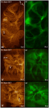
For the first time, we present co-registered autofluorescence imaging and optical coherence tomography (AF/OCT) of excised human palatine tonsils to evaluate the capabilities of OCT to visualize tonsil tissue components. Despite limited penetration depth, OCT can provide detailed structural information about tonsil tissue with much higher resolution than that of computed tomography, magnetic resonance imaging, and Ultrasound. Different tonsil tissue components such as epithelium, dense connective tissue, lymphoid nodules, and crypts can be visualized by OCT. The co-registered AF imaging can provide matching biochemical information. AF/OCT scans may provide a non-invasive tool for detecting tonsillar cancers and for studying the natural history of their development.
Conflict of interest statement
Figures


Similar articles
-
Coregistered autofluorescence-optical coherence tomography imaging of human lung sections.J Biomed Opt. 2014 Mar;19(3):36022. doi: 10.1117/1.JBO.19.3.036022. J Biomed Opt. 2014. PMID: 24687614
-
Histological characteristics and stereological volume assessment of the ovine tonsils.Vet Immunol Immunopathol. 2007 Dec 15;120(3-4):124-35. doi: 10.1016/j.vetimm.2007.07.010. Epub 2007 Jul 25. Vet Immunol Immunopathol. 2007. PMID: 17727965
-
Investigation of optical coherence micro-elastography as a method to visualize micro-architecture in human axillary lymph nodes.BMC Cancer. 2016 Nov 9;16(1):874. doi: 10.1186/s12885-016-2911-z. BMC Cancer. 2016. PMID: 27829404 Free PMC article.
-
Optical coherence tomography in dermatology.G Ital Dermatol Venereol. 2015 Oct;150(5):603-15. Epub 2015 Jul 1. G Ital Dermatol Venereol. 2015. PMID: 26129683 Review.
-
Choroidal imaging using optical coherence tomography: techniques and interpretations.Jpn J Ophthalmol. 2022 May;66(3):213-226. doi: 10.1007/s10384-022-00902-7. Epub 2022 Feb 16. Jpn J Ophthalmol. 2022. PMID: 35171356 Review.
Cited by
-
Dual-modality optical diagnosis for precise in vivo identification of tumors in neurosurgery.Theranostics. 2019 Apr 13;9(10):2827-2842. doi: 10.7150/thno.33823. eCollection 2019. Theranostics. 2019. PMID: 31244926 Free PMC article.
-
Imaging Biomarkers of Oral Dysplasia and Carcinoma Measured with In Vivo Endoscopic Optical Coherence Tomography.Cancers (Basel). 2024 Aug 2;16(15):2751. doi: 10.3390/cancers16152751. Cancers (Basel). 2024. PMID: 39123478 Free PMC article.
-
In Vivo Endoscopic Optical Coherence Tomography of the Healthy Human Oral Mucosa: Qualitative and Quantitative Image Analysis.Diagnostics (Basel). 2020 Oct 15;10(10):827. doi: 10.3390/diagnostics10100827. Diagnostics (Basel). 2020. PMID: 33076312 Free PMC article.
-
Improved secondary caries resistance via augmented pressure displacement of antibacterial adhesive.Sci Rep. 2016 Mar 1;6:22269. doi: 10.1038/srep22269. Sci Rep. 2016. PMID: 26928742 Free PMC article.
References
-
- Suzumoto M, Hotomi M, Fujihara K, Tamura S, Kuki K, et al. (2006) Functions of tonsils in the mucosal immune system of the upper respiratory tract using a novel animal model, Suncus murinus. Acta Oto-Laryngologica 126(11):1164–1170. - PubMed
-
- Frisch M, Goodman MT (2000) Human papillomavirus-associated carcinomas in Hawaii and the mainland US. Cancer 88(6):1464–1469. - PubMed
-
- Frisch M, Hjalgrim H, Jaeger AB, Biggar RJ (2000) Changing pattern of tonsillar squamous cell carcinoma in the United States. Cancer Causes Control 11(6):489–95. - PubMed
-
- Begum S, Cao D, Gillison M, Zahurak M, Westra WH (2005) Tissue distribution of human papillomavirus 16 DNA integration in patients with tonsillar carcinoma. Clinical Cancer Research 11(16):5694–5699. - PubMed
Publication types
MeSH terms
LinkOut - more resources
Full Text Sources
Other Literature Sources

