. 2015 Aug;26(8):1889-904.
doi: 10.1681/ASN.2014040350. Epub 2014 Dec 26.

Vascular Endothelial Growth Factor-A165b Is Protective and Restores Endothelial Glycocalyx in Diabetic Nephropathy

Affiliations

Vascular Endothelial Growth Factor-A165b Is Protective and Restores Endothelial Glycocalyx in Diabetic Nephropathy

Sebastian Oltean et al. J Am Soc Nephrol. 2015 Aug.

Abstract

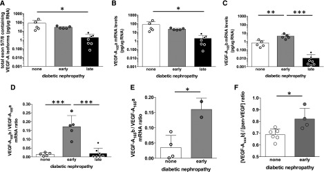
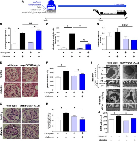
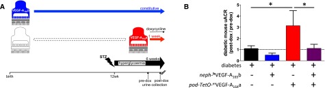
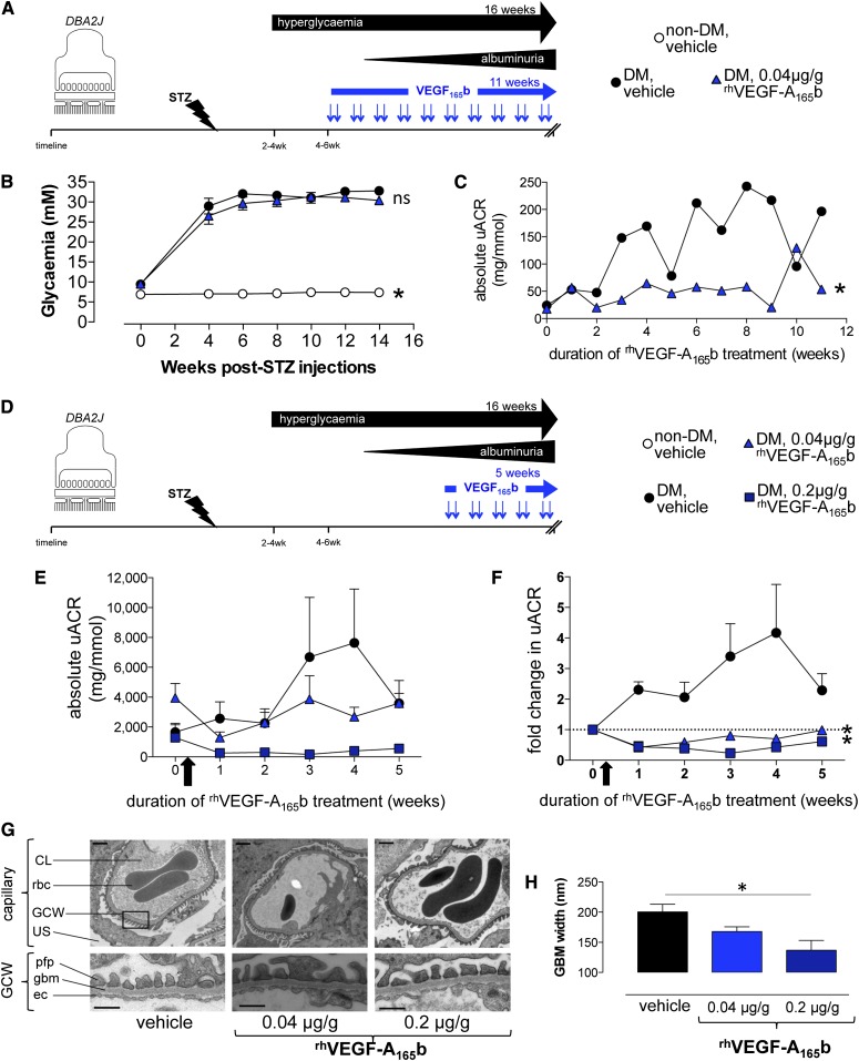
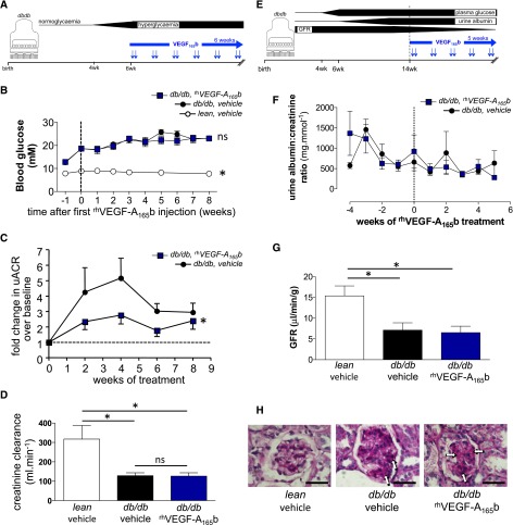
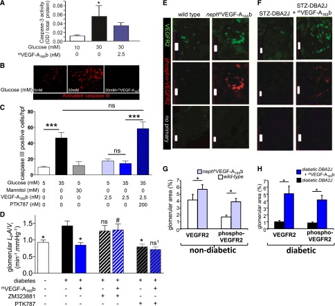
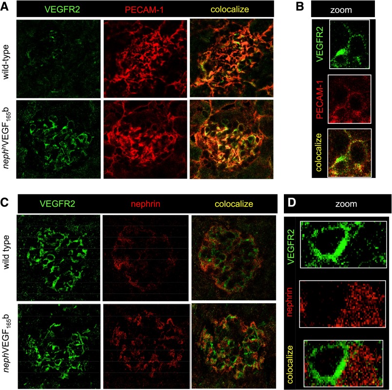
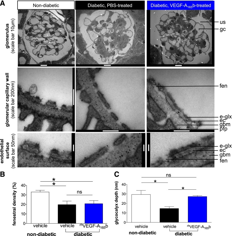
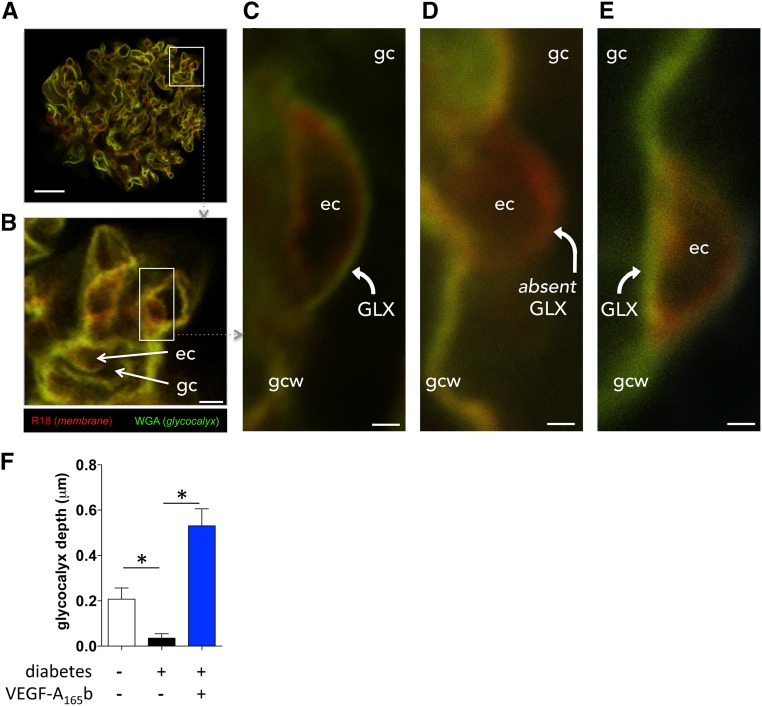
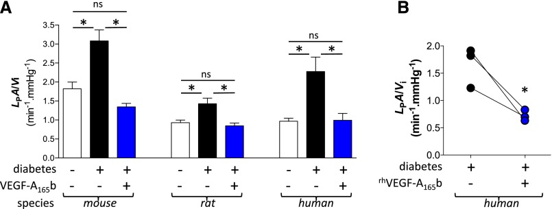
Diabetic nephropathy is the leading cause of ESRD in high-income countries and a growing problem across the world. Vascular endothelial growth factor-A (VEGF-A) is thought to be a critical mediator of vascular dysfunction in diabetic nephropathy, yet VEGF-A knockout and overexpression of angiogenic VEGF-A isoforms each worsen diabetic nephropathy. We examined the vasculoprotective effects of the VEGF-A isoform VEGF-A165b in diabetic nephropathy. Renal expression of VEGF-A165b mRNA was upregulated in diabetic individuals with well preserved kidney function, but not in those with progressive disease. Reproducing this VEGF-A165b upregulation in mouse podocytes in vivo prevented functional and histologic abnormalities in diabetic nephropathy. Biweekly systemic injections of recombinant human VEGF-A165b reduced features of diabetic nephropathy when initiated during early or advanced nephropathy in a model of type 1 diabetes and when initiated during early nephropathy in a model of type 2 diabetes. VEGF-A165b normalized glomerular permeability through phosphorylation of VEGF receptor 2 in glomerular endothelial cells, and reversed diabetes-induced damage to the glomerular endothelial glycocalyx. VEGF-A165b also improved the permeability function of isolated diabetic human glomeruli. These results show that VEGF-A165b acts via the endothelium to protect blood vessels and ameliorate diabetic nephropathy.

Keywords: VEGF; VEGF-A; albuminuria; diabetes; diabetic nephropathy; endothelial glycocalyx; glomerulus; glycocalyx; permeability.


Figures

Figure 1.
Figure 1.
VEGF-A165b is upregulated in humans with early diabetic nephropathy and well preserved kidney function. (A) mRNA expression of exon 5/7/8 containing VEGF-A isoforms (i.e., those coding for 165 amino-acid proteins) determined by RT-qPCR relative to total RNA extracted in whole renal cortical tissue from kidneys unsuitable for transplantation from deceased donors without diabetes (“none”), donors with early diabetic nephropathy with well preserved kidney function (“early”), and kidney biopsy specimens from patients with diabetes and advanced nephropathy (“late”) (note logarithmic scale). (B) VEGF-A165a mRNA expression (note logarithmic scale). (C) VEGF-A165b expression (note logarithmic scale). (D) VEGF-A isoform expression ratio calculated as VEGF-A165b mRNA per unit total mRNA divided by VEGF-A165a mRNA per unit total mRNA. (E) Ratio of VEGF-A165a to VEGF-A165b mRNA in control and early diabetic samples in which it was possible to analyze mRNA from single glomeruli isolated by differential sieving. (F) Isoform-specific VEGF-A165b protein determined by ELISA as a proportion of total VEGF-A in whole kidney cortical tissue in kidney donors with diabetes and early nephropathy (“early”) and those without diabetes (“none”). Error bars, SEM. Multiple comparisons: one-way ANOVA. Two-group comparisons: unpaired t test. *P<0.05; **P<0.01; ***P<0.005, throughout.
Figure 2.
Figure 2.
Podocyte-specific overexpression of VEGF-A165b ameliorates STZ-induced diabetic nephropathy. (A) Transgenic mice constitutively overexpressing human VEGF-A165b in podocytes under control of the nephrin promoter (blue; nephhVEGF-A165b) received injections of STZ or vehicle at 12 weeks of age. Plasma glucose (B), urine albumin-to-creatine ratio (C), and creatinine clearance (D) measured in nondiabetic wild-type, diabetic wild-type, nondiabetic transgenic, and diabetic transgenic mice 6 weeks after diabetes induction. n=5–10 mice per group. Error bars, SEM. *P<0.05, one-way ANOVA. (E and F) Glomerular area assessed in hematoxylin and eosin–stained kidney sections from 21 to 55 glomeruli from 3 mice per group. Scale bars: 20 µm. Error bars, SEM. *P<0.05, one-way ANOVA. (G and H) Periodic acid Schiff–stained kidney sections from each group of mice. Mesangial matrix expansion (arrows) measured as a proportion of total glomerular area in 45–60 glomeruli from 3 to 4 mice per group. Scale bars: 20 µm. Error bars, SEM. *P<0.05, one-way ANOVA. (I) Low- and high-magnification electron micrographs (scale bars: 1 µm and 250 nm, respectively) of glomerular capillaries and the glomerular capillary wall (GCW) from each group of mice. CL, capillary lumen; ec, endothelial cell; gbm, glomerular basement membrane; pfp, podocyte foot process; rbc, red blood cell; US, urinary space. (J) Glomerular basement membrane width calculated from 20 to 60 measurements in 8–16 glomerular capillaries per group. Error bars, SEM. *P<0.05, one-way ANOVA.
Figure 3.
Figure 3.
Podocyte-specific overexpression of VEGF-A165b reduces albuminuria in the dual insult of VEGF-A164a overexpression and STZ-induced diabetic nephropathy. (A) Heterozygous mice overexpressing human VEGF-A165b in podocytes (blue: nephhVEGF-A165b) were crossed with mice in which murine VEGF-A164a (mVEGF-A164a) overexpression in podocytes could be induced with doxycycline (dox): heterozygous podocin-rtTA:TetO-mVEGF-A164a mice (red: pod-TetO-mVEGF-A164a), to generate mice co-overexpressing mVEGF-A164a and hVEGF-A165b in podocytes. Offspring were treated with STZ at 12 weeks of age to induce diabetes in all groups. After 5 weeks of diabetes, all mice received doxycycline, inducing mVEGF-A164a overexpression in the two groups carrying the podocin-rtTA:TetO-mVEGF-A164a transgene. After 1 week of doxycycline treatment, urinary albumin-to-creatine ratio (uACR) was measured and compared with uACR obtained before doxycycline administration. (B) Change in uACR after 7 days of doxycycline treatment in diabetic mice with no transgenes, diabetic mice overexpressing hVEGF-A165b alone, diabetic mice overexpressing mVEGF-A164a alone, and diabetic mice co-overexpressing mVEGF-A164a and hVEGF-A165b. Dashed line represents no change in albuminuria during the 1-week doxycycline treatment period. n=3–6 mice per group. Error bars, SEM. *P<0.05, one-way ANOVA.
Figure 4.
Figure 4.
Systemic treatment with VEGF-A165b blocks progression of albuminuria and glomerular basement membrane thickening in STZ-induced diabetic nephropathy. (A) Diabetes was induced in wild-type DBA2J mice with STZ injection (nondiabetic control mice received buffer injection alone). After 2 consecutive weeks of hyperglycemia, mice received an 11-week course of biweekly intraperitoneal injections (blue arrows) of recombinant human VEGF-A165b (rhVEGF-A165b) (n=16) or vehicle (n=16). (B) Plasma glucose measured repeatedly in nondiabetic control, diabetic vehicle-treated, and 0.04 µg/g rhVEGF-A165b–treated mice. *P<0.05 versus both diabetic groups; "ns" indicates P>0.05 between diabetic groups; one-way ANOVA for both. (C) Urine albumin-to-creatinine ratio (uACR) was measured repeatedly in diabetic DBA2J mice receiving injections of vehicle (n=4) or 0.04 µg/g rhVEGF-A165b (n=4) for up to 11 weeks. *P<0.05, paired t test. (D) Diabetes was induced in wild-type DBA2J mice with STZ injection and vehicle or rhVEGF-A165b treatment after the onset of albuminuria. (E) uACR measured repeatedly in mice receiving biweekly injections of vehicle (n=10), 0.04 µg/g rhVEGF-A165b (n=10), and 0.2 µg/g rhVEGF-A165b (n=6) before organ harvest. Error bars, SEM. (F) Fold change in uACR (uACR at indicated time points, divided by uACR before initiation of treatment [arrow]). Dashed line represents no change in albuminuria after start of injections. Error bars, SEM. *P<0.05, two-way ANOVA. (G) Low- and high-magnification electron micrographs (scale bars: 1 μm and 500 nm, respectively) of glomerular capillaries and the glomerular capillary wall (GCW). CL, capillary lumen; ec, endothelial cell; gbm, glomerular basement membrane; pfp, podocyte foot process; rbc, red blood cell; US, urinary space. (H) Glomerular basement membrane (GBM) width measurements in indicated groups of diabetic mice. Error bars, SEM. *P<0.05, one-way ANOVA.
Figure 5.
Figure 5.
Systemic treatment with VEGF-A165b blocks early albuminuria but not later features of diabetic nephropathy in a genetic model of type 2 diabetic nephropathy. (A) After the onset of hyperglycemia at 6 or 7 weeks of age, db/db mice received biweekly intraperitoneal injections of recombinant human VEGF-A165b (rhVEGF-A165b) or vehicle (blue arrows) for 8 weeks. (B) Blood glucose measurements in 6 lean and 20 db/db mice before and after receipt of twice-weekly vehicle or rhVEGF-A165b (0.2 µg/g) injections. Dashed line indicates start of intraperitoneal injections. Error bars, SEM. *P<0.05 compared with both groups of diabetic mice; "ns" indicates P>0.05 between groups of diabetic mice. (C) Urinary albumin-to-creatinine ratio (uACR) determined repeatedly during the 8-week treatment period, divided by uACR in the same animal before treatment (“baseline”) to calculate fold change in uACR. Dashed line represents no fold change in albuminuria since start of treatment. Error bars, SEM. *P<0.05, paired t test. (D) Creatinine clearance measured at the end of the 8-week injection period. Error bars, SEM. *P<0.05; "ns" indicates P>0.05; one-way ANOVA for both. (E) In a separate cohort of older mice, 5 weeks of biweekly intraperitoneal injections (blue arrows) of rhVEGF-A165b or vehicle were initiated in 14-week-old lean controls (n=5) and db/db mice (n=14) with long-standing hyperglycemia, established albuminuria, and decreased GFR. (F) Serial measurement of uACR in these older vehicle- and rhVEGF-A165b–treated db/db mice. Dashed line indicates start of intraperitoneal injections. Error bars, SEM. *P<0.05, paired t test. (G) At the end of the 5-week injection period, GFR measurements in these older vehicle- and rhVEGF-A165b–treated db/db and lean mice. Error bars, SEM. *P<0.05; "ns" indicates P>0.05; one-way ANOVA for both. (H) At the end of the 5-week injection period, mesangial matrix expansion (arrows) in periodic acid Schiff–stained kidney sections from each group of mice. Scale bar: 50 μm.
Figure 6.
Figure 6.
VEGF-A165b reduces apoptosis, decreases glomerular permeability, and acts via VEGFR-2. (A) Apoptosis (caspase-3 activity) was determined in human podocytes in vitro exposed to normal (10 mM) or high (30 mM) cell culture glucose concentrations with or without recombinant human VEGF-A165b (rhVEGF-A165b). Experiments were done in triplicate. OD, optical density. Error bars, SEM. *P<0.05, one-way ANOVA. (B) Caspase-3 staining performed on human microvascular endothelial cells exposed to normal (5 mM) or high (30 mM) cell culture glucose concentrations in the presence or absence of rhVEGF-A165b. (C) Number of caspase-3–positive cells per high-powered field (hpf) compared between conditions. Experiments were done in quadruplicate. Error bars, SEM. ***P<0.001; "ns" indicates P>0.05; one-way ANOVA for both. (D) Glomerular water permeability (volume-corrected ultrafiltration coefficient: LPA/Vi) measured in glomeruli isolated from vehicle-injected nondiabetic (open bar), and STZ-injected diabetic rats (filled bars). Glomeruli were incubated for 1 hour in rhVEGF-A165b (1 nM), VEGF-A receptor-2 antagonist ZM323881 (10 µM), pan-VEGF-A receptor blocker PTK787 (100 nM), saline, or combinations thereof before LPA/Vi measurement. Error bars, SEM. Compared with diabetic: *P<0.05; "ns" indicates P>0.05. Compared with diabetic+rhVEGF-A165b: #P<0.05; "ns1" indicates P>0.05. One-way ANOVA for all. (E and F) Total and phosphorylated VEGFR-2 in glomeruli of nondiabetic wild-type and nephhVEGF-A165b mice (E) and in glomeruli from STZ-induced diabetic DBA2J mice treated with vehicle or rhVEGF-A165b (F). Controls without primary antibody are also shown. Scale bars: 20 μm. VEGFR-2 and phosporylated-VEGFR-2 by area relative to the whole glomerulus quantified for nondiabetic nephhVEGF-A165b versus littermates (G) and rhVEGF-A165b–treated diabetic mice versus vehicle-treated diabetic mice (H). Error bars, SEM. *P<0.05, one-way ANOVA.
Figure 7.
Figure 7.
VEGF-A165b stimulates VEGFR-2 in glomerular endothelial cells in vivo. (A) Sections of kidney tissue from wild-type and nephhVEGF-A165b mice showing glomeruli costained for VEGFR-2 (green), and the endothelial cell marker PECAM-1 (red), and overlaid images showing colocalization (yellow). (B) High magnification of a single glomerular capillary loop showing VEGFR-2 expression and endothelial cell PECAM-1 colocalize. (C) Sections of kidney tissue from wild-type and nephhVEGF-A165b mice showing glomeruli costained for VEGFR-2 (green) and the podocyte marker nephrin (red). (D) High magnification of a single glomerular capillary loop showing that VEGFR-2 expression and podocyte nephrin do not colocalize.
Figure 8.
Figure 8.
VEGF-A165b restores the glomerular endothelial glycocalyx in early STZ-induced diabetic nephropathy. Six weeks after STZ (or buffer) injections to induce diabetes (or normoglycemia) in DBA2J mice, mice received biweekly intraperitoneal injections of vehicle or rhVEGF-A165b for 4 weeks. Animals were cardiac perfusion fixed and renal cortices were processed for transmission electron microscopy. (A) Glomeruli were imaged at low magnification, the glomerular capillary wall at higher magnification, and endothelial cell surface at high magnification using transmission electron microscopy. e-glx, endothelial glycocalyx; ec, endothelial cell; fen, endothelial fenestra; gbm, glomerular basement membrane; gc, glomerular capillary; pfp, podocyte foot process; us, urinary space. (B) Endothelial fenestral density and (C) glycocalyx depth covering the glomerular endothelial cell surface quantified in electron microscopic images from vehicle-treated nondiabetic, vehicle-treated diabetic, and rhVEGF-A165b–treated diabetic animals. Error bars, SEM. n=3 mice per group. *P<0.05; "ns" indicates P>0.05; one-way ANOVA for both.
Figure 9.
Figure 9.
VEGF-A165b restores glomerular endothelial glycocalyx acutely in diabetic rat glomeruli. Kidneys were perfused in vivo with cell membrane label (R18; red) and glycocalyx label (Alexa Fluor-488–wheat germ agglutinin lectin; green), and then glomeruli were isolated and treated with vehicle or VEGF-A165b for 1 hour (exactly as per physiology experiments) before imaging with confocal microscopy. (A) whole glomerulus; scale bar: 20 μm. (B) Glomerular capillaries (gc) with endothelial cell (ec) bodies; scale bar: 5 μm. (C) High-magnification image (scale bar: 1 μm) of a vehicle-treated glomerular capillary from a healthy animal, demonstrating glomerular endothelial glycocalyx (GLX) lining luminal surface of endothelial cell body. gcw, glomerular capillary wall. (D) High-magnification image (scale bar: 1 μm) of a vehicle-treated glomerular capillary from a diabetic animal. Note absent glomerular endothelial glycocalyx (absent GLX) lining the luminal surface of endothelial cell body. (E) High-magnification image (scale bar: 1μm) of a VEGF-A165b–treated glomerular capillary from a diabetic animal. Note restoration of glomerular endothelial glycocalyx. (F) Mean±SEM glomerular endothelial glycocalyx depth in five glomeruli in each of three animals per group. *P<0.05, one-way ANOVA.
Figure 10.
Figure 10.
VEGF-A165b normalizes permeability of diabetic mouse, rat, and human glomeruli. (A) Glomeruli were harvested from 6 diabetic (STZ-injected) wild-type, 4 nondiabetic wild-type, and 8 diabetic (STZ) nephhVEGF-A165b mice (102 glomeruli); from 3 diabetic (STZ) and 3 nondiabetic rats (74 glomeruli); and from untransplantable kidneys from 3 nondiabetic and 3 diabetic human kidney donors (54 glomeruli). Glomerular water permeability (volume-corrected ultrafiltration coefficient: LPA/Vi) was measured in individual glomeruli following exposure to constitutively overexpressed hVEGF-A165b (mice) or 1-hour incubation in vehicle or rhVEGF-A165b (rat and human glomeruli). Error bars, SEM. *P<0.05; "ns" indicates P>0.05; one-way ANOVA for both. (B) Glomerular LPA/Vi was measured twice in 3 diabetic human glomeruli, before (black) and after (blue) 1-hour incubation in rhVEGF-A165b. *P<0.05, paired t test.

References

    1. Gilbertson DT, Liu J, Xue JL, Louis TA, Solid CA, Ebben JP, Collins AJ: Projecting the number of patients with end-stage renal disease in the United States to the year 2015. J Am Soc Nephrol 16: 3736–3741, 2005 - PubMed
    1. Frank RN: Diabetic retinopathy. N Engl J Med 350: 48–58, 2004 - PubMed
    1. Abbott CA, Malik RA, van Ross ER, Kulkarni J, Boulton AJ: Prevalence and characteristics of painful diabetic neuropathy in a large community-based diabetic population in the U.K. Diabetes Care 34: 2220–2224, 2011 - PMC - PubMed
    1. Forbes JM, Cooper ME: Mechanisms of diabetic complications. Physiol Rev 93: 137–188, 2013 - PubMed
    1. Rask-Madsen C, King GL: Kidney complications: Factors that protect the diabetic vasculature. Nat Med 16: 40–41, 2010 - PubMed

Publication types

MeSH terms

Substances