Review
. 2015 Apr:42:19-41.
doi: 10.1016/j.mam.2014.12.002. Epub 2014 Dec 24.

Natural history of β-cell adaptation and failure in type 2 diabetes

Affiliations
Review

Natural history of β-cell adaptation and failure in type 2 diabetes

Emilyn U Alejandro et al. Mol Aspects Med. 2015 Apr.

Abstract

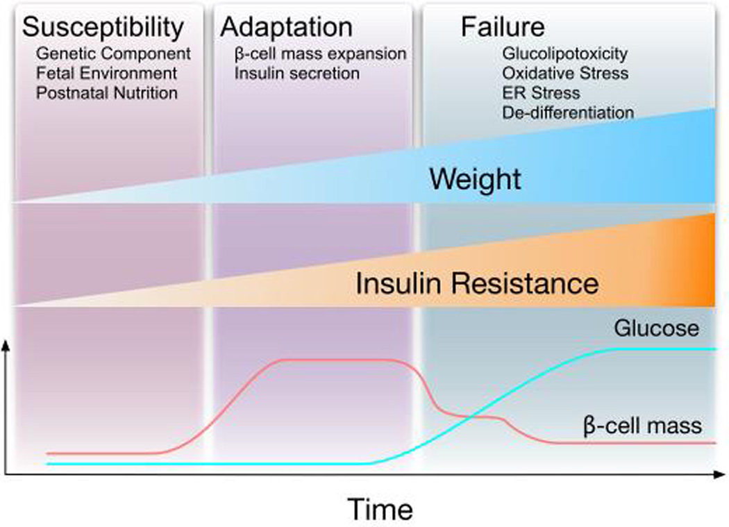
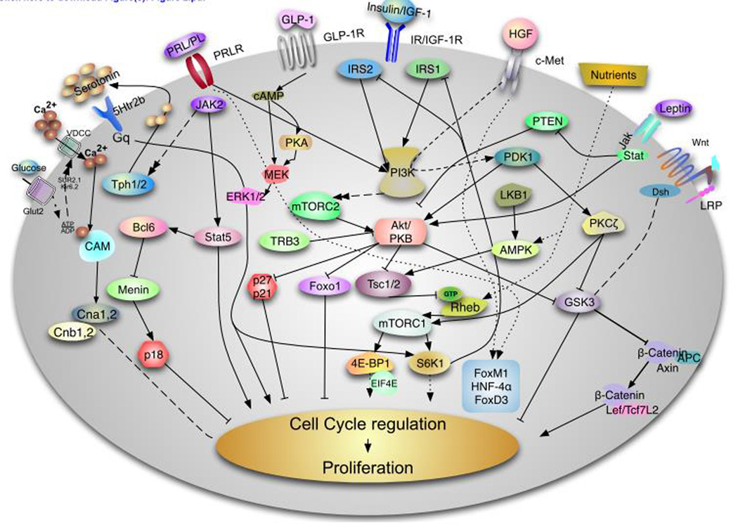
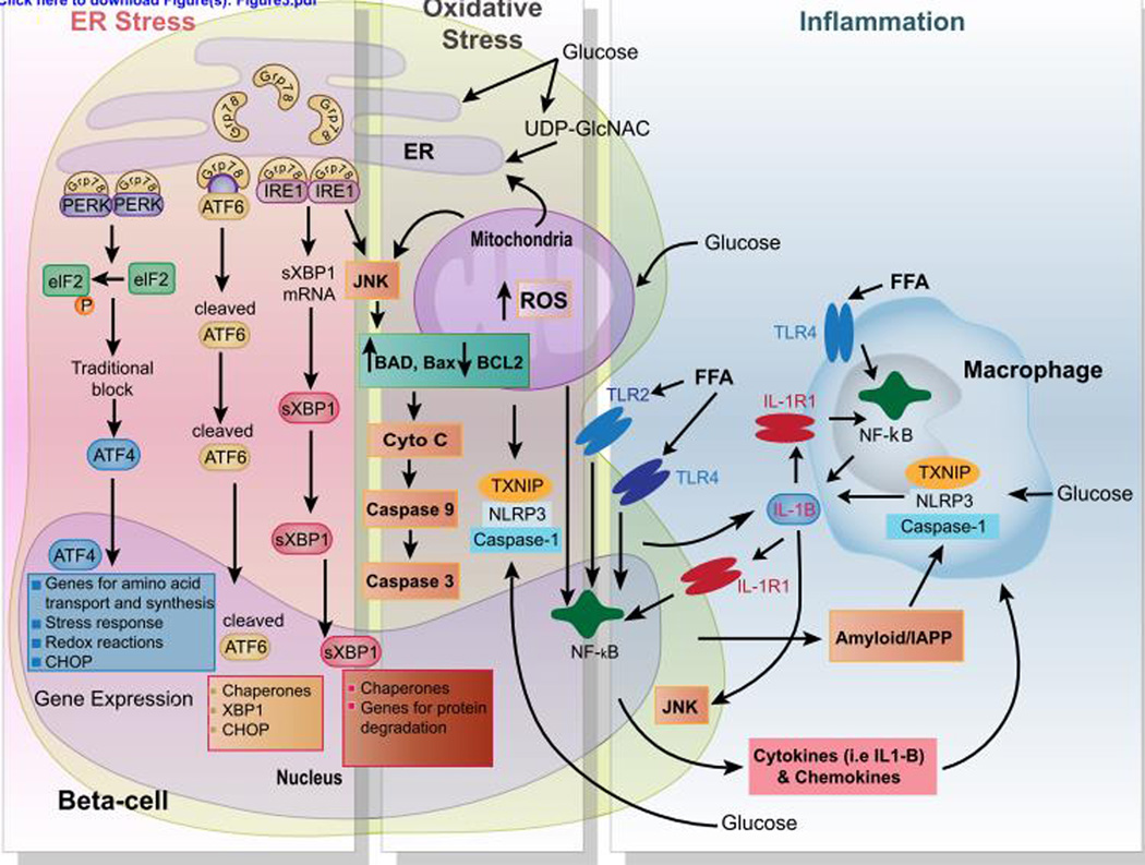
Type 2 diabetes mellitus (T2D) is a complex disease characterized by β-cell failure in the setting of insulin resistance. The current evidence suggests that genetic predisposition, and environmental factors can impair the capacity of the β-cells to respond to insulin resistance and ultimately lead to their failure. However, genetic studies have demonstrated that known variants account for less than 10% of the overall estimated T2D risk, suggesting that additional unidentified factors contribute to susceptibility of this disease. In this review, we will discuss the different stages that contribute to the development of β-cell failure in T2D. We divide the natural history of this process in three major stages: susceptibility, β-cell adaptation and β-cell failure, and provide an overview of the molecular mechanisms involved. Further research into mechanisms will reveal key modulators of β-cell failure and thus identify possible novel therapeutic targets and potential interventions to protect against β-cell failure.

Keywords: Glucolipotoxicity; Insulin resistance; Islet biology; β-cell development; β-cell failure; β-cell programming.


Figures

Figure 1
Figure 1. Natural history of the adaptation of β-cells to obesity and diabetes
It is clear that the majority of obese individuals develop insulin resistance. The β-cells adapt to insulin resistance by increasing mass and the function. The β-cells compensate appropriately in the majority of obese individuals and hyperinsulinemia is a common finding in obesity. However, in a fraction of obese subjects β-cells fail to compensate appropriately with development of hyperglycemia and diabetes. After the development of hyperglycemia, glucose acts synergistically with other factors to induce β-cell failure. The evolution of β-cells in this process can be divided in three major stages: 1. Individuals with high risk of diabetes are born with increase susceptibility by genetic component, the fetal environment and the nutrient environment during the first years of life. 2. As individuals gain weight there is a phase of adaptation. 3. Finally, individuals with increase susceptibility develop β-cell failure by the interaction of different process.
Figure 2
Figure 2. Signaling pathways involved in Rodent β-cell proliferation
This diagram describes some of the major pathways responsible for rodent β-cell proliferation. This field is evolving and we have attempted to provide a summary of the major pathways involve this process. The insulin/insulin-like growth factor I (IGF1) receptor, hepatocyte growth factor and glucagon like peptide 1 (GLP1) signal through the IRS/PI3 kinase pathway to regulate Akt by modulating PDK1. Complete activation of Akt requires the phosphorylation of this kinase by mTORC2. The serine threonine kinase has been demonstrate to play a major role in regulation of β-cell proliferation, survival and cell size by acting on multiple downstream targets including FoxO1, tuberous sclerosis complex (TSC) 1 and 2, Glycogen synthase kinase (GSK) 3 and the cell cycle inhibitors p27cip and p21 among others. Akt activity is negatively regulated directly and indirectly by TRB3 and PTEN. Downstream of Akt, the TSC/mTORC1 integrates signals from growth factors and nutrients. Nutrients can modulate this pathway directly by acting on the protein Rheb or indirectly by regulating the activity of the AMPK pathway, which is also regulated by LKB1. mTORC1 regulates protein translation and cell size by regulating S6K and 4E-BPs. HGF binding to its tyrosine kinase receptor, c-Met, results PKCζ activation which in turns inactivates GSK3β and increases phosphorylation and activation of mTORC1. Activation of the prolactin receptor induces Jak2/Stat5 signaling and/or Jak2/Bcl6/Menin to induce β-cell proliferation. Recent data shows that lactogens induce proliferation by activation of tryptophan hydroxylases (Tph1/2 with generation of serotonin. Serotonin is secreted and act in paracrine fashion to assumed to act via the HTR2b receptor to modulate intracellular calcium and PKC or PI3K members. Glucose is transported by the glucose transporter Glut2 and metabolizes to increase the ATP ratio, which subsequently inhibits the Kir6.2 channel resulting depolarization and calcium influx. Intracellular calcium signaling is a major driver of β-cell proliferation by regulating multiple signaling pathways including Calmodulin (Cam) Calcineurin (Cna1,2 and Cnb1,2) and NFAT transcription. In parallel, glucose activates ChREBP and cMyc to induce cell cycle progression (not shown). Upon binding to GLP-1 GLP-1 receptor results in elevation of cAMP, protein kinase A (PKA) activation and this ultimately results in which can phosphorylation of β-catenin by MEK/ERK1/2 pathway. Leptin acts via the JAK-STAT pathway induces Akt signaling by inhibiting PTEN. The Wnt/frizzled pathway acts by inhibiting GSK-3β and blocking phosphorylation of β-catenin modulate transcription of Lef/Tcf7L2 dependent genes such as cyclin D2 and potentially cyclin D1 and cMyc and activates cell cycle progression. It is important to note that our understanding of human β-cell proliferation is notably limited compared to that of rodents.
Figure 3
Figure 3. Multifactorial signaling events in β-cell failure
Chronic glucolipotoxic conditions induce prolonged ER and oxidative stress, and islet inflammation that cause β-cell death. Once β-cells are committed to death as a result of unresolved ER stress under glucolipotoxic conditions (high levels of glucose, free fatty acids (FFA), and OGlcNac) several well-characterized mechanisms that involve the PERK, ATF6, and IRE1 branches of the UPR act to mediate β-cell apoptosis. These involve association of Bax/Bad with IRE1 leading to the activation of JNK and an ATF6 and PERK-mediated increase in CHOP levels and activity. CHOP can then activate expression of proteins involve in pro-apoptotic pathways. High concentrations of glucose promote the activation of the inflammasome complex consisting of NLRP3, TXNIP, and Caspase1 in both β-cells and macrophages. FFA and islet-derived islet amyloid polypeptide (IAPP) also directly trigger the NLRP3/TXNIP/Caspase1 inflammasome complex. Subsequently, pro-IL-1β is processed by the inflammasome complex and secreted in the microenvironment. In turn, IL-1β sustains autocrine and paracrine activation of both β-cells and macrophages, exacerbating the chronic inflammatory responses in the islets. Furthermore, FFA binds to their cognate receptors (i.e., Toll-like receptor 2 and 4 (TLR2-4)), leading to NF-κB activation and the production of various proinflammatory chemokines and cytokines, including the proform of IL-1β.

References

    1. Mokdad AH, et al. The continuing epidemics of obesity and diabetes in the United States. JAMA : the journal of the American Medical Association. 2001;286:1195–1200. - PubMed
    1. Must A, et al. The disease burden associated with overweight and obesity. JAMA : the journal of the American Medical Association. 1999;282:1523–1529. - PubMed
    1. Narayan KM, Boyle JP, Thompson TJ, Gregg EW, Williamson DF. Effect of BMI on lifetime risk for diabetes in the U.S. Diabetes care. 2007;30:1562–1566. - PubMed
    1. Meier JJ, et al. Beta-cell replication is the primary mechanism subserving the postnatal expansion of beta-cell mass in humans. Diabetes. 2008;57:1584–1594. - PMC - PubMed
    1. Perl S, et al. Significant human beta-cell turnover is limited to the first three decades of life as determined by in vivo thymidine analog incorporation and radiocarbon dating. The Journal of clinical endocrinology and metabolism. 2010;95:E234–E239. - PMC - PubMed

Publication types