. 2015 Jan 8;57(1):191-201.
doi: 10.1016/j.molcel.2014.11.021. Epub 2014 Dec 24.

The strength and cooperativity of KIT ectodomain contacts determine normal ligand-dependent stimulation or oncogenic activation in cancer

Affiliations

The strength and cooperativity of KIT ectodomain contacts determine normal ligand-dependent stimulation or oncogenic activation in cancer

Andrey V Reshetnyak et al. Mol Cell. .

Abstract

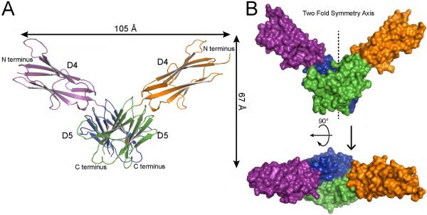
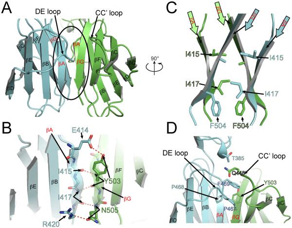
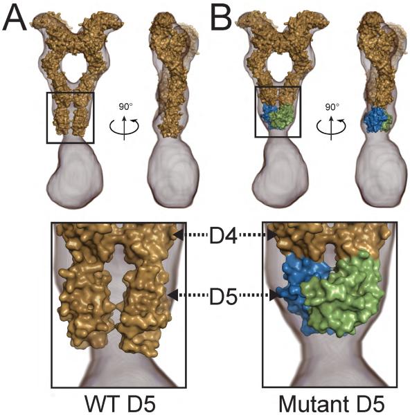
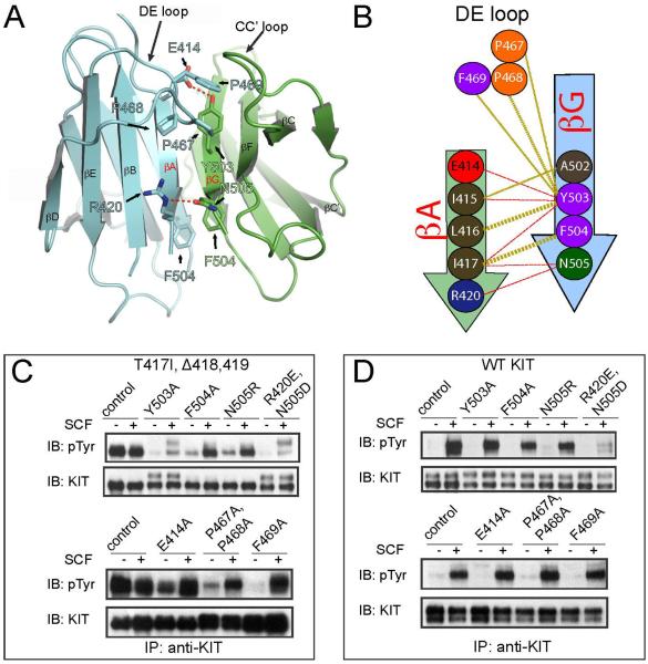
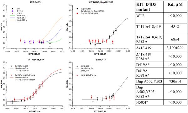
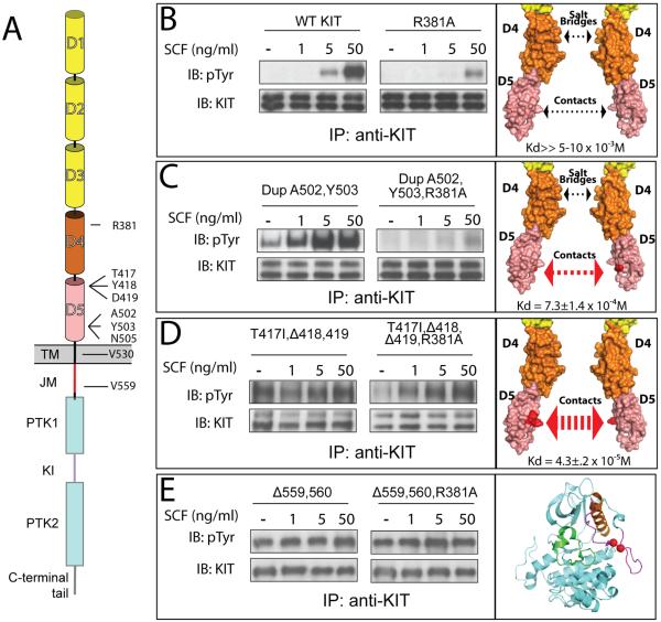
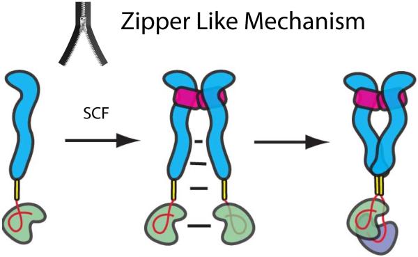
The receptor tyrosine kinase KIT plays an important role in development of germ cells, hematopoietic cells, and interstitial pacemaker cells. Oncogenic KIT mutations play an important "driver" role in gastrointestinal stromal tumors, acute myeloid leukemias, and melanoma, among other cancers. Here we describe the crystal structure of a recurring somatic oncogenic mutation located in the C-terminal Ig-like domain (D5) of the ectodomain, rendering KIT tyrosine kinase activity constitutively activated. The structural analysis, together with biochemical and biophysical experiments and detailed analyses of the activities of a variety of oncogenic KIT mutations, reveals that the strength of homotypic contacts and the cooperativity in the action of D4D5 regions determines whether KIT is normally regulated or constitutively activated in cancers. We propose that cooperative interactions mediated by multiple weak homotypic contacts between receptor molecules are responsible for regulating normal ligand-dependent or oncogenic RTK activation via a "zipper-like" mechanism for receptor activation.


Figures

Figure 1
Figure 1. Crystal structure of T417I,Δ418,419 KIT D4D5 fragment
(A) Ribbon diagram of T417I,Δ418,419 KIT D4D5 dimer. D4 and D5 of one protomer are colored in purple and blue, and D4, D5 of another protomer are in orange and green respectively. N and C termini are labeled. Ile 417, an oncogenic mutation, is marked with a red balls. (B) Surface representation of T417I,Δ418,419 KIT D4D5 dimer. Upper panel shows a view following 90° rotation along the horizontal axis of the view shown in the bottom panel. Color coding is the same as in panel A. 2 fold symmetry axis is shown as vertical line passing through the middle of “V”.
Figure 2
Figure 2. D5:D5 dimerization interface in the crystal structure of the oncogenic T417I,Δ418,419 KIT D4D5 mutant
Ribbon representation of two D5 domains forming dimer. D5 from one protomer is colored with blue and D5 from another is in green. Secondary structure elements are labeled according to IgSF nomenclature. βA and βG – the hot spots for oncogenic mutation are highlighted in red. D4D5 dimer is rotated ~90° around vertical axis relative to the orientation of D4D5 dimer in Figure 1A. (A) Overview of D5:D5 dimerization interface. (B) Detailed view of contacts between βA of one protomer and βG of another protomer. Hydrogen bonds are shown with red dotted lines. Side chains are shown only for those residues, which form hydrogen bonds. (C) The same orientation as D4D5 dimer in Figure 1A and 90° rotated relative to orientation in A. Only strands A and G from each protomer are represented. Side chains involved in van der Waals interactions are represented with sticks model. (D) Contacts between DE loop of one protomer and strand G and CC' loop are depicted. Side chains involved in van der Waals interactions are shown with sticks and labeled.
Figure 3
Figure 3. Fitting of KIT ectodomain crystal structure into electron microscopy EM 3D reconstruction
(A) Crystal structure of KIT ectodomain in complex with SCF (PDB ID 2E9W, Yuzawa et al., 2007) was fitted into the volume of the 3D EM reconstruction (EMD-2648, Opatowsky et al, 2014). KIT:SCF crystal structure is surface represented and colored with beige; 3D EM reconstruction is represented with gray mesh (EMD-2648, Opatowsky et al, 2014). Right panel is 90° rotated along vertical axis relative to the left panel. The homotypic D5 contacts seen in the crystal structure do not fit into the volume of the 3D EM reconstruction (depicted in the lower panel). (B) D1–D4 domains from KIT:SCF complex (PDB ID 2E9W, Yuzawa et al., 2007) and D5 dimer from our current structure of T417I,Δ418,419 KIT mutant are grafted together and fitted into 3D EM reconstruction (EMD-2648, Opatowsky et al, 2014). Color code for D1–D4 of KIT and SCF is the same as in panel A. D5:D5 dimer from T417I,Δ418,419 structure are colored with blue (one protomer) and green (second protomer).
Figure 4
Figure 4. Rescue of ligand dependency of the oncogenic T417I,Δ418,419 KIT mutant
(A) Cartoon representation of D5D5 interface. Residues, which were mutated in (C and D) are shown with sticks and labeled. Color code is the same as in Figure 2. (B) Schematic representation of D5:D5 interface contacts for T417I,Δ418,419 mutant. One protomer is colored by blue and another protomer by green. Beta strands are represented with arrows. Hydrogen bonds are shown with red dashes and hydrophobic interactions with golden dashes. Residues involved into the dimer interface formation are shown with circles and numbered according to WT KIT. Interface contacts were calculated using the PDBsum server (Laskowski, 2009). Only β strands A and G, and DE loop are highlighted, a full scheme of 2D secondary structure of T417I,Δ418,419 mutant is represented in Figure S5. (C) Ligand dependent activity of T417I,Δ418,419 oncogenic KIT mutant can be fully or partially restored by point mutations of residues involved in the D5:D5 dimerization interface. (D) The same point mutations in the context of wild-type KIT (WT KIT) receptor do not interfere with tyrosine kinase activities except for the double mutation of R420E and N505D. 3T3 cells transiently expressing full length KIT receptor variants (as indicated in the upper panel) were stimulated with 25 ng/ml SCF for 6 min at 37°C. (C) Residues indicated in the upper panel were mutated in the context of T417I,Δ418,419 oncogenic KIT mutant. (D) Residues indicated in the upper panel were mutated in the context of WT KIT.
Figure 5
Figure 5. Multiangle light scattering (MALS) experiment demonstrating dimerization state of KIT D4D5 fragments (WT and oncogenic mutants)
Different concentrations of KIT D4D5 fragments were injected on to HPLC system equipped with light scattering (LS) detector and differential refractometer (RI). Weight-average molar mass was calculated based on LS and RI signals. The weight average molar masses were plotted as a function of concentration (dots on the graph). Solid line represents simulation of dimerization curves (top left panel) or nonlinear regression fit of the data to the model (Equation 1). Dimerization constants calculated by nonlinear regression fit are presented in the table (right panel).
Figure 6
Figure 6. In vivo autophosphorylation of WT or oncogenic KIT mutants
(A) Schematic representation of full length KIT receptor. D1 – D5 are Ig-like domains. D1–D3 represented with yellow, D4 and D5 with orange and pink cylinders respectively. TM is transmembrane (black), JM - juxtamembrane (red), PTK - protein tyrosine kinase (blue), KI - kinase insert (purple) domains. Oncogenic mutations and Arg381 are labeled. Tyrosine kinase activity was compared for WT – wild type KIT receptor (B); DupA502,Y503 – duplication of Ala 502 and Tyr 503 (C); T417I ΔY418D419 – substitution of Thr 417 to Ile and deletion of Tyr 418 and Asp 419 (D), and Δ559,560 – deletion of Val 559 and 560 (E). WT or oncogenic KIT mutants (left panel) were compared to those harboring an additional mutation of an Arg 381 (middle panel) - residue responsible for D4:D4 homotypic contacts. NIH-3T3 cells stably expressing WT KIT or oncogenic mutants were stimulated with increasing concentration of SCF (as indicated in the upper panel) for 6 minutes at 37°C. Lysates of unstimulated or SCF-stimulated cells were subject for immunoprecipitation (IP) using anti-KIT antibodies followed by SDS-PAGE and immunoblotting (IB) with anti-pTyr (IB: pTyr) or anti-KIT (IB: KIT) antibodies (as indicated). Right panel – surface representation of membrane proximal Ig-like domains (B–D) or cartoon representation of the kinase domain (E), oncogenic mutations are highlighted with red. The strength of D5:D5 contacts and impact of D4 salt bridge schematically represented with arrows. Dimerization constants for D4D5 fragment are shown in the bottom of the right panel.
Figure 7
Figure 7. A “zipper-like” mechanism for ligand-dependent or oncogenic KIT activation
SCF binding brings two KIT monomers together. The increase in local concentration of KIT upon ligand induced dimerization enables formation of multiple low affinity associations between different regions of neighboring KIT molecules by means of a “zipper-like” process that propagates in a cooperative manner from the SCF binding region to D4, D5, TM and to the cytoplasmic region leading to formation of asymmetric KIT dimers. We propose that the asymmetric kinase arrangements represent snapshots of molecular interactions taking place between two tyrosine kinase domains poised towards trans autophosphorylation of tyrosine residues located in different parts of the cytoplasmic region in an orderly manner. In this process the membrane proximal cis JM autoinhibited kinase may function as a substrate (green) and the membrane distal kinase will function as an active enzyme (purple). After trans autophosphorylation the roles of the two kinase molecules are switched. Ectodomain of KIT depicted in blue, TM in yellow, JM region is shown as a red loop and SCF in magenta.

References

    1. Adams PD, Afonine PV, Bunkóczi G, Chen VB, Davis IW, Echols N, Headd JJ, Hung L-W, Kapral GJ, Grosse-Kunstleve RW, et al. PHENIX: a comprehensive Python-based system for macromolecular structure solution. Acta Crystallogr. D. Biol. Crystallogr. 2010;66:213–221. - PMC - PubMed
    1. Anderson DM, Williams DE, Tushinski R, Gimpel S, Eisenman J, Cannizzaro LA, Aronson M, Croce CM, Huebner K, Cosman D. Alternate splicing of mRNAs encoding human mast cell growth factor and localization of the gene to chromosome 12q22-q24. Cell Growth Differ. 1991;2:373–378. - PubMed
    1. Ashman LK. The biology of stem cell factor and its receptor C-kit. Int. J. Biochem. Cell Biol. 1999;31:1037–1051. - PubMed
    1. Ashman LK, Griffith R. Therapeutic targeting of c-KIT in cancer. Expert Opin. Investig. Drugs. 2012:1–13. - PubMed
    1. Bae JH, Schlessinger J. Asymmetric tyrosine kinase arrangements in activation or autophosphorylation of receptor tyrosine kinases. Mol. Cells. 2010;29:443–448. - PubMed

Publication types

MeSH terms

Associated data