

Macular findings obtained by spectral domain optical coherence tomography in retinopathy of prematurity
- PMID: 25544895
- PMCID: PMC4269090
- DOI: 10.1155/2014/468653
Macular findings obtained by spectral domain optical coherence tomography in retinopathy of prematurity
Abstract
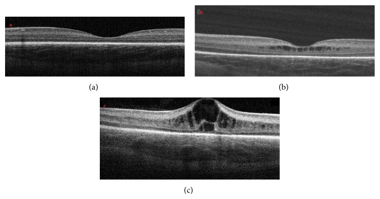
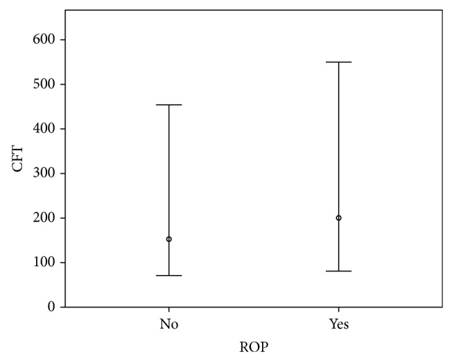
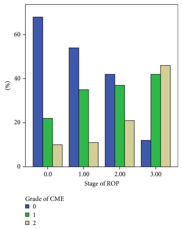
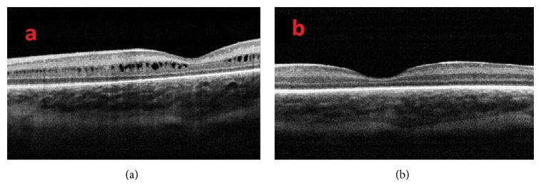
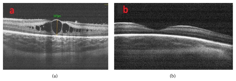
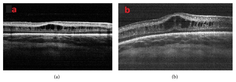
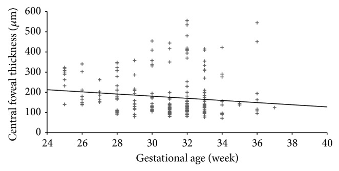
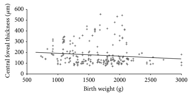
Purpose. To examine the macular findings obtained with spectral domain optical coherence tomography (SD OCT) in infants with retinopathy of prematurity (ROP). Materials and Methods. The macular SD OCT images of 190 premature infants were analyzed. Data regarding central foveal thickness (CFT), cystoid macular edema (CME), and cyst grading were compared. The relationships of CFT with gestational age and birth weight were investigated. Results. The results were obtained from 358 eyes of 179 infants (81 females and 98 males) of a mean gestational age of 30.9 ± 2.7 weeks and a mean birth weight of 1609 ± 477 g. ROP was diagnosed in 126 eyes and CME in 139 eyes. A significantly greater percentage of eyes with ROP were found to have CME (54%) compared to eyes without ROP (31%; P = 0.001). The incidence of CME was 46.3% for stage 1 ROP, 57.1% for stage 2, and 87.5% for stage 3. There was a weakly inverse correlation between CFT, gestational age, and birth weight (P = 0.025, r = -0.227; P = 0.002, r = -0.182, resp., Spearman correlation test). Conclusions. High-quality SD OCT images can be obtained from premature infants using the iVue system. Severity and frequency of CME in premature infants increase as stage of ROP increases.
Figures
References
-
- Dobson V., Quinn G. E., Summers C. G., Hardy R. J., Tung B. Visual acuity at 10 years in cryotherapy for retinopathy of prematurity (CRYO-ROP) study eyes: effect of retinal residua of retinopathy of prematurity. Archives of Ophthalmology. 2006;124(2):199–202. doi: 10.1001/archopht.124.2.199. - DOI - PubMed
-
- Early Treatment for Retinopathy of Prematurity Cooperative Group, Good W. V., Hardy R. J., Dobson V., Palmer E. A., Phelps D. L., Tung B., Redford M. Final visual acuity results in the early treatment for retinopathy of prematurity study. Archives of Ophthalmology. 2010;128(6):663–671. doi: 10.1001/archophthalmol.2010.72. - DOI - PMC - PubMed
-
- Fierson W. M., Palmer E. A., Biglan A. W., Flynn J. T., Petersen R. A., Phelps D. L. Retinopathy of prematurity guidelines. Pediatrics. 1998;101(6, article 1093) - PubMed
-
- International Committee for the Classification of Retinopathy of Prematurity The international classification of retinopathy of prematurity revisited. Archives of Ophthalmology. 2005;123(7):991–999. - PubMed
LinkOut - more resources
Full Text Sources
Other Literature Sources
