. 2014 Oct 2;5(4):228-290.
doi: 10.1080/21501203.2014.963720. Epub 2014 Oct 14.

Hymenoscyphus fraxineus vs. Hymenoscyphus albidus - A comparative light microscopic study on the causal agent of European ash dieback and related foliicolous, stroma-forming species

Affiliations
Free PMC article

Hymenoscyphus fraxineus vs. Hymenoscyphus albidus - A comparative light microscopic study on the causal agent of European ash dieback and related foliicolous, stroma-forming species

Hans-Otto Baral et al. Mycology. .
Free PMC article

Abstract

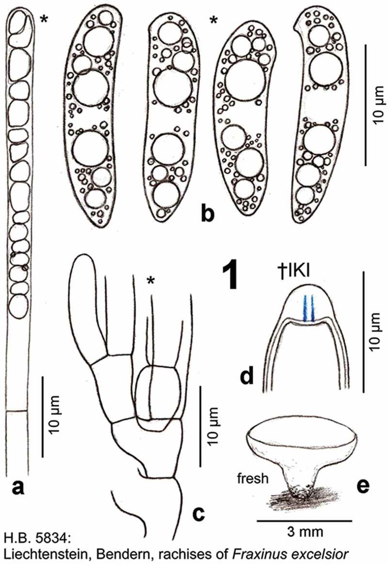
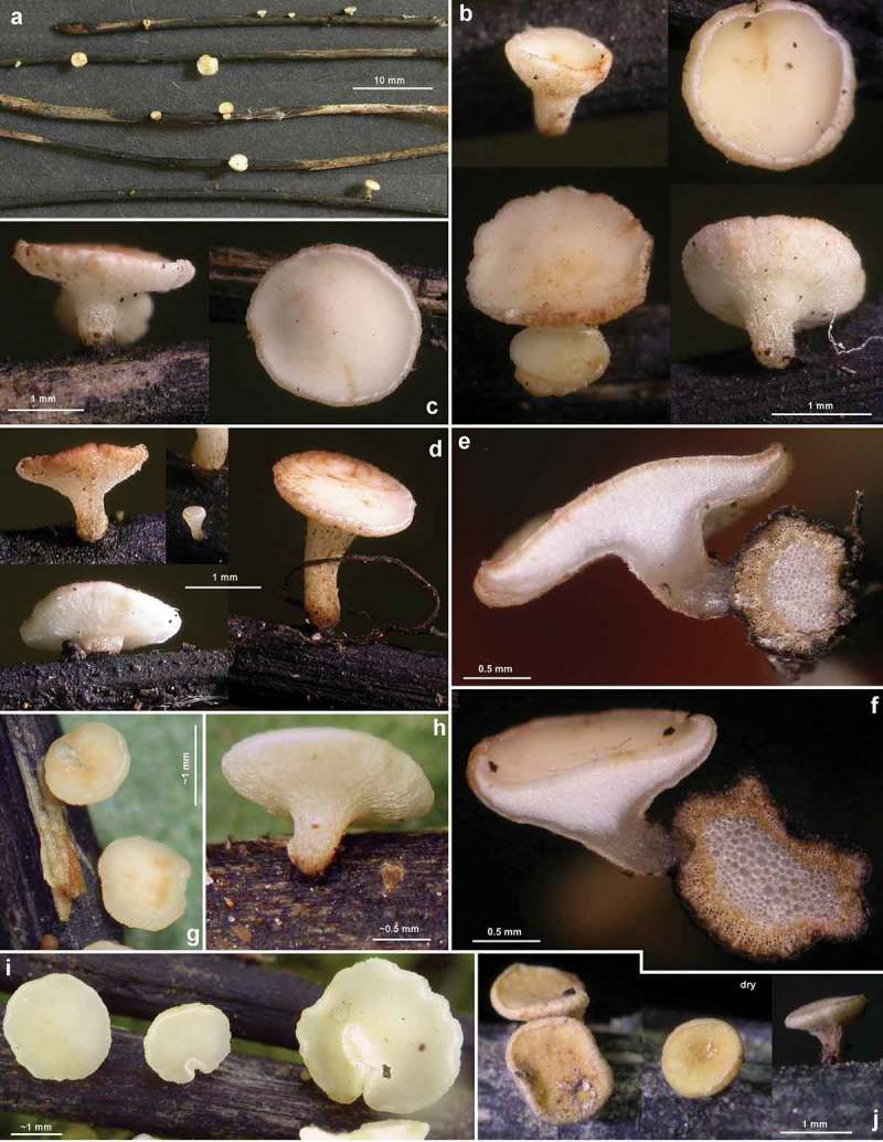
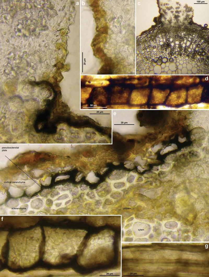
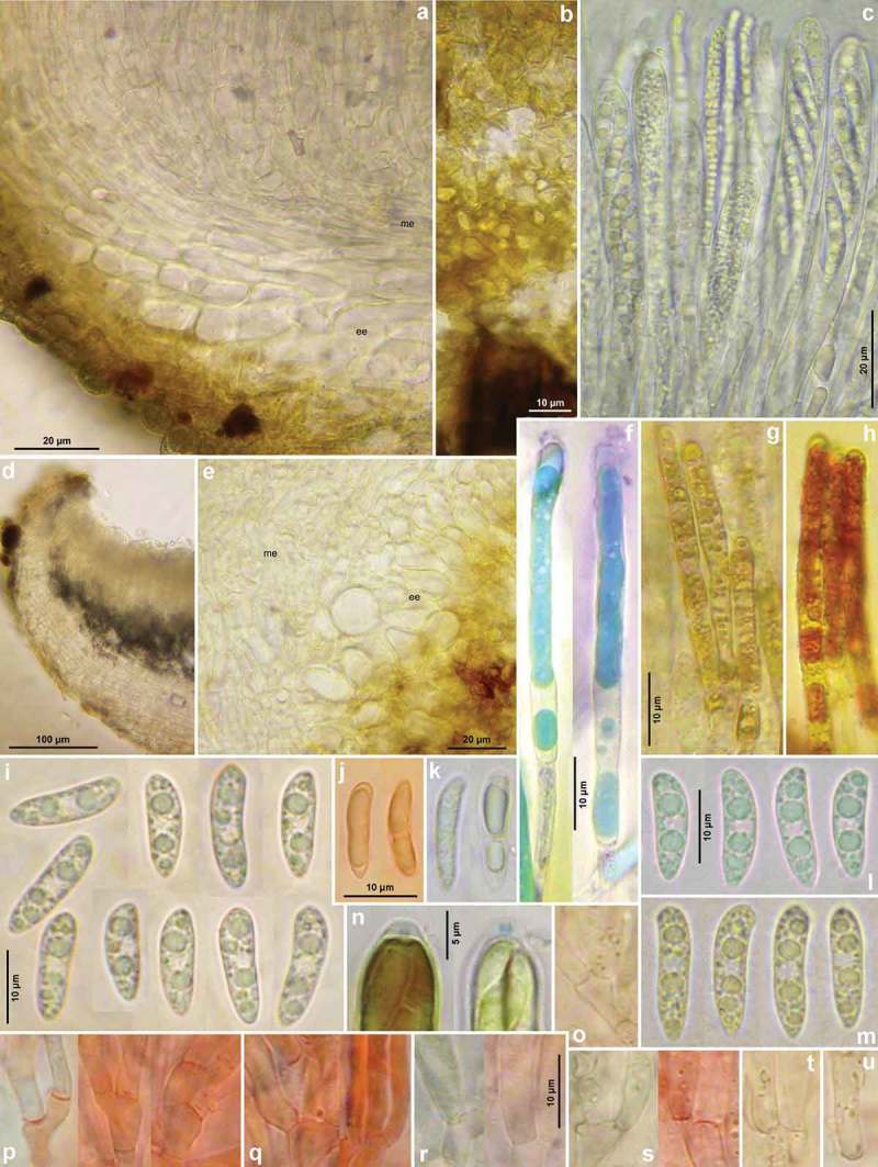
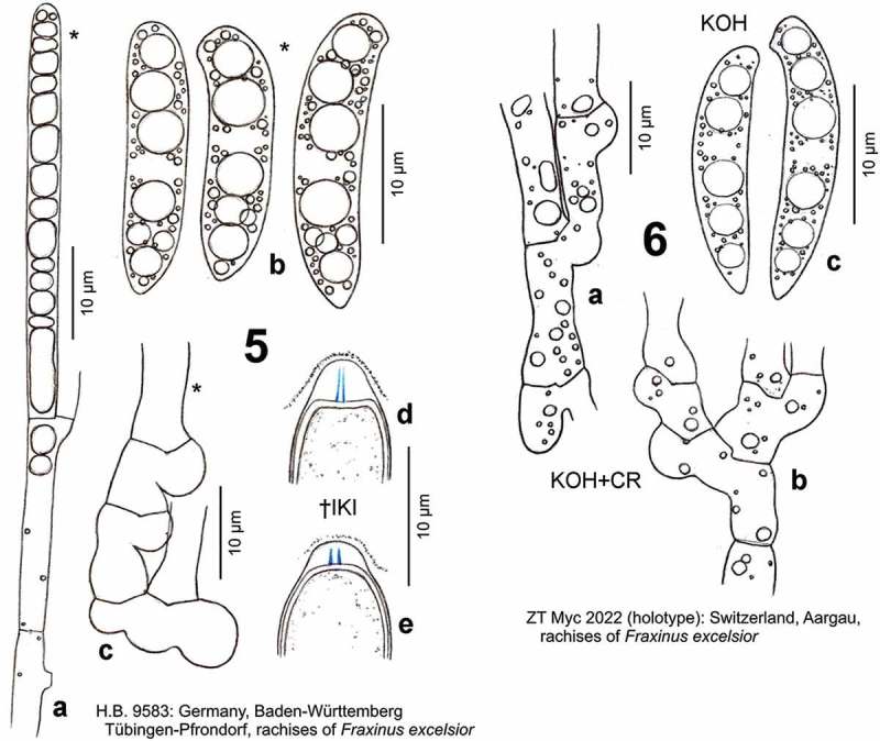
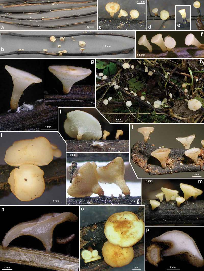
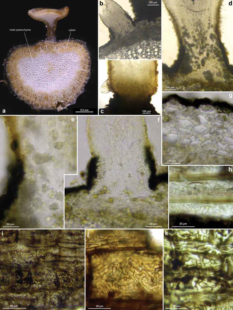
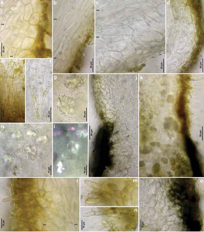
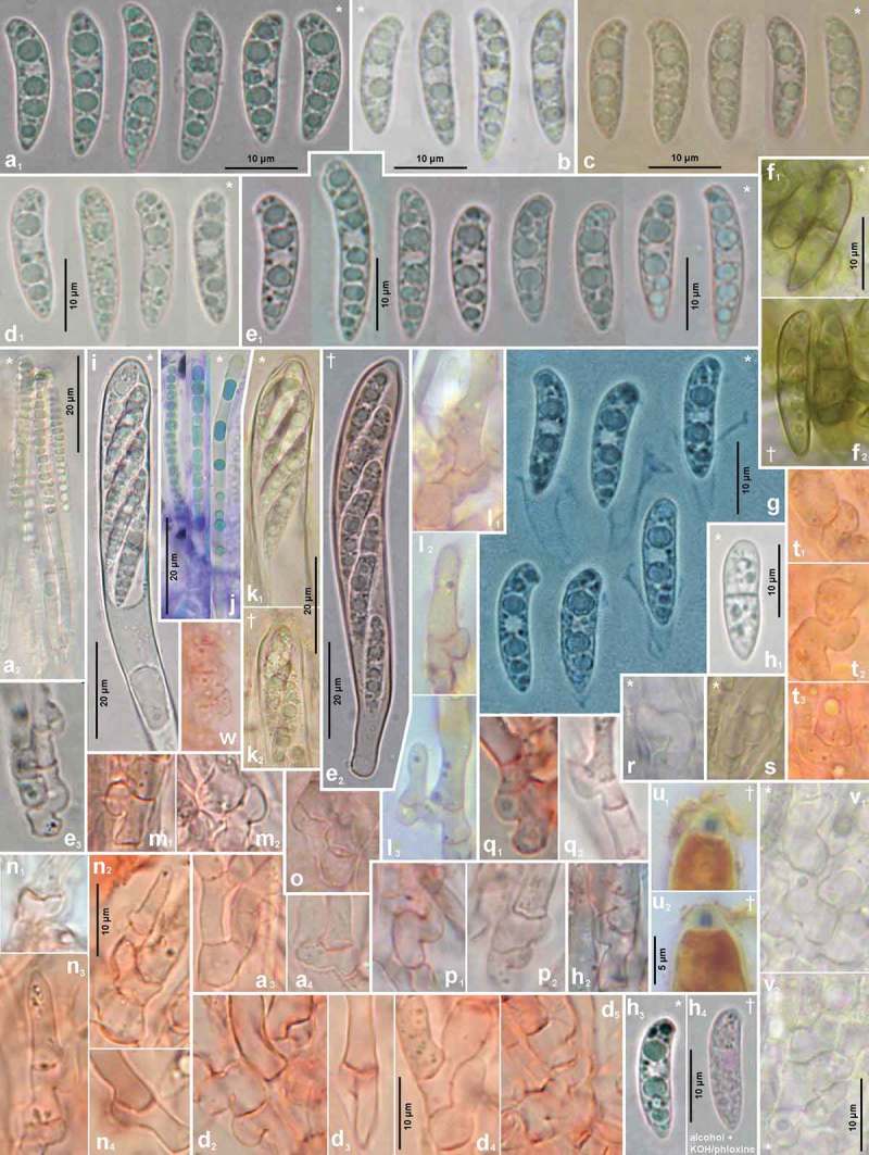
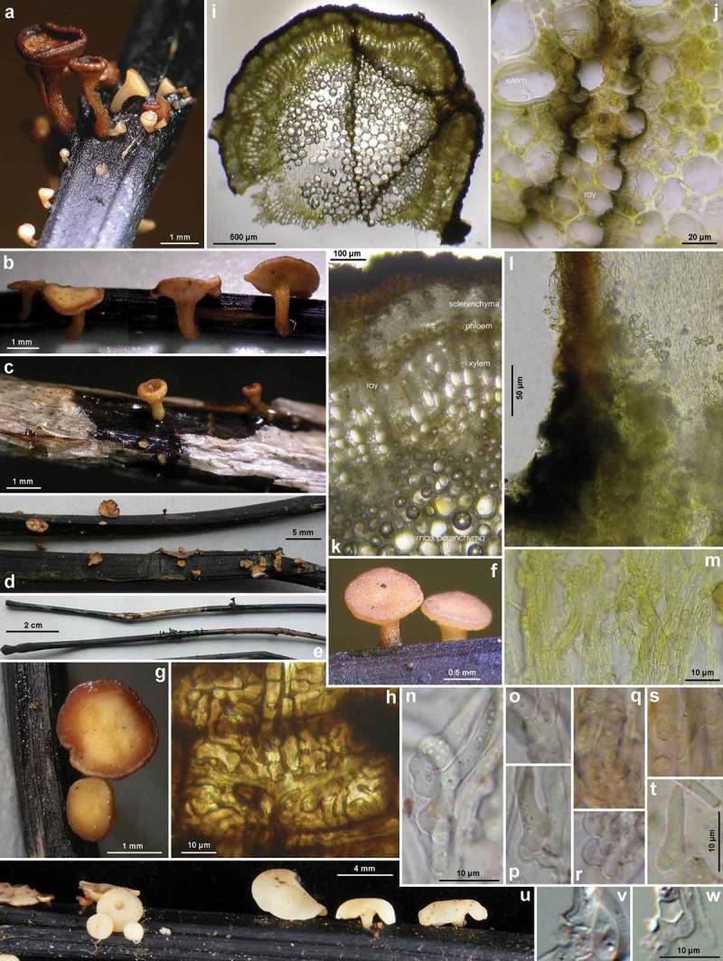
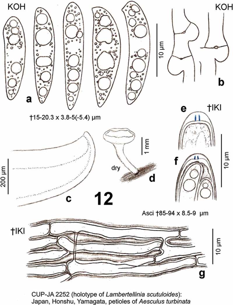
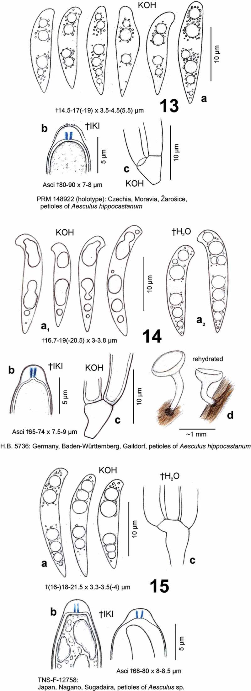
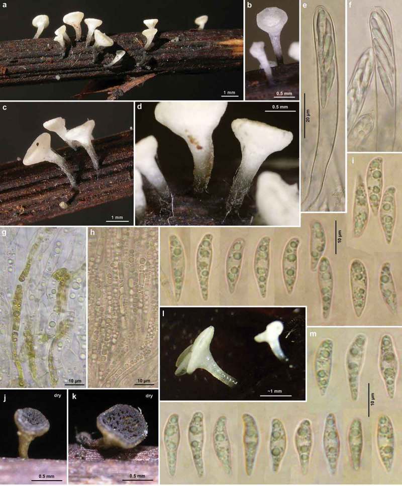
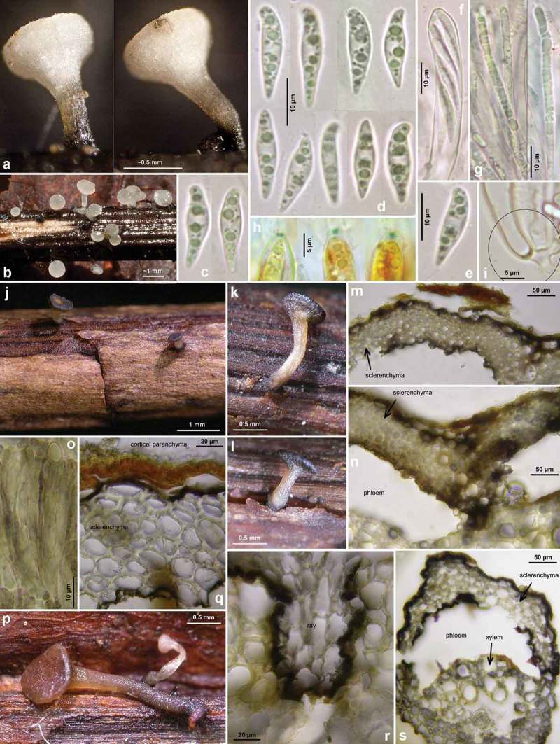
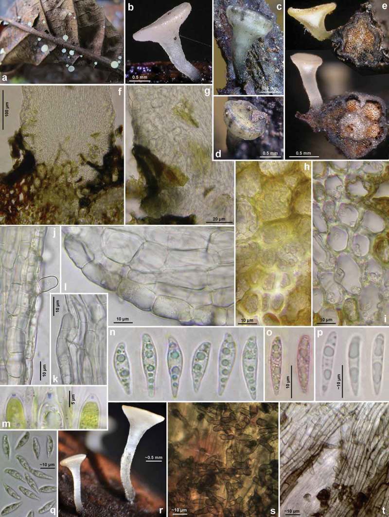
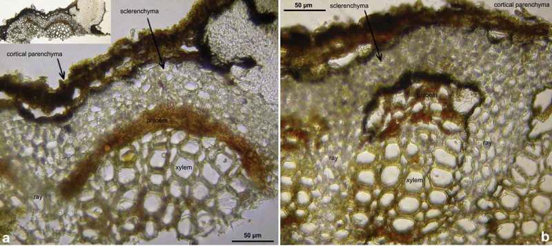
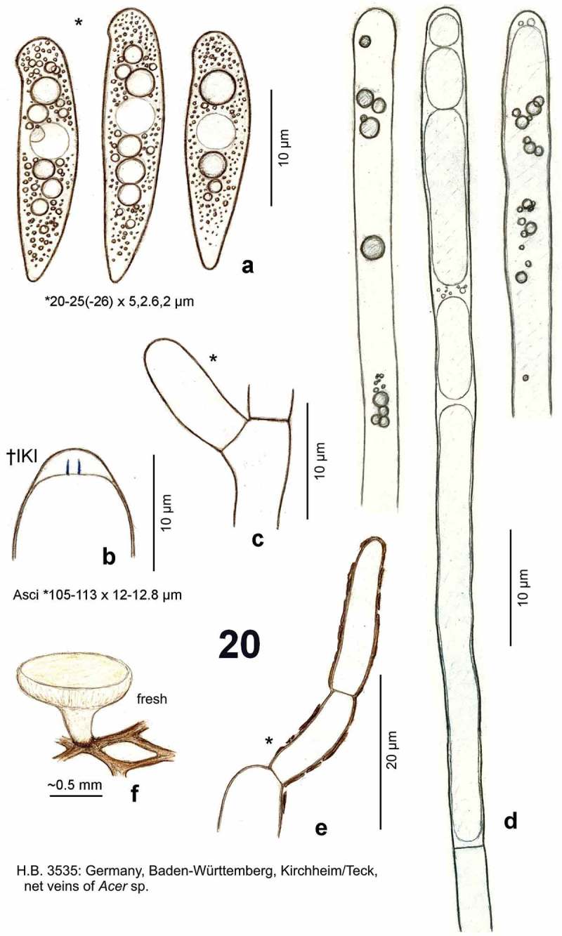
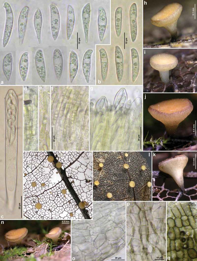
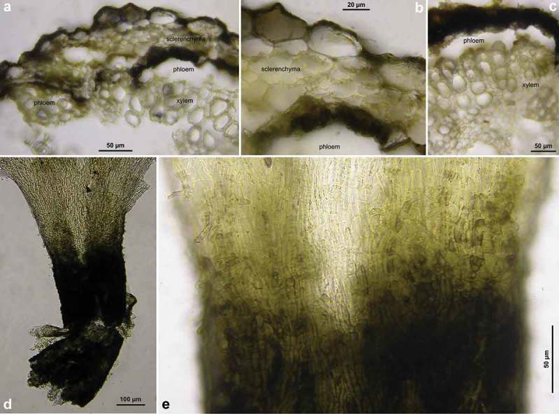
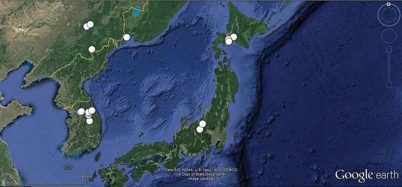
Five species of Hymenoscyphus that fruit on black stromatized parts of dead leaves of deciduous trees are presented, giving details on their morphological and ecological characteristics. Several of these species have previously been misplaced in rutstroemiaceous genera because of the presence of a substratal stroma. However, the heteropolar, scutuloid ascospores with an often hook-like lateral protrusion at the rounded apex and the ascus apical ring of the Hymenoscyphus-type represent two reliable morphological characteristics that, together with molecular data, provide clear evidence for their placement in the genus Hymenoscyphus (Helotiaceae). Among the species treated is Hymenoscyphus fraxineus (=Hymenoscyphus pseudoalbidus), the causal agent of the European ash dieback disease. Since 1992 this species started within Europe to replace the rather uncommon Hymenoscyphus albidus, which is likewise confined to leaves of Fraxinus. Hy. fraxineus has been recorded already since 1990 in Eastern Asia (Japan, Korea, northeast of China), where it had been initially misidentified as Lambertella albida (≡Hy. albidus). In these regions, it occurs as a harmless saprotroph on Fraxinus mandshurica and Fraxinus rhynchophylla, suggesting that those populations are native while the European ash dieback disease has a recent Eastern Asiatic origin. The distinctly higher genetic diversity found in Japanese Hy. fraxineus in contrast to European Hy. fraxineus supports this view. Genetic similarities between Japanese Hy. fraxineus and European Hy. albidus suggest that also Hy. albidus might be a descendant of Asian Hy. fraxineus, though having invaded Europe much earlier. However, consistent genetic deviation between European and Asian Hy. fraxineus at two nucleotide positions of the ITS region indicates that the European ash disease originates from a region different from the presently known areas in Eastern Asia. Our results underline the importance of detailed morphological studies in combination with molecular work. Hy. fraxineus was described from Europe as a cryptic species that differed from Hy. albidus by molecular data alone. However, the Hy. albidus/Hy. fraxineus species complex represents one of many examples within the ascomycetes in which subtle microscopic differences between closely related species, in this case the presence or absence of croziers at the ascus base, are strictly correlated with molecular characteristics. Two species that closely resemble Hy. albidus and Hy. fraxineus form pseudosclerotia in Aesculus leaves and again differ from each other mainly in the ascus base: Hymenoscyphus aesculi on Aesculus hippocastanum from Europe lacks croziers, whereas Hymenoscyphus honshuanus from Japan on Aesculus turbinata possesses croziers. Other taxa treated here include Hymenoscyphus vacini, a European species growing on stromatized net veins of skeletonized leaves of Acer, and Hymenoscyphus torquatus, a Chinese species on unidentified herbaceous stems. An equivalent stroma-forming North American species on leaves of Fraxinus, Rutstroemia longipes (Rutstroemiaceae), is discussed and compared. A key to the Hymenoscyphus species that form a dark stroma on leaves of Acer, Aesculus, Fraxinus, and Picrasma is provided.

Keywords: Acer; Aesculus; Fraxinus; Helotiaceae; Hy. honshuanus nom. nov. (for Lambertellinia scutuloides); Hy. torquatus comb. nov. (for Lambertella torquata); Hymenoscyphus aesculi comb. nov. (for Helotium/Lanzia aesculi); Rutstroemiaceae; croziers; homothallism; invasive species; molecular markers; morphology; pseudosclerotium; simple septa.


Figures

Figure 1.
Figure 1.
Hymenoscyphus albidus: a. paraphysis containing vacuolar bodies (=VBs); b. freshly ejected ascospores, containing oil drops (=LBs); c. simple-septate ascogenous hyphae without crozier formation; d. ascus apex with euamyloid apical ring (Hymenoscyphus-type); e. apothecium. – living state (except for d).
Figure 2.
Figure 2.
Hymenoscyphus albidus: a–j. apothecia emerging from black pseudosclerotia in rachises of Fraxinus excelsior (e–f: in median section). – a–i: fresh state, j: dry state. – a–f. H.B. 9699 (FR-PC, Granzay-Gript); g–h. 21.VII.2007 (CH-ZG, Unterägeri); i. 24.VI.2007 (FR-PC, Poitevin); j. H.B. 9454 (CH-LU, Aesch). – Phot. g–h: U. Graf, i: M. Hairaud.
Figure 3.
Figure 3.
Hymenoscyphus albidus: a–c, e. median section of stipe base (with internal crystals) and cross section of pseudosclerotium in ash rachis (the black demarcation line is restricted to the border between cortical parenchyma and sclerenchyma, hyphae present in all tissues of petiole); d, f–g. external view on pseudosclerotial plate (tangential section of rachis surface), cells of cortical parenchyma and sclerenchyma densely filled with subhyaline hyphae (textura epidermoidea). – All in living state. – a–g. H.B. 9699 (FR-PC, Granzay-Gript).
Figure 4.
Figure 4.
Hymenoscyphus albidus: a, d. median section of receptacle (ee = ectal excipulum, me = medullary excipulum); b. surface view on bright yellowish-ochraceous exudate covering the stipe base; c. paraphyses containing guttular VBs, immature and mature asci; e. median section of ectal excipulum at transition of stipe and receptacle; f–h. paraphyses (containing ± elongate VBs; f: under vital staining, g–h: VBs distorted in dead state); i–m. ascospores (containing large and small LBs; j–k: dead shrunken spores showing confluent LBs); n. ascus apices with euamyloid apical ring; p–u. ascus bases arising from simple septa. – Living state: a–d, f (in CRB), i, l–m; dead state: g (in H2O), h (in IKI), n (in IKI), j–k, p–u (in KOH or KOH+CR). – a–b, d–j, l. H.B. 9699 (FR-PC, Granzay-Gript); c, m. 16.VI.2007 (ibid.); k. H.B. 9611 (BE-LUX, Florenville); n. LUX 047699 (LU, Kehlen); o. ZT 3295 (CH-TI, Aquarossa); p. 21.VII.2007 (CH-ZG, Unterägeri); q. H.B. 9454 (CH-LU, Aesch); r. H.B. 1950 (DE-BW, Stockach); s. ZT 3299 (CH-BE, Eymatt); t. ZT 3294 (CH-TI, Lavorgo); u. ZT 3293 (FR-BN, Bellême). – Phot. c, l–m: M. Hairaud.
Figures 5–6.
Figures 5–6.
Hymenoscyphus fraxineus. 5a. paraphysis containing VBs; 5b. freshly ejected ascospores containing LBs, 6c. do., from inside asci; 5d–e. ascus apices with euamyloid apical ring of the Hymenoscyphus-type; 5c, 6a–b. ascogenous hyphae with crozier formation (in 6a–b containing LBs). – Living state: 5a–c; dead state: 5d–e, 6a–c.
Figure 7.
Figure 7.
Hymenoscyphus fraxineus (Europe). Apothecia emerging from black pseudosclerotia in rachises and veins of F. excelsior (n, p: in median/longitudinal/cross section). – All in fresh state. – a–b. H.B. 9700 (DE-BW, Tübingen); c, e. 23.VI.2011 (DE-BW, Heidelberg, Ziegelhausen); d. 28.V.2011 (ibid.), f. 17.VI.2011 (Tübingen); g. H.B. 9560 (DE-BW, Reutlingen); h. 30.VI.2012 (BE-LUX, Arlon, phot. G. Marson); i. 23.VI.2012 (Heidelberg, Lobbach); j, m. 18.VII.2011 (DE-TH, Sonneberg, phot. I. Wagner); k. 15.VII.2011 (Ziegelhausen); l, n, p. H.B. 9698 (Tübingen); o. 10.VII.2011 (Heidelberg, Boxberg).
Figure 8.
Figure 8.
Hymenoscyphus fraxineus (Europe): a. apothecium in median and rachis in cross section (the black demarcation line covers the outer face of the sclerenchyma), b–f. apothecial stipe in median section (with internal crystals), g. rachis in cross section (with external demarcation line, cells of sclerenchyma containing hyphae), h–k. external view on pseudosclerotial plate (tangential section of rachis surface); cells of cortical parenchyma densely filled with hyaline or ± brown hyphae (textura epidermoidea). – All in living state. – a. H.B. 9596 (DE-BW, Tübingen), b, f–g. H.B. 9589 (DE-BY, Amberg), c–e. H.B. 9593 (DE-SN, Chemnitz), h–k. H.B. 9588 (DE-BY, Hirschau).
Figure 9.
Figure 9.
Hymenoscyphus fraxineus (Europe): a–b, j–o. median section of stipe, showing golden-yellow external exudate and black basal stroma (ee = ectal excipulum, me = medullary excipulum); c–d. median section of receptacle at flanks (c) and margin (d); e–f. surface view of stipe; g–i, k. crystals in medullary excipulum near stipe base; k–n. hairs at stipe base. – All in living state (i: under polarized light). – a, g, k, l–n. H.B. 9593 (DE-SN, Chemnitz); b. H.B. 9698 (DE-BW, Tübingen); c, h–i. H.B. 9700 (Tübingen); d. H.B. 9571 (CH-LU, Hitzkirch); e–f. H.B. 9583 (Tübingen); j, o. H.B. 9589 (DE-BY, Amberg).
Figure 10.
Figure 10.
Hymenoscyphus fraxineus (Europe). a1, b, c, d1, e1, g, h3. ascospores (freshly ejected, containing LBs); h4. do. (LBs deteriorated); a2, j. paraphyses (containing VBs); e2, i, k. mature asci; f, h1. overmature ascospores (septate, partly with olive-brown wall); u. ascus apices with euamyloid apical ring; remaining figures: ascogenous hyphae with crozier formation. – Living state: a1–e1, a2, f1, g (in Waterman blue-black ink), h1, h3, i, j (in CRB), k1, r–s, v; dead state: k2 (in H2O), a3–a4, d2–d4, h2 (in CR); e2–e3, l–n, p–q (in CRSDS), f2 (in KOH), o, t (in KOH+CR), h4 (in ethanol + KOH + phloxine), u1–u2 (in IKI). – a. 28.V.2011 (DE-BW, Heidelberg, Ziegelhausen), b. H.B. 9572 (CH-LU, Emmenbrücke), c. H.B. 8220 (DE-ST, Chemnitz), d. 31.VII.2011 (Heidelberg, Schönbrunn), e. 10.VII.2011 (Heidelberg, Boxberg), f. H.B. 9593 (Chemnitz), g. 14.VII.2012 (Heidelberg), h. 23.VII.2011 (DE-BW, Speyer, Böhl-Iggelheim), i. 15.VII.2011 (Ziegelhausen), j. H.B. 9599 (DE-BW, Tübingen), k. H.B. 8509 (DK-Sjaelland, Sorø), l. 18.VII.2011 (DE-TH, Sonneberg), m. 2.VII.2011 (Heidelberg, Handschuhsheim), n. 5.VII.2011 (Ziegelhausen), o. ZT 3298 (CH-BE, Belp), p. 23.VI.2011 (Ziegelhausen), q. 23.VI.2012 (Heidelberg, Lobbach), r. H.B. 9698 (Tübingen), s. H.B. 9700 (Tübingen), t. ZT 3297 (CH-BE, Burgdorf), u. 8.VII.2011 (Sonneberg), v. H.B. 9583 (Tübingen). – Phot. l1–3, u1–2: I. Wagner.
Figure 11.
Figure 11.
Hymenoscyphus fraxineus (Japan). a–g, u. Apothecia emerging from black pseudosclerotia in rachises of Fraxinus mandshurica (u: fresh; a–g: rehydrated); i–k. pseudosclerotium in cross section (i–j: several stromata in one rachis, delimited by black demarcation lines that occur in the xylem along radial rays and through the mark parenchyma); l. median section of stipe base (with black basal surface and internal crystals); m. surface view on ectal excipulum near margin (with yellow exudate); n–t, v–w. ascogenous hyphae with croziers. – Dead state (h–k, m in H2O; l in KOH; n–t KOH+CR; v–w in phloxine B). – Nagano: a, e, k. TNS-F-40074 (Sugadaira); b, g. TNS-F-12761 (ibid.); f, h. TNS-F-17817 (ibid.); v. TNS-F-12503 (Yuzawa); Hokkaido: c, m, s–t. TNS-F-52060 (Tomakomai); d, l, n–r, u. TNS-F-40043 (ibid.); i, j, w. TNS-F-52061 (Sapporo). – Phot. u: YJ Zhao.
Figure 12.
Figure 12.
Hymenoscyphus honshuanus (holotype of Lambertellinia scutuloides): a. ascospores (partly from inside asci, containing LBs); b. ascogenous hyphae with crozier formation; c. median section of receptacle; d. apothecium; e–f. ascus apices with euamyloid apical ring (Hymenoscyphus-type); g. ectal excipulum at flanks (median section), with gelatinized walls. – All in dead state.
Figures 13–15.
Figures 13–15.
Hymenoscyphus aesculi. a. ascospores (partly from inside asci, containing LBs); b. ascus apices with euamyloid apical ring (Hymenoscyphus-type); c. ascus bases arising from simple septa; d. apothecia. – All in dead state.
Figure 16.
Figure 16.
Hymenoscyphus aesculi: a–d, l. fresh apothecia; j–k. dry apothecia (phot. over 3 years after collection); g. cortical ectal excipulum of receptacle in surface view (VBs in living cells, distorted brass-yellow cytoplasm of scattered external hyphae); h. paraphyses in squash mount (containing VBs); e–f. turgescent mature asci; i, m. ascospores (freshly ejected, containing LBs). – All in living state (except for j–k, brass-yellow cells in g). – a–f, h–i. H.B. 8914 (DE-NI, Hamburg); g. H.B. 9843 (DE-BW, Tübingen); l–m. H.B. 9417 (DE-SN, Chemnitz). – Phot. l: B. Mühler.
Figure 17.
Figure 17.
Hymenoscyphus aesculi: a–b. fresh apothecia; j–l, p. rehydrated apothecia (colour was originally white except for base of stipe); f–g, o. asci and paraphyses(o: with olive secondary pigment especially in paraphyses); h. ascus apices with euamyloid apical ring; i: simple-septate ascus base; c–e. ascospores; m–n, q–s. cross section through cortical region of petioles, with black demarcation line surrounding the sclerenchyma on outer and inner face (cortical parenchyma above and phloem beneath being entirely decomposed). – Living state: c–g; dead state: h (in IKI), o (in H2O). – a–i. E.R.D. 5685 (ES-Asturias, Somiedo); j–o. H.B. 5736 (DE-BW, Gaildorf); p–s. TNS-F-12758 (JP-Honshu, Nagano, Sugadaira). – Phot. a–i: E. Rubio.
Figure 18.
Figure 18.
Hymenoscyphus aesculi: a–b, e, r. fresh apothecia (e: in median section, petioles in cross section); c–d. senescent apothecia (with dark bluish-olive patches on receptacle); f–g. stipe base in median section, showing erumpent growth and absence of crystals; h. cortical parenchyma and sclerenchyma below insertion of apothecial stipe, densely filled with intracellular hyphae; i. sclerenchyma forming a radial ray, loosely filled with hyphae; j–l. ectal excipulum at stipe and (l) at lower flanks in median section, showing VBs in cortical cells; m. ascus apices with euamyloid apical ring; n–q. ascospores (n–o, q: freshly ejected); s–t. surface view on stipe base, showing brown cortical cells and hairs. – Living state, except for m (in KOH+IKI), o (in KOH+CRB), p (in H2O?). – a. 31.VII.2011 (DE-HS, Darmstadt), b–p. H.B. 9701 (Darmstadt), q–t. C.Y. F/2194 (UK-Yorkshire, Halifax). – Phot. a, p: H. Lotz, q–t: C. Yeates.
Figure 19.
Figure 19.
Hymenoscyphus aesculi: a–b. petiole in median section, showing black stroma and erumpent apothecial growth; pseudosclerotial plate occurring between cortical parenchyma and sclerenchyma but also between sclerenchyma and vascular bundle (phloem/xylem). – a–b. H.B. 9701 (DE-HS, Darmstadt).
Figure 20.
Figure 20.
Hymenoscyphus vacini: a. ascospores (freshly ejected, containing large and small LBs); b. ascus apex with euamyloid apical ring (Hymenoscyphus-type); c. simple-septate ascus base; d. paraphyses containing elongate, low-refractive vacuoles, partly with scattered internal globose VBs of high refractivity; e. cortical hypha of ectal excipulum covered by olive-brown exudate; f. apothecium emerging from net veins of Acer leaf. – Living state (except for b).
Figure 21.
Figure 21.
Hymenoscyphus vacini (on leaves of Acer): a–b. freshly ejected ascospores (containing LBs); c. mature turgescent ascus; d–f. paraphyses (containing pale yellowish VBs); g. mature turgescent asci projecting beyond paraphyses; h–n. fresh apothecia; o. ectal excipulum at lower flanks in median section; p. ectal excipulum at mid flanks in surface view, with olive-brown exudate; q. do., towards base of stipe. – All in living state. – a, c–k, m–q. H.B. 9590 (DE-ST, Merseburg); b, l: H.B. 8296 (DE-BY, München).
Figure 22.
Figure 22.
Hymenoscyphus vacini: a–c. cross section through cortical region of net vein (in H2O), showing black demarcation line surrounding the sclerenchyma on outer and inner face (cortical parenchyma above and phloem beneath being entirely decomposed); fungal hyphae visible inside cells; d–e. apothecial stipe in surface view (slightly squashed, in KOH). – a–c. H.B. 9590 (DE-ST, Merseburg); d–e. H.B. 456 (DE-BW, Stuttgart).
Figure 23.
Figure 23.
Distribution of Hymenoscyphus albidus within Europe according to trustable records known to us (those for which data about the host and the presence of a stroma were not available are omitted, also all those doubtful records which fall in the period of invasion by Hy. fraxineus).
Figure 24.
Figure 24.
Known records of Hymenoscyphus fraxineus in Eastern Asia.

References

    1. Allescher A. Verzeichnis in Süd-Bayern beobachteter Pilze, IV. Abteilung: Hysteriaceae, Hiscomycetaceae et Tuberaceae. Bericht des botanischen Vereins in Landshut. 1898;15:1–138.
    1. Arendholz W-R. Hamburg: Universität Hamburg; 1979. Morphologisch-taxonomische Untersuchungen an blattbewohnenden Ascomyceten aus der Ordnung der Helotiales. dissertation.
    1. Arnolds E, Kuyper ThW, Noordeloos ME, editors. Overzicht van de paddestoelen in Nederland. 2nd revised ed. Wijster: Nederlandse Mycologische Vereniging; 1999.
    1. Baier U, Thiel J, Stürtz M, Völker L. Waldschutzbericht für 2010 und prognostische Hinweise für 2011. [cited 2013 Nov 6;];2010 http://www.thueringenforst.de/imperia/md/content/folder/waldschutz/walds...
    1. Bakys R. Uppsala: Swedish University of Agricultural Sciences; 2013. Dieback of Fraxinus excelsior in the Baltic Sea region: associated fungi, their pathogenicity and implications for silviculture. thesis.

LinkOut - more resources