. 2015 Feb;18(2):157-65.
doi: 10.1089/jmf.2014.3187. Epub 2014 Dec 29.

Deer bone extract prevents against scopolamine-induced memory impairment in mice

Affiliations

Deer bone extract prevents against scopolamine-induced memory impairment in mice

Chun Nan Du et al. J Med Food. 2015 Feb.

Abstract

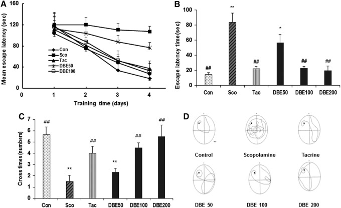
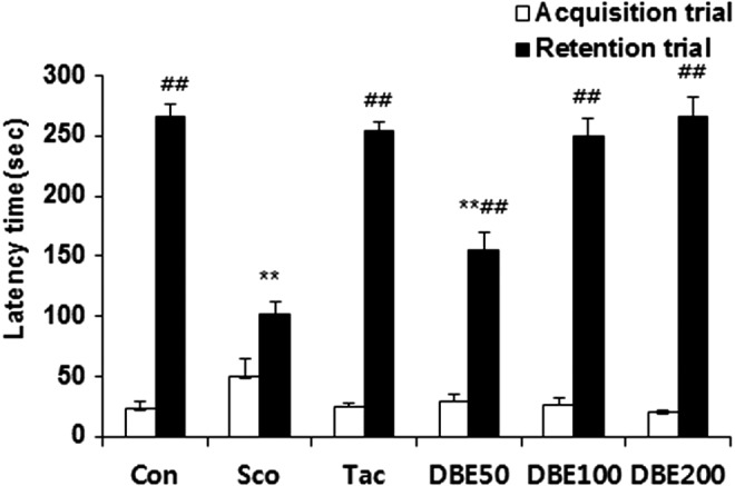
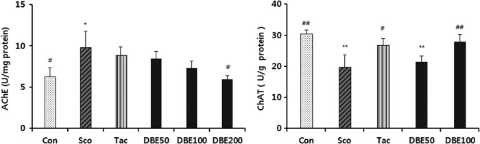
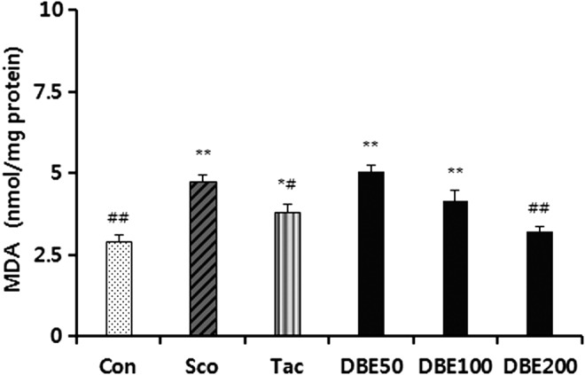
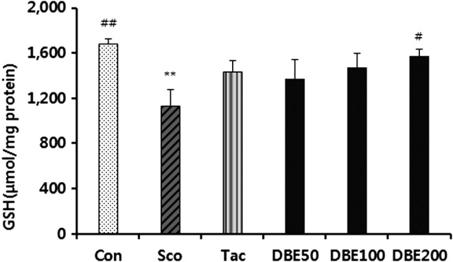
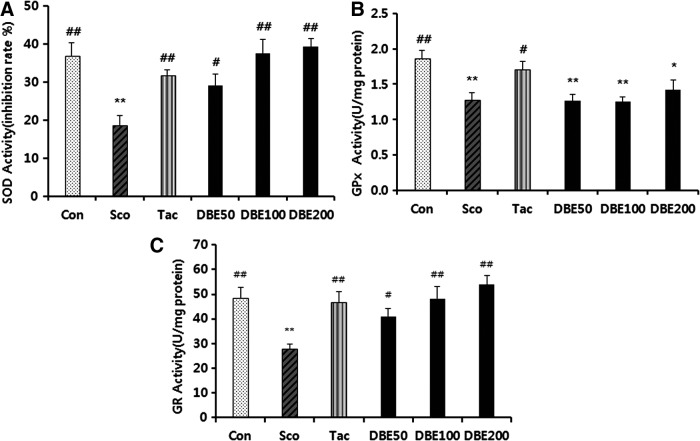
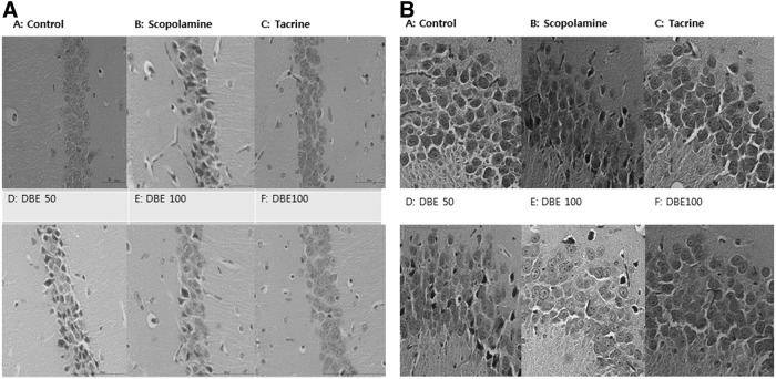
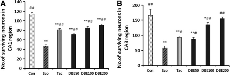
Deer bone has been used as a health-enhancing food as well as an antiaging agent in traditional Oriental medicine. Recently, the water extract of deer bone (DBE) showed a neuroprotective action against glutamate or Aβ1-42-induced cell death of mouse hippocampal cells by exerting antioxidant activity through the suppression of MAP kinases. The present study is to examine whether DBE improves memory impairment induced by scopolamine. DBE (50, 100 or 200 mg/kg) was administered orally to mice for 14 days, and then scopolamine (2 mg/kg, i.p.) was administered together with DBE for another 7 days. Memory performance was evaluated in the Morris water maze (MWM) test and passive avoidance test. Also, brain acetylcholinesterase (AChE) and choline acetyltransferase (ChAT) activity, biomarkers of oxidative stress and the loss of neuronal cells in the hippocampus, was evaluated by histological examinations. Administration of DBE significantly restored memory impairments induced by scopolamine in the MWM test (escape latency and number of crossing platform area), and in the passive avoidance test. Treatment with DBE inhibited the AChE activity and increased the ChAT activity in the brain of memory-impaired mice induced by scopolamine. Additionally, the administration of DBE significantly prevented the increase of lipid peroxidation and the decrease of glutathione level in the brain of mice treated with scopolamine. Also, the DBE treatment restored the activities of antioxidant enzymes such as superoxide dismutase, glutathione peroxidase, and glutathione reductase to control the level. Furthermore, scopolamine-induced oxidative damage of neurons in hippocampal CA1 and CA3 regions were prevented by DBE treatment. It is suggested that DBE may be useful for memory improvement through the regulation of cholinergic marker enzyme activities and the suppression of oxidative damage of neurons in the brain of mice treated with scopolamine.

Keywords: antioxidant defense system; cholinergic enzymes; deer bone extract; memory; scopolamine.


Figures

<b>FIG. 1.</b>
FIG. 1.
Effect of deer bone extract (DBE) on mean latency time (A), probe trial (B), and number of crossing platform area (C) in trial sessions of the Morris water maze test. DBE (50, 100, or 200 mg/kg, p.o.), tacrine (10 mg/kg, p.o.) or saline was administered to the mice. Thirty minutes later, the mice of DBE, tacrine, or control groups were treated with scopolamine (Sco, 2 mg/kg, i.p.) and subjected to the Morris water maze test. Probe trial sessions were performed for 120 s. Representative swimming paths of mice from each group subjected to Morris water maze test on the fifth training trial day (D). Data represent mean±standard error of the mean (SEM) (n=9/group); *P<.05, and **P<.01, compared with the control group. ##P<.01, compared with the scopolamine-treated group.
<b>FIG. 2.</b>
FIG. 2.
Effect of DBE on the step-through passive avoidance test. At 1 h before the test, DBE (50, 100 or 200 mg/kg, p.o.) or tacrine (10 mg/kg, p.o.) was administered to the mice. Thirty minutes later, the mice were treated with scopolamine (Sco, 2 mg/kg, i.p.) and tested for passive avoidance. To assess the effect of DBE on passive avoidance, DBE (50, 100 or 200 mg/kg, p.o.) was administered to mice 60 min before the tests. Data represent mean±SEM (n=9/group). **P<.01 compared with the control group. ##P<.01 compared with the scopolamine-treated group.
<b>FIG. 3.</b>
FIG. 3.
Effects of DBE on acetylcholinesterase (AChE) and choline acetyltransferase (ChAT) activities in brains of mice with memory impairment induced by scopolamine. Animals were decapitated 60 min after the passive avoidance test, and the brain was homogenized to assay AChE and ChAT activities. Data represent mean±SEM (n=6/group). *P<.05 and **P<.01 compared with the control group. #P<.05 and ##P<.01 compared with the scopolamine-treated group.
<b>FIG. 4.</b>
FIG. 4.
Effects of DBE on malondialdehyde (MDA) level in brains of mice with memory impairment induced by scopolamine. Animals were decapitated 60 min after the passive avoidance test, and the brain was homogenized to assay TBA reactive substances activity. Data represent mean±SEM (n=6/group). *P<.05 and **P<.01 compared with the control group. #P<.05 and ##P<.01 compared with the scopolamine-treated group.
<b>FIG. 5.</b>
FIG. 5.
Effects of DBE on glutathione (GSH) level in brains of mice with memory impairment induced by scopolamine. Animals were decapitated 60 min after the passive avoidance test, and the brain was homogenized to assay GSH level. Data represent mean±SEM (n=6/group). **P<.01 compared with the control group. #P<.05 and ##P<.01 compared with the scopolamine-treated group.
<b>FIG. 6.</b>
FIG. 6.
Effect of DBE on the activities of antioxidant enzymes (A) SOD, (B) GPx, and (C) GR in the brains of mice with memory impairment induced by scopolamine. Data represent mean±SEM (n=6/group). *P<.05 and **P<.01 compared with the control group. #P<.05 and ##P<.01 compared with the scopolamine-treated group.
<b>FIG. 7.</b>
FIG. 7.
Effect of DBE on hippocampal CA1 (A) and CA3 (B) regions of brains from mice treated with scopolamine. Histological sections of the brain tissue showing neurological lesions (A–F). A part of the brain tissue was fixed in 10% neutral formalin solution, and the formalin-fixed brain tissues were processed and embedded in paraffin. Serial coronal sections (4 μm in thickness) were obtained, and stained with Hematoxylin and Eosin. The histopathological examination was assessed under a light microscope (a 400-fold-magnification).
<b>FIG. 8.</b>
FIG. 8.
The number of surviving neurons in hippocampal CA1 (A) and CA3 (B) regions. Data represent mean±SEM (n=6/group). *P<.05 and **P<.01 compared with the control group. #P<.05 and ##P<.01 compared with the scopolamine-treated group.

References

    1. Coyle JT, Puttfarcken P: Oxidative stress, glutamate, and neurodegenerative disorders. Science 1993;262:689–695 - PubMed
    1. Halliwell B: Oxidative stress and neurodegeneration: Where are we now. J Neurochem 2006;97:1634–1658 - PubMed
    1. Francis PT, Palmer AM, Snape M, et al. : The cholinergic hypothesis of Alzheimer's disease: A review of progress. J Neurol Neurosurg Psychiatry 1999;66:137–147 - PMC - PubMed
    1. Kwon SH, Ma SX, Joo HJ, et al. : Inhibitory effects of Eucommia ulmoides Oliv. bark on scopolamine-induced learning and memory deficits in mice. Biomol Ther 2013;21:462–469 - PMC - PubMed
    1. Collerton D: Cholinergic function and intellectual decline in Alzheimer's disease. Neuroscience 1986;19:1–28 - PubMed

Publication types

MeSH terms

LinkOut - more resources