Review
. 2015 Jun;27(6):487-97.
doi: 10.1111/jne.12252.

Neuroendocrine-autonomic integration in the paraventricular nucleus: novel roles for dendritically released neuropeptides

Affiliations
Review

Neuroendocrine-autonomic integration in the paraventricular nucleus: novel roles for dendritically released neuropeptides

J E Stern. J Neuroendocrinol. 2015 Jun.

Abstract

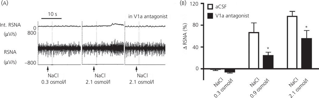
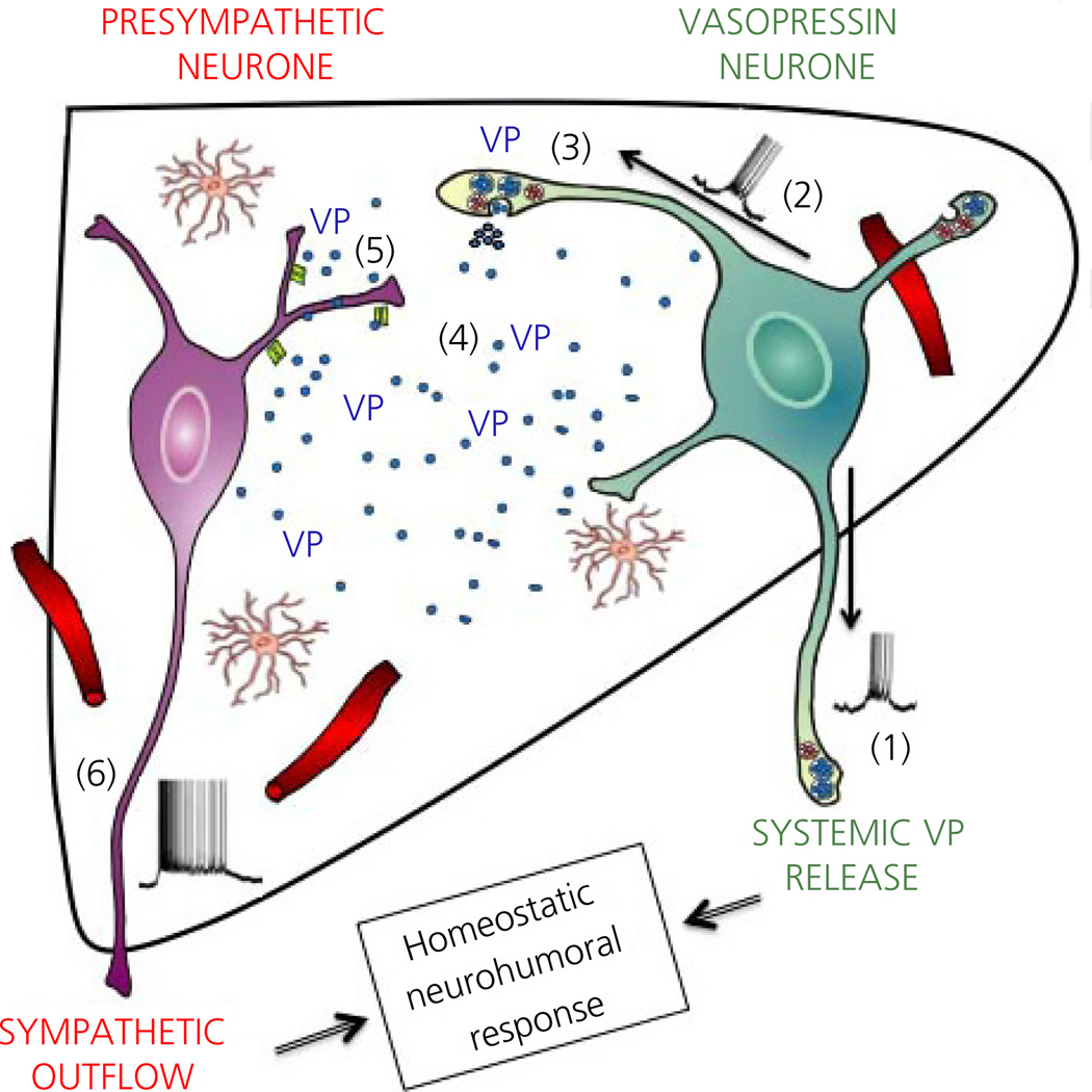
Communication between pairs of neurones in the central nervous system typically involves classical 'hard-wired' synaptic transmission, characterised by high temporal and spatial precision. Over the last two decades, however, knowledge regarding the repertoire of communication modalities used in the brain has notably expanded to include less conventional forms, characterised by a diffuse and less temporally precise transfer of information. These forms are best suited to mediate communication among entire neuronal populations, now recognised to be a fundamental process in the brain for the generation of complex behaviours. In response to an osmotic stressor, the hypothalamic paraventricular nucleus (PVN) generates a multimodal homeostatic response that involves orchestrated neuroendocrine (i.e. systemic release of vasopressin) and autonomic (i.e. sympathetic outflow to the kidneys) components. The precise mechanisms that underlie interpopulation cross-talk between these two distinct neuronal populations, however, remain largely unknown. The present review summarises and discusses a series of recent studies that have identified the dendritic release of neuropeptides as a novel interpopulation signalling modality in the PVN. A current working model is described in which it is proposed that the activity-dependent dendritic release of vasopressin from neurosecretory neurones in the PVN acts in a diffusible manner to increase the activity of distant presympathetic neurones, resulting in an integrated sympathoexcitatory population response, particularly within the context of a hyperosmotic challenge. The cellular mechanism underlying this novel form of intercellular communication, as well as its physiological and pathophysiological implications, is discussed.

Keywords: hypothalamus; neuroendocrine; osmotic; sympathetic; vasopressin.


Figures

Fig. 1
Fig. 1
Compartmentalised distribution of functionally distinct neurona populations within the hypothalamic paraventricular nucleus (PVN). Graph of the PVN showing its distinct anatomical subdivisions containing neurones that project to the median eminence (grey colour; mpd, medial parvocellular subdivision dorsal; pv, periventricular subnucleus), posterior pituitary [red and green colours; containing primarily oxytocin (OT) and vasopressin (VP) neurones, respectively; pml, posterior magnocellular lateral; pmm, posterior magnocellular medial] and brainstem/spinal cord (light blue colour; dp, dorsal parvocellular subnucleus; lp, lateral parvocellular subnucleus; mpv, media parvocellular ventral subnucleus). Modified from Hatton (70). The inset shows a coronal section of the PVN (bregma = −1.80) showing immunoreactive OT (red), VP (green) and presympathetic retrogradely labelled neurones that innervate the rostral ventrolateral medulla in the brainstem. Scale bar = 100 µm. 3V, third ventricle.
Fig. 2
Fig. 2
Close anatomical inter-relationships among the dendrites of magnocellular neurosecretory and presympatehtic paraventricular nucleus (PVN) neurones. (a1) Photomicrograph showing the topographical segregation between immunoreactive magnocellular vasopressin (VP) neurones (VP, green) and retrogradely labelled presympathetic PVN-rostral ventrolateral medulla neurones. Note the clear anatomical segregation between the somata of these two PVN neurona populations. In (a2) and (a3), the squared regions are shown at progressively higher magnification. Note in (a2) and (a3) that thick and varicose immunoreactive VP dendrites extend beyond the compartment of VP neurones, into the compartment containing presympathetic neurones, becoming in close apposition with somata and dendrites of the latter. Vertical and horizontal bars in (a1) point dorsally and medially, respectively. Modified from Brussaard et al. (56).
Fig. 3
Fig. 3
Expression of functional vasopressin (VP) V1a receptors in presympathetic paraventricular nucleus (PVN) neurones. (a1) Representative photomicrograph showing a couple of retrogradely-labelled PVN-rostral ventrolateral medulla (RVLM) neurones (blue) that have dense V1a receptor immunoreactivity (green, a2). In (a3), the squared area in (a1) is magnified to better depict dendritic V1a immunoreactive clusters (arrows). (b) Single-cell V1a mRNA expression in identified PVN-RVLM and enhanced green fluorescent protein (eGFP) -VP neurones. A nontemplate negative control is shown on the right, and a small piece of a DNA ladder is shown on the left. Scale bars: (a1, a2) = 20 µm and (a3) = 2.5 µm. (C1) VP (VP, 1 µM) puffed directly onto a presympathetic PVN-RVLM neurone evokes a burst of action potentials. (C2) In the presence of tetrodotoxin (0.5 µM), puffs of VP (1 µM) of decremental durations evoke membrane depolarisation in a proportionally decremental manner. The arrow indicates an evoked Ca2+ spike. (C3) Summary data showing that the increased action potential firing evoked by VP was completely blocked by a V1a receptor antagonist. **P < 0.01 and ***P < 0.001. Modified from Brussaard et al. (56). aCSF, artificial cerobrospinal fluid.
Fig. 4
Fig. 4
Dendritically-released vasopressin (VP) mediates cross-talk between magnocelluar neurosecretory and the presympathetic paraventricular nucleus (PVN) neurones. (a) Hypothalamic slice obtained from an enhanced green fluorescent protein (eGFP)-VP rat that received an injection of the retrograde tracer rhoda-mine beads in the rostral ventrolateral medulla (RVLM). Magnocellular VP neurones are shown in green and presympathetic PVN neurones are shown in red The inset shows eGFP-stained fibers in the posterior pituitary. Vertical and horizontal bars point dorsally and medially, respectively. (b1) Sample of another hypothalamic slice showing a patched presympathetic PVN-RVLM neurone (red, asterisk) and neighbouring eGFP-VP neurones (green, 1–4). (b2) Laser photolysis of caged NMDA onto eGFP-VP neurones (1,2, orange flashes) resulted in a delayed (approximately 5 s) membrane depolarisation and increased firing activity in the patched presympathetic neurone. (c1) Sample pair of intracellularly labelled (Alexa 633, blue, arrows) PVN neurones during simultaneous dual-patch recordings. The neurone at the left (single arrow) was identified as an eGFP-VP (cyan), whereas the neurone to the right (double arrow) was a retrogradely-labelled PVN-RVLM (purple). (c2) Bursts of action potentials evoked in the eGFP-VP neurone via current injection through the patch pipette resulted in a delayed membrane depolarisation and increased firing discharge in the paired PVN-RVLM neurone. Modified from Brussaard et al. (56). 3V, third ventricle.
Fig. 5
Fig. 5
Dendritically-released vasopressin (VP) contributes to osmotieally-driven renal sympathetic nerve activity (RSNA). (a) Recordings or RSNA after intracar-otid infusions of an isosmotic (NaCl 0.3 osmol/l) or hyperosmotic (NaCl 2.1 osmol/l) solution, in the absence or presence of bilateral microinjections of the V1a receptor antagonist (0.4 mg/ml) into the paraventricular nucleus (PVN). (b) Summary data showing a dose-dependent increase of RSNA after intracarotid infusions of NaCI. Note the blunted sympathetic response after an intra-PVN microinjection of the V1a receptor antagonist [*P < 0.0001 versus respective artificial cerobrospinal fluid (aCSF)]. Modified from Brussaard et al. (56).
Fig. 6
Fig. 6
Neurosecretory-autonomic cross-talk mediated by dendritically-released vasopressin (VP). (1) Activation of neurosecretory VP neurones (e.g. osmotic stimulus, NMDA receptor activation) leads to a burst of action ootentials that propagates anterogradely to depolarise axonal terminals at the neurohypophysis, resulting in the systemic release of VP. (2) In addition, action potentials back-propagate into dendritic segments resulting (3) in the dendritic, intranuclear release of VP. (4) VP passively diffuses in the extracel-lular space in a volume-transmission manner. (5) Binding of VP to V1a receptor subtypes in presympathetic paraventricular nucleus (PVN) neurones evokes membrane depolarisation and increased firing activity, leading in turn to increased sympathetic outflow to peripheral organs (e.g. kidneys). Taken together, we propose that the concerted activation of neurosecretory VP and presympathetic PVN neurones contributes to a proper multimodal homeostatic neurohumoral response, such as that required during an osmotic challenge.

References

    1. Galarreta M, Hestrin S. Spike transmission and synchrony detection in networks of GABAergic interneurons. Science. 2001;292:2295–2299. - PubMed
    1. Abbott LF, Regehr WG. Synaptic computation. Nature. 2004;431:796–803. - PubMed
    1. Lisman JE, Raghavachari S, Tsien RW. The sequence of events that underlie quantal transmission at central glutamatergic synapses. Nat Rev Neurosci. 2007;8:597–609. - PubMed
    1. Mody I, De Koninck Y, Otis TS, Soltesz I. Bridging the cleft at GABA synapses in the brain. Trends Neurosci. 1994;17:517–525. - PubMed
    1. Rothstein JD, Dykes-Hoberg M, Pardo CA, Bristol LA, Jin L, Kuncl RW, Kanai Y, Hediger MA, Wang Y, Schielke JP, Welty DF. Knockout of glutamate transporters reveals a major role for astroglial transport in excitotoxicity and clearance of glutamate. Neuron. 1996;16:675–686. - PubMed

LinkOut - more resources