Review
. 2015 Aug;103(8):438-48.
doi: 10.1002/bip.22601.

Review toward all RNA structures, concisely

Affiliations
Review

Review toward all RNA structures, concisely

Kevin M Weeks. Biopolymers. 2015 Aug.

Abstract

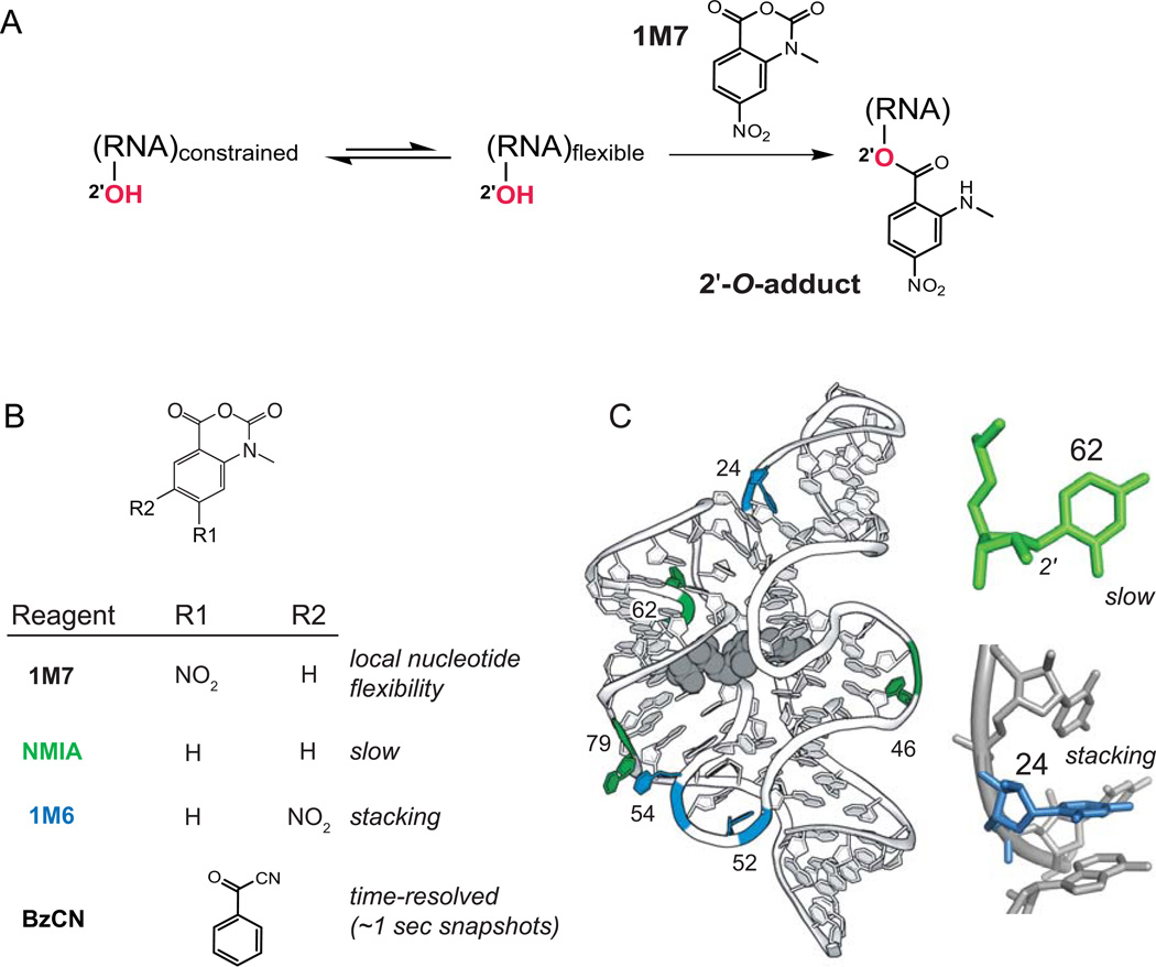
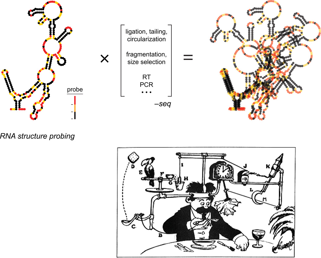
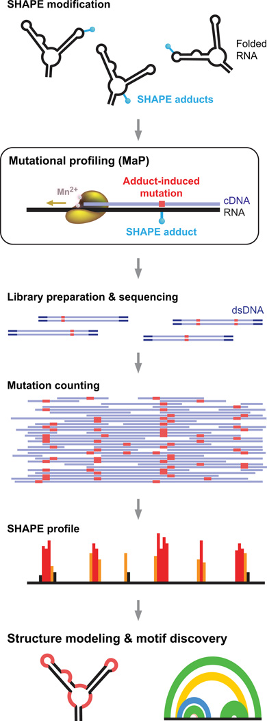
Profound insights regarding nucleic acid structure and function can be gleaned from very simple, direct, and chemistry-based strategies. Our approach strives to incorporate the elegant physical insights that Don Crothers instilled in those who trained in his laboratory. Don emphasized the advantages of focusing on direct and concise experiments even when the final objective was to understand something complex-potentially including the large-scale architectures of the genomes of RNA viruses and the transcriptomes of cells. Here, the author reviews the intellectual path, and a few detours, that led to the development of the SHAPE-MaP and RING-MaP technologies for interrogating RNA structure and function at large scales. The author also argues that greater attention to creating direct, less inferential experiments will convert "omics" investigations into lasting and definitive contributions to our understanding of biological function.

Keywords: RNA structure; chemical probing; concision; structure modeling.


Figures

Figure 1
Figure 1
Representative Crothers’ laboratory work that emphasized simple and direct physical ideas and experimentation to understand biological macromolecules. (A) The Zimm-Crothers viscometer, (B) model for understanding the effect of polyvalency on antibody-ligand interactions, (C) model for drug-DNA intercalation based on measurements using equilibrium dialysis, and (D) the Fried-Crothers electrophoretic mobility shift assay.
Figure 2
Figure 2
SHAPE chemistry monitors local nucleotide dynamics and non-canonical interactions. (A) Mechanism of RNA SHAPE chemistry to form 2'-O-adducts at flexible nucleotides. (B) Core set of useful SHAPE reagents. (C) Structural contexts for nucleotides exhibiting NMIA (green) and 1M6 (blue) enhancements superimposed on a model for the TPP riboswitch aptamer domain; the bound ligand shown in gray. Structure illustrations for the TPP riboswitch are based on 2gdi.
Figure 3
Figure 3
SHAPE reactivities used to understand the structure and ligand-induced folding of the TPP riboswitch aptamer domain. (A) Superposition of absolute SHAPE reactivities. (B) Changes in local nucleotide dynamics induced upon binding by the TPP ligand.
Figure 4
Figure 4
Accuracy of SHAPE-directed modeling of secondary structure. Secondary structure modeling accuracies reported as a function of sensitivity (sens) and positive predictive value (ppv) for calculations performed without experimental constraints (no data), with SHAPE data obtained with the 1M7 reagent, or with three-reagent data. Results are colored on a scale to reflect low (red) to high (green) modeling accuracy.
Figure 5
Figure 5
Illustration of the challenges in melding a structure probing experiment with readout by massively parallel sequencing. The many intervening steps (in brackets) impose selection on the final data that, in essence, blur and conflate the results of the original experiment. Lower panel shows a comparative example of a complex process used to complete a task. Illustration is "Professor Butts and the Self-Operating Napkin" by Rube Goldberg.
Figure 6
Figure 6
SHAPE-MaP overview. RNA is treated with a SHAPE reagent that reacts at conformationally dynamic nucleotides. Specialized reverse transcription conditions allow the polymerase to read through chemical adducts in the RNA and to record the site as a nucleotide non-complementary to the original sequence (red) in the cDNA. The resulting cDNA is subjected to massively parallel sequencing to create a mutational profile. Sequencing reads are then converted to a SHAPE reactivity profile. SHAPE reactivities can be used to model secondary structures, visualize competing and alternative structures, discover functional RNA motifs, and quantify any process that modulates local nucleotide RNA dynamics. Figure reprinted from Ref. .
Figure 7
Figure 7
SHAPE-MaP analysis of the HIV-1 RNA genome (NL4-3 strain). (A) SHAPE reactivities. Reactivities are shown as the centered 55-nt median window, relative to the global median. (B) Shannon entropies. (C) Pairing probabilities. Arcs representing base pairs are colored by their pairing probabilities, with green arcs indicating highly probable helices. Areas with overlapping arcs have multiple potential structures. Black arcs indicate pseudoknots (PK). (D) RNA regions with known biological functions. Bars and blue shading enclose low SHAPE (highly structured) and low Shannon entropy regions; these regions overlap with known RNA functional motifs much more frequently than expected by chance. (E) Secondary structure models for regions identified de novo. Names of known structures are given. Figure adapted from Ref. .
Figure 8
Figure 8
RING-MaP overview. (A) Detection of through-space structural communication. RNA molecules experience local structural variations in which nucleotides become reactive to a chemical probe in a correlated way. Statistical association analysis detects these interdependencies. (B) Analysis of multiple coexisting RNA conformations based on clustering analysis. Initial RING-MaP experiments were performed with the RNA-modifying reagent dimethyl sulfate (DMS).
Figure 9
Figure 9
Through-space RNA structural relationships revealed by single-molecule correlated chemical probing. Direct, through-helix, and global internucleotide interactions are illustrated on both (A) secondary structure and (B) three-dimensional models of the RNase P catalytic domain. Each class of interaction is indicated by a line of a distinct color. Conventional tertiary interactions are red; non-canonical base pairs are magenta; through-helix interactions are yellow and orange; coupled tertiary interactions are blue; and helix packing is green. The RNase P structure is from model 3dhs.
Figure 10
Figure 10
Multiple in-solution conformations for TPP riboswitch in the absence of TPP ligand. (A) Spectral clustering analysis. There are two clusters in the no-ligand state. The major cluster (red) reflects an unstructured state with few internucleotide interactions. The minor cluster (blue) is more highly structured than the major cluster specifically in the region of the thiamine binding pocket (blue closed circles). (B, C) Nucleotides that are more structured in the minor cluster – which flank the thiamine-binding pocket – are emphasized in blue on RNA secondary and tertiary structure models. The thiamine moiety is gray in panel C.

References

    1. Zimm BH, Crothers DM. Simplified rotating cylinder viscometer for DNA. Proc Natl Acad Sci U S A. 1962;48:905–911. - PMC - PubMed
    1. Citation numbers were retrieved from Google Scholar on 1 Oct 2014.

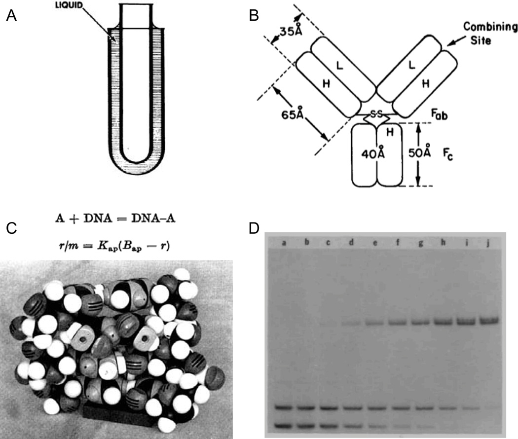
    1. Crothers DM, Metzger H. The influence of polyvalency on the binding properties of antibodies. Immunochemistry. 1972;9:341–357. - PubMed
    1. Müller W, Crothers DM. Studies of the binding of actinomycin and related compounds to DNA. J Mol Biol. 1968;35:251–290. - PubMed
    1. Chaires JB, Dattagupta N, Crothers DM. Studies on interaction of anthracycline antibiotics and deoxyribonucleic acid: equilibrium binding studies on interaction of daunomycin with deoxyribonucleic acid. Biochemistry. 1982;21:3933–3940. - PubMed

Publication types

LinkOut - more resources