

Tetrakis(p-carboranylthio-tetrafluorophenyl)chlorin (TPFC): application for photodynamic therapy and boron neutron capture therapy
- PMID: 25546823
- PMCID: PMC4415589
- DOI: 10.1002/jps.24317
Tetrakis(p-carboranylthio-tetrafluorophenyl)chlorin (TPFC): application for photodynamic therapy and boron neutron capture therapy
Abstract
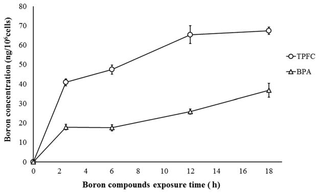
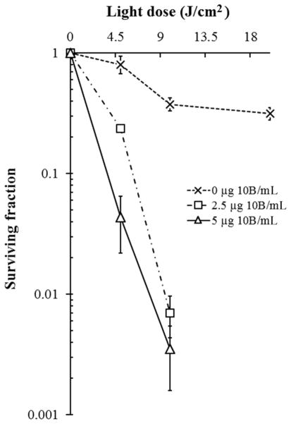
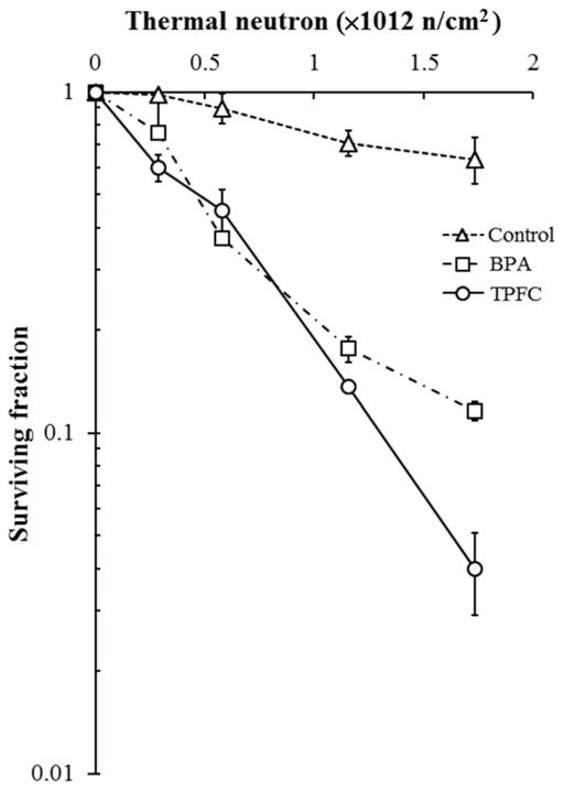
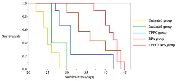
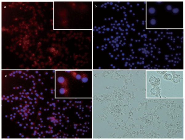
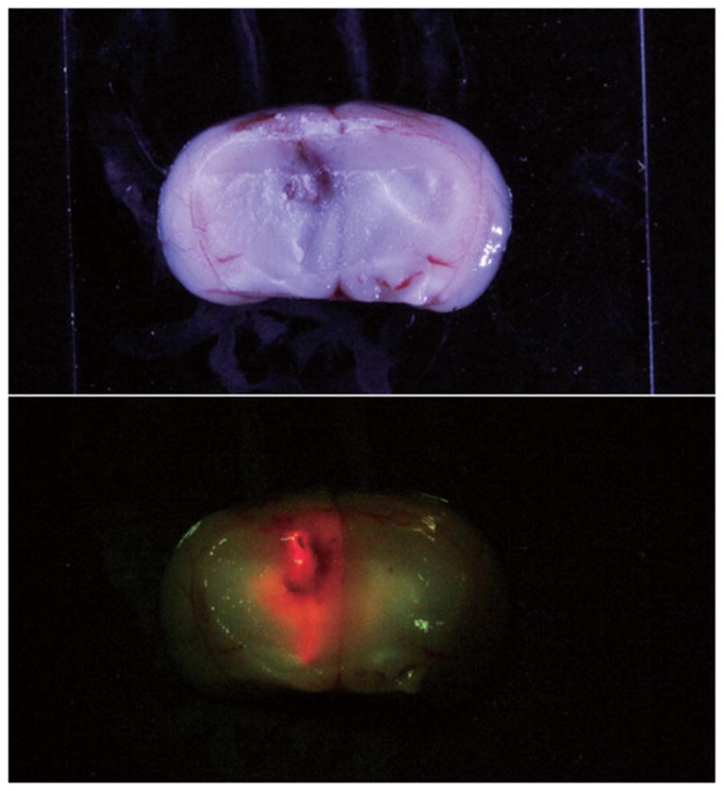
Carboranyl-containing chlorins have emerged as promising dual sensitizers for use in both photodynamic therapy (PDT) and boron neutron capture therapy (BNCT), by virtue of their known tumor affinity, low cytotoxicity in dark conditions, and their strong absorptions in the red region of the optical spectrum. Tetrakis(p-carboranylthio-tetrafluorophenyl)chlorin (TPFC) is a new synthetic carboranyl-containing chlorin of high boron content (24% by weight). To evaluate TPFC's applicability as sensitizer for both PDT and BNCT, we performed an in vitro and in vivo study using F98 rat glioma cells and F98 rat glioma-bearing brain tumor models. For the in vivo BNCT study, we used boronophenylalanine (BPA), which is currently used in clinical BNCT studies, via intravenous administration (i.v.) and/or used TPFC via convection-enhanced delivery (CED), a method for local drug infusion directly into the brain. In the in vitro PDT study, the cell surviving fraction following laser irradiation (9 J/cm(2) ) was 0.035 whereas in the in vitro BNCT study, the cell surviving fraction following neutron irradiation (thermal neutron = 1.73 × 10(12) n/cm(2) ) was 0.04. In the in vivo BNCT study, the median survival time following concomitant administration of BPA (i.v.) and TPFC (CED) was 42 days (95% confidence interval; 37-43 days).
Keywords: BNCT; CED; CNS; PDT; TPFC; absorption; blood-brain barrier; boron-containing chlorin; cell culture; osmotic pumps.
© 2014 Wiley Periodicals, Inc. and the American Pharmacists Association.
Figures
References
-
- Stupp R, Hegi ME, van den Bent MJ, Mason WP, Weller M, Mirimanoff RO, Cairncross JG. Changing paradigms—An update on the multidisciplinary management of malignant glioma. Oncologist. 2006;11(2):165–180. - PubMed
-
- Origitano TC, Caron MJ, Reichman OH. Photodynamic therapy for intracranial neoplasms. Literature review and institutional experience. Mol Chem Neuropathol. 1994;21(2–3):337–352. - PubMed
-
- Kostron H, Obwegeser A, Jakober R. Photodynamic therapy in neurosurgery: A review. J Photochem Photobiol B. 1996;36(2):157–168. - PubMed
-
- Barth RF, Soloway AH, Fairchild RG, Brugger RM. Boron neutron capture therapy for cancer. Realities and prospects. Cancer. 1992;70(12):2995–3007. - PubMed
-
- Barth RF, Coderre JA, Vicente MG, Blue TE. Boron neutron capture therapy of cancer: Current status and future prospects. Clin Cancer Res. 2005;11(11):3987–4002. - PubMed
Publication types
MeSH terms
Substances
Grants and funding
LinkOut - more resources
Full Text Sources
Other Literature Sources
Medical
