. 2015 Jan 15;29(2):157-70.
doi: 10.1101/gad.251785.114. Epub 2014 Dec 29.

Poly-ADP ribosylation of PTEN by tankyrases promotes PTEN degradation and tumor growth

Affiliations

Poly-ADP ribosylation of PTEN by tankyrases promotes PTEN degradation and tumor growth

Nan Li et al. Genes Dev. .

Abstract

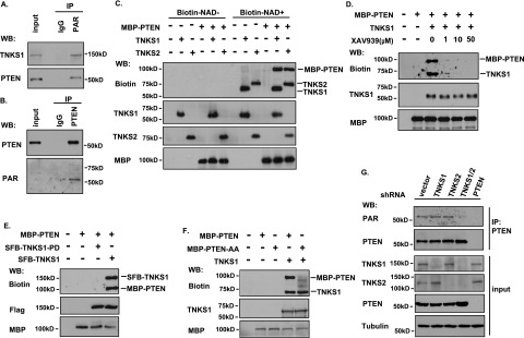
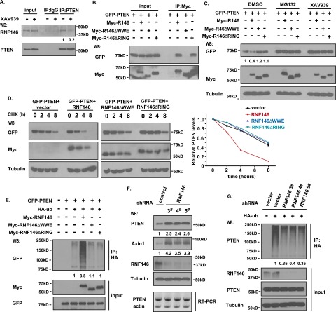
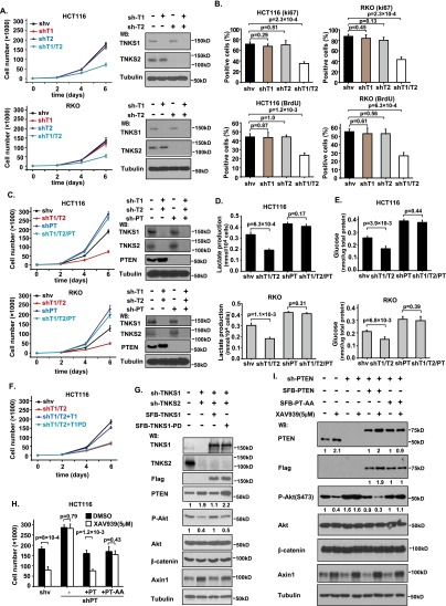
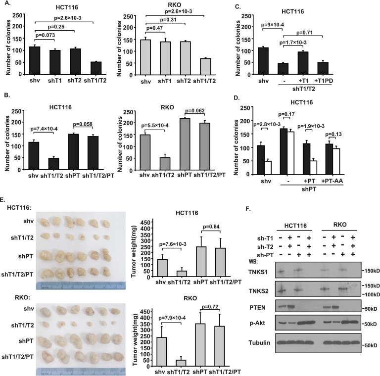
PTEN [phosphatidylinositol (3,4,5)-trisphosphate phosphatase and tensin homolog deleted from chromosome 10], a phosphatase and critical tumor suppressor, is regulated by numerous post-translational modifications, including phosphorylation, ubiquitination, acetylation, and SUMOylation, which affect PTEN localization and protein stability. Here we report ADP-ribosylation as a new post-translational modification of PTEN. We identified PTEN as a novel substrate of tankyrases, which are members of the poly(ADP-ribose) polymerases (PARPs). We showed that tankyrases interact with and ribosylate PTEN, which promotes the recognition of PTEN by a PAR-binding E3 ubiquitin ligase, RNF146, leading to PTEN ubiquitination and degradation. Double knockdown of tankyrase1/2 stabilized PTEN, resulting in the subsequent down-regulation of AKT phosphorylation and thus suppressed cell proliferation and glycolysis in vitro and tumor growth in vivo. Furthermore, tankyrases were up-regulated and negatively correlated with PTEN expression in human colon carcinomas. Together, our study revealed a new regulation of PTEN and highlighted a role for tankyrases in the PTEN-AKT pathway that can be explored further for cancer treatment.

Keywords: PARsylation; PTEN; RNF146; tankyrase; ubiquitination.


Figures

Figure 1.
Figure 1.
PTEN is a tankyrase-binding protein. (A) Schematic representation of wild-type and the double point mutation of PTEN. The putative tankyrase-binding motif RYQEDG is indicated. (B) Alignment of the N terminus of PTEN in different species. The identical RYQEDG motif is indicated in red. (C) TNKS1 and TNKS2 interact with PTEN. 293T cells were transfected with plasmids encoding GFP-tagged PTEN together with vector or plasmids encoding myc-tagged NEDD4, WWP2, TNKS1, or TNKS2. Immunoprecipitation (IP) was carried out using anti-myc antibody and then subjected to Western blotting using the indicated antibodies. (D,E) Interaction between endogenous PTEN and TNKS1. Lysates from HCT116 cells were subjected to immunoprecipitation/Western analysis using the indicated antibodies. (F) The tankyrase-binding motif of PTEN is required for TNKS1/PTEN interaction. 293T cells were cotransfected with constructs encoding myc-tagged TNKS1/TNKS2 together with vector alone or construct encoding SFB-tagged wild-type or the tankyrase-binding motif point mutant of PTEN (PTEN-AA). Immunoprecipitation/Western analysis was conducted using the indicated antibodies.
Figure 2.
Figure 2.
PTEN is ADP-ribosylated by tankyrases in vitro and in vivo. (A,B) PTEN is PARylated in vivo. (A) HCT116 cells were lysed with NETN buffer containing PARG inhibitor ADP-HPD (5 μM) and protease inhibitor. Lysates were then immunoprecipitated using control or anti-PAR antibodies and immunoblotted using the indicated antibodies. (B) HCT116 cells were lysed with NETN denaturing buffer containing PARG inhibitor ADP-HPD (5 μM) and protease inhibitor. Lysates were then immunoprecipitated using control or anti-PTEN antibodies followed by Western blotting as indicated. (C) Ribosylation of PTEN by TNKS1 and TNKS2 in vitro. Recombinant TNKS1, TNKS2, and PTEN were subjected to in vitro ribosylation assays in the absence or presence of biotin-labeled NAD+. The recombinant proteins were detected by the indicated antibodies, and the ribosylated proteins were determined with anti-biotin antibody. (D) The ribosylation of PTEN by TNKS1 is diminished by tankyrase inhibitor XAV939. The recombinant MBP-PTEN and TNKS1 were subjected to an in vitro ribosylation reaction as described above in the absence or presence of the indicated concentrations of XAV939. (E) The catalytic activity of TNKS1 is required for PTEN ribosylation. The MBP-PTEN and immunoprecipitated SFB-tagged wild-type or the catalytically inactive mutant of TNKS1 (TNKS1-PD) were subjected to an in vitro ribosylation reaction followed by Western blotting as indicated. (F) The tankyrase-binding motif of PTEN is required for its ribosylation by TNKS1. The recombinant MBP-PTEN, MBP-PTEN-AA, and TNKS1 were subjected to an in vitro ribosylation assay and analyzed by Western blotting as indicated. (G) Only double knockdown of TNKS1/2 diminishes the ribosylation of PTEN in vivo. HCT116-derived cells with stable knockdown of TNKS1, TNKS2, TNKS1/2, or PTEN were collected and immunoprecipitated using anti-PTEN antibody. The input and immunoprecipitated proteins were analyzed by Western blotting using the indicated antibodies, and the ribosylation of endogenous PTEN was detected by anti-PAR antibody.
Figure 3.
Figure 3.
Tankyrases regulate PTEN protein stability through a ubiquitination-dependent pathway. (A) TNKS1 and TNKS2 promote the degradation of PTEN. 293T cells were transfected with constructs encoding GFP vector, GFP-tagged PTEN, and myc-tagged TNKS1 or TNKS2. Twenty-four hours after transfection, cells were treated with DMSO or 10 μM protease inhibitor MG132 for 6 h. Cell lysates were immunoblotted with the indicated antibodies. (B) TNKS1-mediated PTEN degradation is abolished by tankyrase inhibitor XAV939. 293T cells were transfected with constructs encoding GFP-tagged PTEN and myc-tagged TNKS1. Twenty-four hours after transfection, cells were treated with DMSO, 10 μM MG132 for 6 h, or 5 μM XAV939 for 6 h, and cell extracts were examined by immunoblotting using antibodies as indicated. (C) TNKS1 catalytic activity is required for PTEN degradation. 293T cells were transfected with constructs encoding Myc-tagged PTEN and SFB-tagged TNKS1 or TNKS1-PD. Cell lysates were subjected to Western blotting with the indicated antibodies. (D) Tankyrase inhibition stabilizes wild-type PTEN but not the tankyrase-binding motif double point mutant of PTEN. 293T cells transfected with constructs encoding myc-tagged TNKS1, PTEN, or PTEN-AA were treated with 5 μM tankyrase inhibitor XAV939 for 6 h. Cell extracts were examined by Western blotting as indicated. (E) Double knockdown of TNKS1/2 increases the protein level of PTEN. TNKS1, TNKS2, TNKS1/2 stable knockdown cells and control HCT116 cells were harvested, and cell lysates were analyzed by immunoblotting using the indicated antibodies. (F) TNKS1 and TNKS2 promote the polyubiquitination of wild-type but not tankyrase-binding motif point mutant PTEN in vivo. 293T cells were transfected with constructs encoding HA-ubiquitin, Myc-PTEN, and Myc-PTEN-AA together with constructs encoding SFB-TNKS1 or TNKS2. Cells were treated with 10 μM MG132 for 6 h. Whole-cell lysates were subjected to immunoprecipitation with anti-HA antibody followed by Western blotting using the indicated antibodies. (G) The catalytic activity of TNKS1 is critical for TNKS1-mediated PTEN ubiquitination. 293T cells were transfected with constructs encoding HA-ubiquitin and Myc-PTEN together with constructs encoding SFB-TNKS1 or TNKS1-PD. Cells were treated with MG132, and cell lysates were subjected to immunoprecipitation/Western analysis using the indicated antibodies. (H) Double knockdown of TNKS1/2 suppresses PTEN ubiquitination in vivo. TNKS1, TNKS2, TNKS1/2 stable knockdown cells and control HCT116 cells were transfected with HA-ubiquitin and treated with 10 μM MG132 for 6 h. Cell lysates were immunoprecipitated with anti-HA antibody, and Western blotting was carried out using the indicated antibodies.
Figure 4.
Figure 4.
The E3 ligase RNF146 is involved in tankyrase-mediated PTEN ubiquitination and degradation. (A) Interaction between PTEN and RNF146. HCT116 cells were treated without or with 5 μM XAV939 for 6 h. Cell lysates were immunoprecipitated with anti-PTEN or control antibody followed by Western blotting using the indicated antibodies. (B) The WWE domain of RNF146 is required for its binding to PTEN. 293T cell were transfected with constructs encoding GFP-tagged PTEN together with vector alone or constructs encoding myc-tagged wild-type RNF146, the RNF146-△WWE mutant, or the RNF146-△RING mutant. Immunoprecipitation reactions were conducted using anti-myc antibody and followed by Western blotting analysis. (C) Both the WWE and RING domains of RNF146 are required for PTEN degradation. 293T cells were transfected with constructs as described in B. Twenty-four hours after transfection, cells were treated with DMSO, 10 μM MG132, or 5 μM XAV939 for 6 h. Cell lysates were examined by Western blotting using the indicated antibodies. (D) Overexpression of RNF146, but not of the RNF146-△WWE mutant or the RNF146-△RING mutant, destabilizes PTEN. 293T transfection was conducted as described in B. Cells were treated with 100 μg/mL cycloheximide (CHX) for the indicated times. Protein levels were analyzed by immunoblotting using antibodies as indicated (left panel) and quantified (right panel). (E) RNF146 ubiquitinates PTEN in vivo. 293T cells were transfected with constructs encoding HA-tagged ubiquitin, GFP-tagged PTEN, and myc-tagged wild-type RNF146, the △WWE mutant of RNF146, or the △RING mutant of RNF146. Cells were treated with MG132 before being collected and analyzed by immunoprecipitation/Western blotting using the indicated antibodies. (F) Depletion of RNF146 stabilizes PTEN in vivo. RNF146 stable knockdown cells and control HCT116 cells were harvested, and cell lysates were examined by Western blotting and RT–PCR. (G) Knockdown of RNF146 suppresses PTEN ubiquitination in vivo. RNF146 stable knockdown cells and control cells were transfected with HA-ubiquitin and treated with 10 μM MG132 for 6 h. Cell lysates were immunoprecipitated with anti-HA antibody, and Western blotting was carried out using the indicated antibodies.
Figure 5.
Figure 5.
Tankyrases regulate tumor cell proliferation in a PTEN-dependent manner. (A,B) Double knockdown of TNKS1/2 suppresses cell proliferation. TNKS1, TNKS2, and TNKS1/2 stable knockdown cells and control cells were examined to determine their rate of cell proliferation in HCT116 and RKO cells. (A) Data are means ± SD (n = 3 independent experiments), and knockdown efficiency was validated by Western blot. (B) Ki67 and BrdU staining of these control and HCT116/RKO knockdown cells was performed (top panel), and the percentages of positive cells were summarized (bottom panel). Data are means ± SD (n = 3 independent experiments). (C) Tankyrases regulate cell proliferation in a PTEN-dependent manner. PTEN, TNKS1/2, and PTEN/TNKS1/2 triple stable knockdown cells and control cells were examined to determine their rate of cell proliferation in HCT116 and RKO cells. Data are means ± SD (n = 3 independent experiments), and knockdown efficiency was validated by Western blot. (D,E) Lactate production (D) and glucose uptake (E) in HCT116 and RKO cells by knockdown of TNKS1/2/PTEN. Data are means ± SD (n = 3 independent experiments). (F) Inhibition of cell proliferation by depletion of TNKS1/2 was restored by the expression of wild-type TNKS1 but not the catalytically inactive mutant of TNKS1. shRNA-resistant inducible TNKS1 and TNKS1-PD were transduced into TNKS1/2-depleted HCT116 cells, and cell proliferation rate was measured. Data are means ± SD (n = 3 independent experiments). (G) PTEN protein level in TNKS1/2 knockdown cells was rescued by expression of TNKS1 but not TNKS1-PD. Protein levels in the cells shown in F were determined by Western blotting as indicated. (H) Resistance of PTEN-depleted cells to XAV939 was reversed by the expression of wild-type PTEN but not the PTEN-AA mutant. shRNA-resistant inducible PTEN and PTEN-AA were transduced into PTEN-depleted HCT116 cells, and cell number was determined at day 6 (n = 3). (I) Protein levels in the cells shown in H were determined by Western blotting.
Figure 6.
Figure 6.
Tankyrases are required for tumor growth in a PTEN-dependent manner. (A) Double knockdown of TNKS1/2 suppresses colony formation of HCT116 and RKO cells. TNKS1, TNKS2, and TNKS1/2 stable knockdown cells and control cells were tested in colony formation assays. Colonies viable after 2 wk were counted and analyzed (n = 3). (B) Tankyrases regulate tumor cell colony formation in a PTEN-dependent manner. PTEN, TNKS1/2, and PTEN/TNKS1/2 stable knockdown cells and control cells were tested in colony formation assays. Colonies viable after 2 wk were counted and analyzed (n = 3). (C) Inhibition of colony formation by depletion of TNKS1/2 was restored by the expression of TNKS1 but not TNKS1-PD. shRNA-resistant inducible TNKS1 and TNKS1-PD were transduced into TNKS1/2-depleted HCT116 cells, and cells were tested in colony formation assays (n = 3). (D) Resistance of PTEN-depleted cells to XAV939 was reversed by the expression of wild-type PTEN but not the PTEN-AA mutant as measured by colony formation assays. shRNA-resistant inducible PTEN and PTEN-AA were transduced into PTEN-depleted HCT116 cells, and colonies viable after 2 wk were measured (n = 3). (E) Tankyrases regulate tumor formation in a PTEN-dependent manner. Nude mice 4 to 6 wk old were injected with PTEN, TNKS1/2, and PTEN/TNKS1/2 stable knockdown cells or control cells. The tumor weights of each group are shown, presented as average weights ± SD (21 d after injection; n = 6 mice per group). (F) Immunoblotting of TNKS1, TNKS2, PTEN, p-Akt, and Tubulin in tumor lysates from E.
Figure 7.
Figure 7.
Tankyrases are up-regulated and negatively correlate with PTEN status in human colon tumors. (A) IHC staining of PTEN, Axin1, TNKS1, and TNKS2 in representative normal colon and colon carcinoma specimens on the US Biomax tissue microarrays. Brown staining indicates positive immunoreactivity. Bars, 50 μm. (B) PTEN, Axin1, TNKS1, and TNKS2 expression status in normal colon tissue and colon carcinoma specimens. (C) IHC staining of PTEN, Axin1, TNKS1, and TNKS2 of representative human colon tumor samples. Bars, 50 μm. (D) Correlation between expression status of PTEN/TNKS1, PTEN/TNKS2, Axin1/TNKS1, and Axin1/TNKS2 in human colon tumor samples.

References

    1. Bassi C, Ho J, Srikumar T, Dowling RJ, Gorrini C, Miller SJ, Mak TW, Neel BG, Raught B, Stambolic V. 2013. Nuclear PTEN controls DNA repair and sensitivity to genotoxic stress. Science 341: 395–399. - PMC - PubMed
    1. Berg M, Danielsen SA, Ahlquist T, Merok MA, Agesen TH, Vatn MH, Mala T, Sjo OH, Bakka A, Moberg I, et al. . 2010. DNA sequence profiles of the colorectal cancer critical gene set KRAS–BRAF–PIK3CA–PTEN–TP53 related to age at disease onset. PLoS ONE 5: e13978. - PMC - PubMed
    1. Chang W, Dynek JN, Smith S. 2003. TRF1 is degraded by ubiquitin-mediated proteolysis after release from telomeres. Genes Dev 17: 1328–1333. - PMC - PubMed
    1. Chiang YJ, Nguyen ML, Gurunathan S, Kaminker P, Tessarollo L, Campisi J, Hodes RJ. 2006. Generation and characterization of telomere length maintenance in tankyrase 2-deficient mice. Mol Cell Biol 26: 2037–2043. - PMC - PubMed
    1. Chiang YJ, Hsiao SJ, Yver D, Cushman SW, Tessarollo L, Smith S, Hodes RJ. 2008. Tankyrase 1 and tankyrase 2 are essential but redundant for mouse embryonic development. PLoS ONE 3: e2639. - PMC - PubMed

Publication types

MeSH terms