. 2014 Dec 25;16(1):307-20.
doi: 10.3390/ijms16010307.

USP22 promotes NSCLC tumorigenesis via MDMX up-regulation and subsequent p53 inhibition

Affiliations

USP22 promotes NSCLC tumorigenesis via MDMX up-regulation and subsequent p53 inhibition

Fangbao Ding et al. Int J Mol Sci. .

Abstract

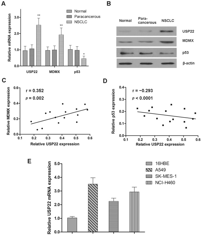
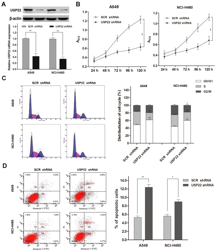
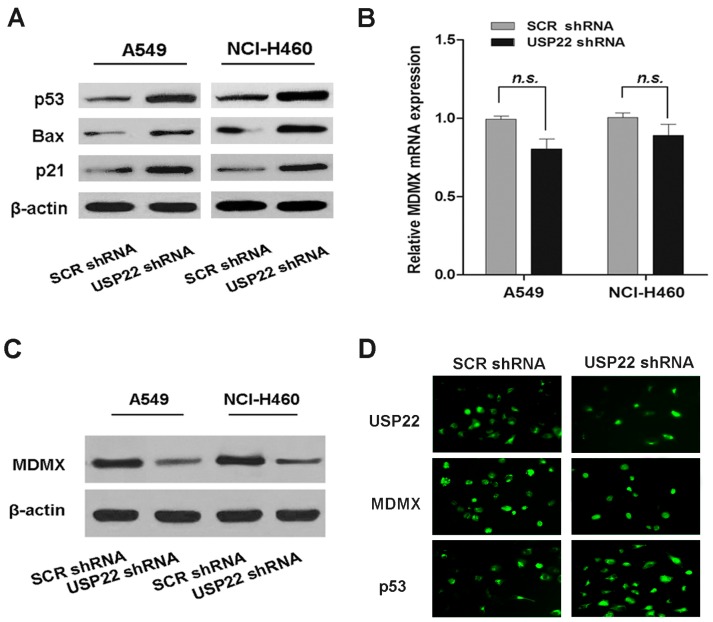
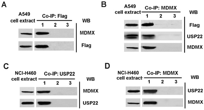
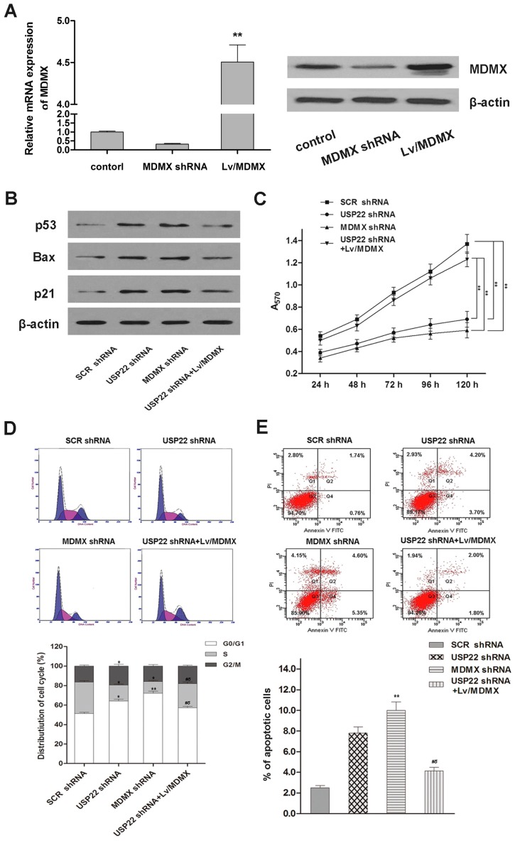
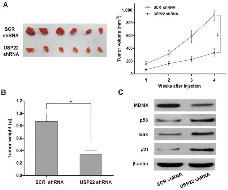
Increasing evidence suggests that ubiquitin-specific protease 22 (USP22) has great clinicopathologic significance in oncology. In this study, we investigated the role of USP22 in human NSCLC tumorigenesis along with the underlying mechanisms of action. First, we determined the expression of USP22 in human NSCLC, as well as normal tissues and cell lines. We then studied the effects of USP22 silencing by shRNA on NSCLC cell growth in vitro and tumorigenesis in vivo, along with the effect on the p53 pathway. We found that USP22 is overexpressed in human NSCLC tissues and cell lines. USP22 silencing by shRNA inhibits proliferation, induces apoptosis and arrests cells at the G0/G1 phases in NSCLC cells and curbs human NSCLC tumor growth in a mouse xenograft model. Additionally, USP22 silencing downregulates MDMX protein expression and activates the p53 pathway. Our co-immunoprecipitation analysis shows that USP22 interacts with MDMX in NSCLC cells. Furthermore, MDMX silencing leads to growth arrest and apoptosis in NSCLC cells, and over-expression of MDMX reverses the USP22 silencing-induced effects. Taken together, our results suggest that USP22 promotes NSCLC tumorigenesis in vitro and in vivo through MDMX upregulation and subsequent p53 inhibition. USP22 may represent a novel target for NSCLC treatment.


Figures

Figure 1
Figure 1
Expression patterns of USP22, MDMX and p53 in NSCLC. (A,B) The mRNA and protein expressions of USP22, MDMX and p53 in normal, paracancerous and NSCLC tissues (n = 15); (C,D) Pearson correlation analysis of the protein expressions between USP22 and MDMX/p53 in NSCLC tissues; (E) the relative mRNA expression of USP22 in 16HBE and NSCLC cell lines (A549, SK-MES-1 and NCl-H460) normalized to β-actin (n = 3). Data are expressed as the means ± SD. * p < 0.05, ** p < 0.01.
Figure 2
Figure 2
USP22 silencing suppresses proliferation and induces apoptosis and cell cycle arrest in NSCLC cells. (A) USP22 mRNA and protein expressions in A549 and NCl-H460 cells transfected with USP22 shRNA or scrambled control shRNA (SCR shRNA) (n = 3); (B) cell proliferation in transfected A549 and NCl-H460 cells by the MTT assay (n = 3); (C) cell cycle distribution by flow cytometry with PI staining (n = 3). Histograms are representative of three independent experiments; (D) Cell apoptosis by flow cytometry with Annexin V-FITC and PI double staining (n = 3). The values of the Q2 plus Q4 quadrant represent apoptosis rates. Data are expressed as the means ± SD. * p < 0.05 vs. SCR shRNA, ** p < 0.01 vs. SCR shRNA.
Figure 3
Figure 3
USP22 silencing activates the p53 pathway, down-regulates MDMX protein expression and interacts with MDMX in NSCLC cells. (A) The levels of p53 pathway proteins in A549 and NCl-H460 cells transfected with USP22 shRNA or SCR shRNA; (B,C) the mRNA and protein expressions of MDMX in A549 and NCl-H460 cells transfected with USP22 shRNA or SCR shRNA (n = 3). β-actin was used as an internal control. Data are expressed as the means ± SD. n.s. indicates no significant difference; (D) Immunofluorescence (IF) analysis of USP22, MDMX and p53 expression in A549 cells transfected with USP22 shRNA or SCR shRNA.
Figure 4
Figure 4
USP22 interacts with MDMX in NSCLC cells. (A) The cell lysates from A549-USP22 were immunoprecipitated with anti-Flag antibody and immunoblotted with anti-Flag and anti-MDMX antibodies, respectively. Lane 1, Flag-immunoprecipitates; Lane 2, isotype control antibody-immunoprecipitates; Lane 3, PBS-immunoprecipitates; (B) The cell lysates from A549-USP22 cells were immunoprecipitated with anti-MDMX antibody and immunoblotted with anti-MDMX, anti-Flag and anti-USP22 antibodies, respectively. Lane 1, MDMX-immunoprecipitates; Lane 2, isotype control antibody-immunoprecipitates; Lane 3, PBS-immunoprecipitates; (C,D) The cell lysates from NCl-H460 were immune-precipitated with anti-USP22 antibody (C) or anti-MDMX antibody (D) and immunoblotted with anti-Flag and anti-MDMX antibodies, respectively. Lane 1, USP22- or MDMX-immunoprecipitates; Lane 2, isotype control antibody-immunoprecipitates; Lane 3, PBS-immunoprecipitates.
Figure 5
Figure 5
Cell proliferation, cell cycle distribution, cell apoptosis and p53 pathway in A549 cells transfected with SCR shRNA, USP22 shRNA, MDMX shRNA or co-transfected with USP22 shRNA and Lv/MDMX. (A) The mRNA and protein expressions of MDMX; (B) levels of p53 pathway proteins in A549 cells by Western blot; (C) cell proliferation by the MTT assay. ** p < 0.01; (D,E) Cell cycle distribution by flow cytometry with PI staining (D) and cell apoptosis by flow cytometry with Annexin V-FITC and PI double staining (E). * p < 0.05 vs. SCR shRNA, ** p < 0.01 vs. SCR shRNA, # p < 0.05 vs. USP22 shRNA, δ p < 0.05 vs. MDMX shRNA. Data are expressed as the means ± SD, n = 3.
Figure 6
Figure 6
USP22 knockdown inhibits NSCLC tumorigenesis and modulates p53 pathway signals in vivo. (A) Growth of xenograft tumors derived from A549 cells transfected with USP22 shRNA or SCR shRNA in nude mice, ** p < 0.01; (B) The weight of xenograft tumors in nude mice. Data are expressed as the means ± SD, n = 6, ** p < 0.01; (C) Levels of MDMX and p53 pathway proteins in xenograft tumor tissues derived from A549 cells transfected with USP22 shRNA or SCR shRNA.

References

    1. Ferlay J., Shin H.R., Bray F., Forman D., Mathers C., Parkin D.M. Estimates of worldwide burden of cancer in 2008: Globocan 2008. Int. J. Cancer. 2010;127:2893–2917. doi: 10.1002/ijc.25516. - DOI - PubMed
    1. Siegel R., DeSantis C., Virgo K., Stein K., Mariotto A., Smith T., Cooper D., Gansler T., Lerro C., Fedewa S., et al. Cancer treatment and survivorship statistics, 2012. CA Cancer J. Clin. 2012;62:220–241. doi: 10.3322/caac.21149. - DOI - PubMed
    1. Herbst R.S., Heymach J.V., Lippman S.M. Lung cancer. N. Engl. J. Med. 2008;359:1367–1380. doi: 10.1056/NEJMra0802714. - DOI - PMC - PubMed
    1. Mascaux C., Shepherd F.A. Adjuvant chemotherapy after pulmonary resection for lung cancer. Thoracic Surg. Clin. 2013;23:401–410. doi: 10.1016/j.thorsurg.2013.04.005. - DOI - PubMed
    1. Binder D., Hegenbarth K. Emerging options for the management of non-small cell lung cancer. Clin. Med. Insights Oncol. 2013;7:221–234. doi: 10.4137/CMO.S10269. - DOI - PMC - PubMed

Publication types

MeSH terms